Comparative Study
. 2015 Jul;35(7):1339-50.
doi: 10.1097/IAE.0000000000000471.

OUTER RETINAL TUBULATION IN ADVANCED AGE-RELATED MACULAR DEGENERATION: Optical Coherence Tomographic Findings Correspond to Histology

Affiliations
Comparative Study

OUTER RETINAL TUBULATION IN ADVANCED AGE-RELATED MACULAR DEGENERATION: Optical Coherence Tomographic Findings Correspond to Histology

Karen B Schaal et al. Retina. 2015 Jul.

Abstract

Purpose: To compare optical coherence tomography (OCT) and histology of outer retinal tubulation (ORT) secondary to advanced age-related macular degeneration in patients and in postmortem specimens, with particular attention to the basis of the hyperreflective border of ORT.

Method: A private referral practice (imaging) and an academic research laboratory (histology) collaborated on two retrospective case series. High-resolution OCT raster scans of 43 eyes (34 patients) manifesting ORT secondary to advanced age-related macular degeneration were compared to high-resolution histologic sections through the fovea and superior perifovea of donor eyes (13 atrophic age-related macular degeneration and 40 neovascular age-related macular degeneration) preserved ≤4 hours after death.

Results: Outer retinal tubulation seen on OCT correlated with histologic findings of tubular structures consisted largely of cones lacking outer segments and lacking inner segments. Four phases of cone degeneration were histologically distinguishable in ORT lumenal walls, nascent, mature, degenerate, and end stage (inner segments and outer segments, inner segments only, no inner segments, and no photoreceptors and only Müller cells forming external limiting membrane, respectively). Mitochondria, which are normally long and bundled within inner segment ellipsoids, were small and scattered within shrunken inner segments and cell bodies of surviving cones. A lumenal border was delimited by an external limiting membrane. Outer retinal tubulation observed in closed and open configurations was distinguishable from cysts and photoreceptor islands on both OCT and histology. Hyperreflective lumenal material seen on OCT represents trapped retinal pigment epithelium and nonretinal pigment epithelium cells.

Conclusion: The defining OCT features of ORT are location in the outer nuclear layer, a hyperreflective band differentiating it from cysts, and retinal pigment epithelium that is either dysmorphic or absent. Histologic and OCT findings of outer retinal tubulation corresponded in regard to composition, location, shape, and stages of formation. The reflectivity of ORT lumenal walls on OCT apparently does not require an outer segment or an inner/outer segment junction, indicating an independent reflectivity source, possibly mitochondria, in the inner segments.


Figures
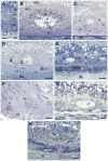
Figure 1
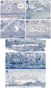
Figure 1. Cellular constituents of ORT in neovascular AMD
Toluidine blue stained, 0.8 μm thick sections of maculas post-fixed by osmium-tannic acid-paraphenylendiamine method. INL, inner nuclear layer; OPL syn, outer plexiform layer, pedicles and spherules; HFL, Henle fiber layer (containing photoreceptor and Müller cell fibers); ONL, outer nuclear layer (R=rods, C=cones); ELM, external limiting membrane; IS, photoreceptor inner segments; my, myoid; el, ellipsoid; OS, outer segments. Bars = 25 μm. A. Photoreceptors at 2.5 mm temporal to the foveal center in a healthy macula. Rod and cone nuclei are distinguishable by size, chromatin patterns, staining, and position relative to ELM. ISmy (-my) is pale. ISel (-el) has darkly stained vertical streaks indicating closely packed, thin mitochondria. M, Müller cell nuclei in the INL. Due to artifactual post-mortem detachment, OS are bent, en masse, in parallel. 85 yr old woman. B. Closed ORT. Cone nuclei (C) surround a lumen delimited by the ELM (white arrowheads). Cone IS, some with OS, protrude into the lumen, maintaining a radial organization with respect to the lumen center. IS have mitochondria-containing ellipsoids and less frequently, myoids (one cell containing both indicated by black arrowheads). One cell with green-staining lipofuscin granules is in the lumen (red arrow). HFL contains darkly stained cone fibers in cross-section. Sc, fibrocellular scar. Green arrowheads, Bruch's membrane, which is breached in this panel.79 yr old man. C. Cone cell bodies (arrowheads), spheroid in shape, are retracted from the lumen yet contain mitochondria. 83 yr old man. D. Apparent rod cell bodies (flanking R) as well as cones (flanking C) in a closed ORT. E, erythrocytes; Sc, fibrocellular scar secondary to CNV. 79 yr old man. E. Three ORT recognizable by ELM border only (arrows) and lacking IS protruding into the lumen; RPE, retinal pigment epithelium; *, persistent basal laminar deposit overlying neo-capillaries; Sc, fibrocellular scar subsequent to type I CNV; 82 yr old woman. F. Three end-stage ORT (arrowheads). Cells contributing to ELM are all Müller cells, as no photoreceptors survived in this desolate macula. 90 yr old woman. G. Cells in ORT lumen are RPE-derived with green-staining lipofuscin granules (right) and non-RPE derived (left) overlying ghost choriocapillaries. Arrowheads, Bruch's membrane; 80 yr old woman. H. Nucleated RPE cell within an ORT lumen; RPE*, RPE-derived cells with spherical melanosomes; Sc, fibrovascular scar; 87 yr old man.
Figure 2
Figure 2. Sub cellular constituents of ORT visible by transmission electron microscopy
Green arrows point to ELM, and red arrowhead point to mitochondria, in all panels. A. Photoreceptors at 2.5 mm temporal to the foveal center in a healthy macula. Electron dense mitochondria (red arrowheads) are thin and tightly packed in the ISel (Inset, magnified box.) 85 yr old woman. B. Degenerate ORT containing macrophage (asterisk) in lumen at 3.2 mm temporal to the foveal center. Mitochondria translocated from ISel to area between ELM (green arrows) and cone nucleus. Cone lipofuscin (yellow arrowheads) are electron dense granules. Inset, mitochondria in the same photoreceptor on an adjacent section. 85 yr old woman. C. Cone in ORT showing mitochondria clustered between ELM and nucleus (between red arrowheads); located 3.2 mm from foveal center. D. ORT denoted by continuous ELM containing Müller cell and photoreceptor cross sections; located at 2.4 mm nasal to foveal center. Inset, magnified box of ELM, Müller cell microvilli in cross-section, and mitochondria. 79 yr old man. Inset scale bars 1 μm. Other scale bars 5 μm.
Figure 3
Figure 3. ORT forms, differentials, and biogenesis by histology
Neovascular AMD, except where noted. Histological preparation details and labeling conventions are described in Figure 1 caption. Bars A-D, 20 μm; bars E-G, 50 μm. A. Closed ORT have circular or oval cross-sections, with an ELM border and photoreceptors that completely encircle the lumen. ORT are distinguishable from cysts, which lack an ELM and an organized arrangement of surrounding cells and contains predominantly fluid. RPE is multilayered and overlies persistent rivulet basal laminar deposits with neovascularization (BLamD/NV). Basal linear deposit is apparent on the surface of Bruch's membrane (arrowheads) 87 yr old woman. B. Open ORT has horizontally elongated cross-sections, curving ELM at the ends, and an absence of photoreceptors on the outer aspect; arrow, cone lipofuscin; 77 yr old woman. C. Asymmetric ORT has photoreceptors on a lateral or outer aspect of the lumen only; C, cone nuclei. Müller cell nuclei in INL are circled. Arrows, cone lipofuscin. Sc, fibrovascular scar; 79 yr old man. D. Open ORT with a lemniscate cross-sectional profile attributed to a branch point. ORT has various cells in lumen and Müller cells with watery cytoplasm. E. Flat, closed ORT with cone nuclei (C) and ISel mitochondria, which have retracted inwardly from the ELM (green arrowhead) and from the IS. Spheroid cones have piled up on the outer aspect. Arrow, cone lipofuscin; 87 yr old woman F. Surviving island of photoreceptors, not considered an ORT because the ELM (small arrow) is not curved at the ends. Yellow circle, intra-retinal RPE; large arrow, cone lipofuscin; RPE, hyper-pigmented RPE-derived cells within sub-retinal fibrocellular scar; B Lam D, persistent basal laminar deposit overlying neovascular membrane; 94 yr old woman. G. An open ORT exhibits different stages of maturity along the lumenal wall, suggesting ORT dynamism. On the right are nearly continuous RPE, IS with myoid and ellipsoids, moderately retracted cone nuclei, and remnant OS. On the left are RPE fragments, IS only with mitochondria retracted behind the ELM, and cone nuclei retracted to a more inward level relative to the right. Persistent basal laminar deposits and neuroglial scar underlie RPE throughout. D, calcified druse. Arrow, cone lipofuscin. 87 yr old man with geographic atrophy.
Figure 4
Figure 4. ORT shapes and differentials vs other outer retinal structures in advanced AMD
All images were obtained in a Spectralis OCT (with signal averaging). All OCT images are shown in the 1:1 pixel mode for better illustration. A. Two circular cross-sections (orange arrows) with internal hypo-reflectivity (lumen) and bounded by a thick hyper-reflective border, located in the outer nuclear layer (ONL) overlying a fibrovascular scar. B. Next to a circular ORT with a hyper-reflective border is a circular cross-section with internal hypo-reflectivity lacking a hyper-reflective border (orange arrow), which is either a cyst or an end-stage ORT. C. A circular cross-section with 360° of hyper-reflective border is a closed ORT (orange arrow). An ovoid cross-section lacking hyper-reflectivity on its outer aspect (adjacent to the scar) is an open ORT (green arrow). D. A large and ovoid cross-section with hyper-reflective material attached to the upper part of the hyper-reflective border and intra-lumenal free floating hyper-reflective material (large ORT). E-F. ORT show different forms. E. A flat and ovoid cross-section with a hyper-reflective border (orange arrow) has internal hyper-reflective material in the lumen. A circular cross-section has a hyper-reflective border (green arrow). F. A cross section of a very small (42 μm in horizontal diameter) ORT with a hyper-reflective border (yellow arrow). G. A cross-section showing a preserved hyper-reflective band (ellipsoid zone, two orange arrows), where the correct orientation of the photoreceptors and their interdigitation with the RPE is still present (photoreceptor island). H. A cross-section showing a forming tubulation (forme fruste ORT) with a free edge to scroll (orange arrow) next to an ovoid cross-section with internal hypo reflectivity lacking a hyper-reflective border, located in the outer plexiform layer (cyst, green arrow). Circular cross-section with a thick hyper-reflective border (pink arrow) in the outer nuclear layer (ORT).
Figure 5
Figure 5
ORT internal contents, associated structures, and formation. A. A cross section with a partial hyper-reflective line (orange arrow; asymmetric ORT). B – D. Cross-sections of well-defined round homogenously hyper-reflective clouds (orange arrow) at a variable distance from ORT. E. A Cross-section with internal hypo-reflectivity bordered by a hyper-reflective border, (no lumenal content). F. A cross-section showing hyper-reflective material attached to the hyper-reflective border at the upper part of the ORT (orange arrow). G – H. Cross-sections showing intra- luminal free floating hyper-reflective material of different sizes and shapes (orange arrows). I – K. Cross sections showing different stages in ORT formation (J and K are scans 77 μm apart in the same patient). I. External limiting membrane (ELM) circles and thickens (free edge scrolling, orange arrow). J. More advanced stage in ORT-formation with further scrolling of the free edge (orange arrow), and visibly separating the lumen from the underlying scar by ‘embedding’ the ORT in the ONL. K. Circular cross-section of an ORT with a visible 360° hyper-reflective band (closed and completely formed ORT). L. En face OCT showing ORT branch. Blue line indicates B-scan shown in M. M. Scan through the ORT branch shown in L (Two lumen are shown).

References

    1. Curcio CA, Medeiros NE, Millican CL. Photoreceptor loss in age-related macular degeneration. Invest Ophthalmol Vis Sci. 1996;37:1236–49. - PubMed
    1. Tsukamoto Y, Masarachia P, Schein SJ, Sterling P. Gap junctions between the pedicles of macaque foveal cones. Vision Res. 1992;32:1809–15. - PubMed
    1. Drasdo N, Millican CL, Katholi CR, Curcio CA. The length of Henle fibers in the human retina and a model of ganglion receptive field density in the visual field. Vision Res. 2007;47:2901–11. - PMC - PubMed
    1. Polyak SL. The Vertebrate Visual System. Chicago: University of Chicago; 1957.
    1. Zweifel SA, Engelbert M, Laud K, et al. Outer retinal tubulation: a novel optical coherence tomography finding. Arch Ophthalmol. 2009;127:1596–602. - PubMed

Publication types