. 2015 Jan 30:6:5962.
doi: 10.1038/ncomms6962.

Endothelial destabilization by angiopoietin-2 via integrin β1 activation

Affiliations

Endothelial destabilization by angiopoietin-2 via integrin β1 activation

Laura Hakanpaa et al. Nat Commun. .

Abstract

Angiopoietins regulate vascular homeostasis via the endothelial Tie receptor tyrosine kinases. Angiopoietin-1 (Ang1) supports endothelial stabilization via Tie2 activation. Angiopoietin-2 (Ang2) functions as a context-dependent Tie2 agonist/antagonist promoting pathological angiogenesis, vascular permeability and inflammation. Elucidating Ang2-dependent mechanisms of vascular destablization is critical for rational design of angiopoietin antagonists that have demonstrated therapeutic efficacy in cancer trials. Here, we report that Ang2, but not Ang1, activates β1-integrin, leading to endothelial destablization. Autocrine Ang2 signalling upon Tie2 silencing, or in Ang2 transgenic mice, promotes β1-integrin-positive elongated matrix adhesions and actin stress fibres, regulating vascular endothelial-cadherin-containing cell-cell junctions. The Tie2-silenced monolayer integrity is rescued by β1-integrin, phosphoinositide-3 kinase or Rho kinase inhibition, and by re-expression of a membrane-bound Tie2 ectodomain. Furthermore, Tie2 silencing increases, whereas Ang2 blocking inhibits transendothelial tumour cell migration in vitro. These results establish Ang2-mediated β1-integrin activation as a promoter of endothelial destablization, explaining the controversial vascular functions of Ang1 and Ang2.


Figures

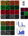
Figure 1
Figure 1. Ang2 reduces endothelial monolayer integrity in Tie2-dependent and -independent manners.
(a) BECs were transduced with scramble (Scr) or Tie2 shRNA lentiviruses, fixed and stained for filamentous actin (F-actin) and VE-cadherin. (b) BECs were transduced with Scr, Tie2, Tie1 or Ang2 shRNA lentiviruses, and the cell lysates were analysed by western blotting using the indicated antibodies. (c) Quantification of the percentage of cells displaying actin stress fibres (% of stress fibre+ cells) (3 microscopic images/experiment, analysis of 60 cells/lentiviral transduction, n=3 independent experiments, P=0.04, Dunnet’s test). (d) Quantification of VE-cadherin area/nuclei in Scr or Tie2-silenced BECs (5 images/experiment, n=3 independent experiments, P=0.01, Dunnet’s test). (e) BECs seeded on fibronectin-coated Transwell inserts were treated with anti-Ang2 antibody or control hIg, and the GFP-expressing LNM-35 cancer cells transmigrated in 5 or 9 h were counted (percentage of transmigrated anti-Ang2 versus control antibody-treated cells, n=3 independent experiments, each performed in triplicate, P=0.05 (5 h), P=0.01 (9 h), Student’s T-test). (f) LLC-GFP cancer cells transmigrated during 9 h through a BEC monolayer transduced with Scr or Tie2 shRNA lentiviruses were quantified as the GFP-positive area (n=3 independent experiments, each in duplicate or triplicate, P=0.007, Student’s T-test). (g) BECs were transduced with Scr, Tie2, Ang2 or Tie2+Ang2 shRNA lentiviruses, fixed and stained for F-actin. Quantification of the percentage of cells displaying actin stress fibres (5–7 microscopic images/experiment, 500 cells/transduction analysed, n=4 independent experiments, P=0.001, Dunnet’s test). (h) Q-RT–PCR analysis of Ang1, Ang2 and Ang4 messenger RNA (mRNA) expression in BECs and HeLa cells. Shown is mRNA copy number in 10 ng total RNA. ND=mRNA not detected. (i) Representative images of Scr, Tie2, Ang2 and Tie2+Ang2-silenced BECs. Mean and s.d. *P<0.05, **P<0.01, ***P<0.005. Scale bars, 20 μm. Nuclear 4′,6-diamidino-2-phenylindole stain. Confocal microscopic images.
Figure 2
Figure 2. Ang2 promotes β1-integrin activation and interferes with fibronectin fibrillogenesis in Tie2-silenced endothelial cells.
(a) BECs were transduced with scramble (Scr), Tie2, Ang2 or Tie2+Ang2 shRNA lentiviruses, fixed and stained for filamentous actin (F-actin), active β1-integrin (12G10) and Tie2. Note the presence of central, active β1-integrin-positive elongated matrix adhesions in Tie2-silenced BECs, and the peripheral localization of active β1-integrin, overlapping with cortical actin in Scr, Ang2 and Tie2+Ang2 double-silenced cells (dashed circle). Magnification of the boxed area is shown in the lowest row. (b) BECs transduced with Scr or Tie2 shRNA lentiviruses were allowed to adhere for 30 min on fibronectin or BSA, fixed and stained for F-actin, active β1-integrin and Tie2. Note the presence of active β1-integrin-positive matrix adhesions in the central half of the total cell area (boxed area, dashed line) in Tie2-silenced, but not in Scr-transduced, BECs. (c) Quantification of β1-integrin-positive matrix adhesion sites normalized to the central 50% of cell area used for quantification (n=3 independent experiments for Scr and Tie2#4, 45 cells/transduction analysed, P=0.01; n=2 for Tie2#5, 30 cells analysed, 10 microscopic fields/experiment analysed, P=0.07). Mean and s.d. *P<0.05. Student’s T-test. (d) BECs on vitronectin were transduced with Scr or Tie2 shRNA lentiviruses in growth media containing fibronectin-depleted serum. The cells were stained for F-actin and fibronectin. Nuclear Hoechst stain. Projections of confocal z-stacks. Scale bars, 20 μm.
Figure 3
Figure 3. Silencing of β1-, but not αv-, integrin rescues cortical actin cytoskeleton in Tie2-silenced BECs.
(a) BECs were transduced with scramble (Scr), Tie2, β1-integrin or Tie2+β1-integrin shRNA lentiviruses, fixed and stained for filamentous actin (F-actin), total β1-integrin and Tie2. β1-integrin-positive adhesions (arrows) are reduced, but not completely abolished in β1-integrin-silenced cells. Diffusely localized and perinuclear β1-integrin (asterisk) is not detected in β1-integrin-silenced cells. (b) Western blot of Scr and β1-integrin-silenced BEC lysates using the indicated antibodies. (c) BECs were transduced with Scr and Tie2 shRNA lentiviruses for 48 h, treated with control or β1-integrin-blocking antibodies (mab13) (10 μg ml−1) during 32–48 h after transduction and stained for F-actin and active β1-integrin. Mab13 was detected with anti-rat secondary antibodies. (d) Integrin expression in BECs using Q-RT–PCR, relative to β1-integrin expression (set as 100). (e) BECs were transduced with Scr, two different αv-integrin shRNA lentiviruses alone and in combination with Tie2 shRNA, fixed and stained for F-actin, αvβ3-integrin and VE-cadherin. (f) Quantification of the percentage of cells displaying actin stress fibres (% of stress fibre+ cells) in e (number of cells analysed/lentiviral transduction: 307/Scr; 229/Tie2; 242/αv-integrin#9; 315/αv-integrin#9+Tie2; 278/αv-integrin#8; 249/αv-integrin#8+Tie2, n=2). Mean and s.d. *P<0.05, ***P<0.005. NS=not significant, Dunnet’s test. Scale bars, 20 μm. Nuclear 4′,6-diamidino-2-phenylindole stain. Projections of confocal z-stacks.
Figure 4
Figure 4. The N-terminal domain of Ang2, but not of Ang1, activates β1-integrin.
(ac) HeLa cells seeded on fibronectin were stimulated with 4 μg ml−1 rhAng2 (a,b) or rhAng1 (c) for 30 min, or pretreated with β1-integrin-blocking antibody (4 μg ml−1 mab13, a,b) or cilengitide (10 μM, b) for 5 min, and then further stimulated for 30 min with rhAng2. The cells were stained for active β1-integrin (12G10) and for His-tag (a,c) or Ang2 (b). (b) Ang2-positive matrix adhesions were analysed from 7 (mAb13) or 10 (cilengitide) microscopic images/experiment (total of 500 cells/mAb13 treatment and 320 cells/cilengitide treatment, n=3 for mAb13, for cilengitide a representative experiment is shown, repeated three times) P=0.03 for mAb13 and P=0.496 for cilengitide, Student’s T-test. (c) Active β1-integrin was quantified from 7 microscopic images/experiment, total of 350 cells/treatment analysed, n=3, P=0.04 for control versus Ang2, P=0.01 for Ang2 versus Ang1, P=0.99 for control versus Ang1, Dunnet’s test. (d) CHO cells were incubated with fluorescently labelled fibronectin fragment (FN7–10) and with various concentrations of rhAng2 or rhAng1, as indicated. FN7–10 binding to CHO cells was quantified using fluorescence-activated cell sorting, and normalized to total α5β1 levels, as explained in the methods. P=0.0004 for 10 μg ml−1 (150 nM) (n=3) and P=0.007 for 5 μg ml−1 (75 nM) (n=3) concentration of rhAng2 versus FN7–10 only, Dunnet’s test. (e) CHO cells were treated with the indicated concentrations of rhAng1 or rhAng2, and integrin activation measured as in d. (f) Binding of Ang2–Flag to biotinylated β1-integrin ectodomain was measured in triplicate using ELISA, a representative experiment of two independent experiments is shown. (g,h) CHO cells transduced with a control plasmid, or plasmids encoding for flag-tagged Ang2, Ang1–Ang2 (Ang1–2) or Ang2–Ang1 (Ang2–1) chimeric proteins (schematic structures are shown in g) were incubated with FN7–10 and, where indicated, with 10 μg ml−1 rhAng2. FN7–10 binding to CHO cells was quantified as in d. P=0.004 for rhAng2, P=0.002 for Ang2–Flag, P=0.001 for Ang2–1–Flag versus FN only, (n=3, Dunnet’s test). Mean and s.d. *P<0.05, **P<0.01, ***P<0.005. Nuclear Hoechst stain. Projections of confocal z-stacks. Scale bar, 20 μm.
Figure 5
Figure 5. Ang2-mediated β1-integrin activation is inhibited by Tie2 in a kinase-independent manner.
(a) BECs were transduced with Scr or Tie2 shRNA lentiviruses, with or without vectors expressing the membrane-bound form of either mouse Tie2 ectodomain (mTie2-ECD), or of GFP as a control. The cells were fixed and stained for F-actin and mouse Tie2, or for F-actin only (GFP-transduced samples). (b) Quantification of the percentage of stress fibre positive, GFP- or mTie2-positive cells (7 microscopic fields, 400 cells/transduction analysed, P=0.002, n=3 independent experiments). (c) CHO cells transduced with a control vector (CHO-Ctrl) or CHO cells expressing the membrane-bound Tie2 ectodomain (CHO-Tie2-ECD) were incubated with fluorescently labelled fibronectin fragment (FN7–10) and the indicated amounts of rhAng2. FN7–10 binding to CHO and CHO-Tie2-ECD cells, detected by staining for the Tie2 ectodomain, was quantified using fluorescence-activated cell sorting (P=0.002 for CHO-Ctrl stimulated with rhAng2 (10 μg ml−1) and P=0.99 for rhAng2 (10 μg ml−1)-stimulated CHO-Tie2-ECD, both compared with FN only control, n=3). (d) Schematic structures of full-length Tie2 and mTie2-ECD; Ang2 ligand in red. Mean and s.d., Dunnet’s test, ***P<0.005. Confocal microscopic images. 4′,6-Diamidino-2-phenylindole staining of nuclei. Scale bar, 20 μm.
Figure 6
Figure 6. Irregular endothelial cell–cell junctions and increased β1-integrin activation in the aortic endothelium of VEC-tTA/Ang2 mice.
(a) Schematic illustration of the mouse aorta and the different areas (1–3) analysed. (b) Representative en face stainings of VE-cadherin in VEC-tTA/Ang2 transgenic or WT littermate mouse aortic endothelium from the areas indicated. (c) Quantification of VE-cadherin. Note the trend of increased VE-cadherin area in the VEC-tTA/Ang2 (Ang2) transgenic mice (statistically significant in area 2. P=0.03, 3 microscopic images/area, n=3 mice/genotype). (d) Representative images of active β1-integrin stained with mab 9EG7 in the aortas of VEC-tTA/Ang2 and WT mice. Note increased central localization of elongated active β1-integrin-positive matrix adhesions in the VEC-tTA/Ang2 aortas when compared with WT aortas (n=4 mice/genotype). Mean and s.d., Student’s T-test, *P<0.05. Projections of confocal z-stacks. Scale bars, 20 μm.
Figure 7
Figure 7. Localization of Tie2 in the aortic endothelium of wild-type and VEC-tTA/Ang2 mice.
Representative en face stainings of VE-cadherin and Tie2 in VEC-tTA/Ang2 transgenic or WT littermate mouse aortic endothelium from the areas indicated, n= 3 VEC-tTA/Ang2 and n=4 WT mice. Projections of confocal z-stacks. Scale bar, 20 μm, in magnification 10 μm.

References

    1. Augustin H. G., Koh G. Y., Thurston G. & Alitalo K. Control of vascular morphogenesis and homeostasis through the angiopoietin-Tie system. Nat. Rev. 10, 165–177 (2009). - PubMed
    1. Eklund L. & Saharinen P. Angiopoietin signaling in the vasculature. Exp. Cell Res. 319, 1271–1280 (2013). - PubMed
    1. Suri C. et al. Requisite role of angiopoietin-1, a ligand for the TIE2 receptor, during embryonic angiogenesis. Cell 87, 1171–1180 (1996). - PubMed
    1. Jeansson M. et al. Angiopoietin-1 is essential in mouse vasculature during development and in response to injury. J. Clin. Invest. 121, 2278–2289 (2011). - PMC - PubMed
    1. Hayashi M. et al. VE-PTP regulates VEGFR2 activity in stalk cells to establish endothelial cell polarity and lumen formation. Nat. Commun. 4, 1672 (2013). - PMC - PubMed

Publication types