. 2015 Jan 30;11(1):e1004604.
doi: 10.1371/journal.ppat.1004604. eCollection 2015 Jan.

Dengue virus RNA structure specialization facilitates host adaptation

Affiliations

Dengue virus RNA structure specialization facilitates host adaptation

Sergio M Villordo et al. PLoS Pathog. .

Abstract

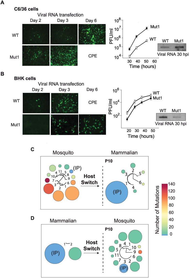
Many viral pathogens cycle between humans and insects. These viruses must have evolved strategies for rapid adaptation to different host environments. However, the mechanistic basis for the adaptation process remains poorly understood. To study the mosquito-human adaptation cycle, we examined changes in RNA structures of the dengue virus genome during host adaptation. Deep sequencing and RNA structure analysis, together with fitness evaluation, revealed a process of host specialization of RNA elements of the viral 3'UTR. Adaptation to mosquito or mammalian cells involved selection of different viral populations harvesting mutations in a single stem-loop structure. The host specialization of the identified RNA structure resulted in a significant viral fitness cost in the non-specialized host, posing a constraint during host switching. Sequence conservation analysis indicated that the identified host adaptable stem loop structure is duplicated in dengue and other mosquito-borne viruses. Interestingly, functional studies using recombinant viruses with single or double stem loops revealed that duplication of the RNA structure allows the virus to accommodate mutations beneficial in one host and deleterious in the other. Our findings reveal new concepts in adaptation of RNA viruses, in which host specialization of RNA structures results in high fitness in the adapted host, while RNA duplication confers robustness during host switching.


Conflict of interest statement

The authors have declared that no competing interests exist.

Figures

Figure 1
Figure 1. Sequence variations at the DENV 3’UTR during experimental host adaptation.
(A) Deep sequencing of viral populations after 10 and 20 passages (P10 and P20) in mosquito and mammalian cells. The area and color of circles represent the frequency and number of mutations of the viral variants, respectively. A schematic fan dendrogram indicating the distance between the variants for each population is also shown. For nucleotide sequences and frequencies of each viral variant also see S1 Fig. (B) Sequence of cloned amplicons corresponding to the complete 3’UTR. For each P10 population obtained, 20 individual clones were sequenced. Cells lines used for adaptation experiments are indicated (mosquito C6/36, mosquito U4.4, and human A549 cells). Mutations are indicated in red and deletions in grey. A conservation plot is presented at the bottom. The three independent experiments in C6/36 cells are indicated on the left (I, II and III).
Figure 2
Figure 2. Fitness parameters and evolution of viral populations after host switch indicate that the DENV 3’UTR is under host-specific selective pressure.
(A) Immunofluorescence and growth kinetics of recombinant virus carrying the 3’UTR of a variant selected in mosquito cells (Mut1) compared with the parental virus (WT) performed in C6/36 cells. Cytopathic effect (CPE) is indicated. Inset shows the accumulation of specific viral RNA. (B) Fitness studies in BHK cells for the two viruses shown in A. (C) and (D) Deep sequencing of viral populations passaged 10 successive times in mammalian or mosquito cells obtained after host switch as indicated. For nucleotide sequences and frequencies of each viral variant also see S4 Fig.
Figure 3
Figure 3. Structural organization of the 3’UTR of dengue virus.
(A) Secondary structure model predicted by conservation and stability. Different DENV type 2 genotypes were analyzed using RNAalifold and RNAz softwares. A plot with the base pairing probability calculated for the A1, A2, A3 and A4 domains (top) and the most stable conserved RNA structures (bottom) are shown. (B) Secondary and tertiary structures assessed by chemical probing. SHAPE reactivity plot (top) and predicted RNA structure model for DENV 3’UTR (bottom) are shown. An unstructured non-conserved region of 23 nucleotides is followed by two SLs (SLI and SLII) and two dumbbell (DB1 and DB2) structures. Four pseudoknots are predicted as indicated (PKI, PKII, PKIII and PKIV). (C) Comparison between structure conservation and SHAPE prediction of DEN-SLI and DEN-SLII. Predicted pseudoknots and additional hybridization at the base of the stem-loops are shown with red and green lines, respectively; indicating the existence of two alternative conformations. (D) Regions with identical sequences in DEN-SLI and DEN-SLII are shown (blue boxes), suggesting a common origin of these two RNA structures.
Figure 4
Figure 4. Nucleotide variations detected in experimentally adapted viruses correlate with mutations found in natural isolates.
(A) Adaptive mutations and deletions were mapped on the RNA structure of the variable region of the viral 3’UTR. Deletions and point mutations rescued from mosquito cell-adapted populations are indicated in grey and red, respectively. Mutation selected in mammalian cells is shown in blue. (B) Conservation plots comparing variable regions of cell adapted viruses and natural isolates. (C) Representation of SL-II RNA structures of DENV genomes from natural isolates corresponding to different serotypes. Sequence alignment plots and secondary RNA structure models are shown for DENV isolates from humans (top) and mosquitoes (bottom).
Figure 5
Figure 5. Requirements of DEN-SLI and DEN-SLII for viral replication in mosquito and human cells.
(A) RNAs of reporter DENVs with deletions ΔSLI, ΔSLII or ΔSLI-II were transfected along with WT and replication impaired NS5M controls into C6/36, BHK and human A549 cells. Normalized luciferase levels are shown using a logarithmic scale at 48 h post transfection. The luciferase values are the mean +/- SD, n = 4. (B) Schematic representation of mutations in the DEN-SLII introduced in the reporter DENV. Red lines indicate substitutions and the circle indicates a deletion. (C) Viral RNA replication as in (A) for DEN-SLII mutants. The luciferase values are the mean +/- SD, n = 4. (D) Competition experiments in A. albopictus mosquitoes indicate a fitness advantage of variants with DEN-SLII mutations. Mosquitoes were injected with a mixture of the two viruses (WT: M132) at a 9:1 ratio and on day 10 individual mosquitoes were collected for viral RNA extraction and sequencing analysis of viral 3’UTRs. Pie charts with frequencies of viruses obtained from each mosquito are shown.
Figure 6
Figure 6. Fitness advantage of RNA structure duplication during DENV host switching.
(A) Normalized luciferase levels expressed by reporter DENV with or without mutations resembling adaptations in mosquito or mammalian cells, respectively, carrying one or two SLs are shown for mammalian cells. The luciferase values are the mean +/- SD, n = 4. (B) The same as in (A) using mosquito cells. (C) Schematic representation of adaptable RNA structures emulating host switching of viruses with single or double SLs. Viruses with a single SL encounter a fitness barrier in the transition from mosquito to mammalian cells due to accumulation of mosquito adaptive mutations, while viruses with double SLs show robustness during host switching.
Figure 7
Figure 7. Models of flavivirus 3’UTR secondary RNA structures.
(A) Comparative analysis of predicted RNA structures of members of the Flavivirus genus. Mosquito-borne, tick-borne, no-know vector and insect-specific flaviviruses are shown. Pseudoknots are indicated with red lines. Common SLs and DB structures are labeled with red and green lines, respectively. Specific yellow fever repeats are indicated with yellow lines and repeats in tick-borne flaviviruses with black and grey lines. See S1 Table for information on sources of sequences used for this analysis. The distance tree was drawn using neighbor joining method and jukes-cantor substitution model. (B) Conserved sequence of SL and DB structures common to all MBFVs are shown.

References

    1. Steinhauer DA, Domingo E, Holland JJ (1992) Lack of evidence for proofreading mechanisms associated with an RNA virus polymerase. Gene 122: 281–288. 10.1016/0378-1119(92)90216-C - DOI - PubMed
    1. Borderia AV, Stapleford KA, Vignuzzi M (2011) RNA virus population diversity: implications for inter-species transmission. Curr Opin Virol 1: 643–648. 10.1016/j.coviro.2011.09.012 - DOI - PubMed
    1. Domingo E, Holland JJ (1997) RNA virus mutations and fitness for survival. Annu Rev Microbiol 51: 151–178. 10.1146/annurev.micro.51.1.151 - DOI - PubMed
    1. Jenkins GM, Rambaut A, Pybus OG, Holmes EC (2002) Rates of molecular evolution in RNA viruses: a quantitative phylogenetic analysis. J Mol Evol 54: 156–165. 10.1007/s00239-001-0064-3 - DOI - PubMed
    1. Woolhouse ME, Taylor LH, Haydon DT (2001) Population biology of multihost pathogens. Science 292: 1109–1112. 10.1126/science.1059026 - DOI - PubMed

Publication types

LinkOut - more resources