Review
. 2015 Jan 28;12(2):1487-547.
doi: 10.3390/ijerph120201487.

Multidrug efflux pumps from Enterobacteriaceae, Vibrio cholerae and Staphylococcus aureus bacterial food pathogens

Affiliations
Review

Multidrug efflux pumps from Enterobacteriaceae, Vibrio cholerae and Staphylococcus aureus bacterial food pathogens

Jody L Andersen et al. Int J Environ Res Public Health. .

Abstract

Foodborne illnesses caused by bacterial microorganisms are common worldwide and constitute a serious public health concern. In particular, microorganisms belonging to the Enterobacteriaceae and Vibrionaceae families of Gram-negative bacteria, and to the Staphylococcus genus of Gram-positive bacteria are important causative agents of food poisoning and infection in the gastrointestinal tract of humans. Recently, variants of these bacteria have developed resistance to medically important chemotherapeutic agents. Multidrug resistant Escherichia coli, Salmonella enterica, Vibrio cholerae, Enterobacter spp., and Staphylococcus aureus are becoming increasingly recalcitrant to clinical treatment in human patients. Of the various bacterial resistance mechanisms against antimicrobial agents, multidrug efflux pumps comprise a major cause of multiple drug resistance. These multidrug efflux pump systems reside in the biological membrane of the bacteria and actively extrude antimicrobial agents from bacterial cells. This review article summarizes the evolution of these bacterial drug efflux pump systems from a molecular biological standpoint and provides a framework for future work aimed at reducing the conditions that foster dissemination of these multidrug resistant causative agents through human populations.


Figures

Figure 1
Figure 1
Bacterial antibiotic resistance mechanisms. Red blocks indicate antibiotics. Yellow channels indicate drug entry ports/porins. Mechanisms of bacterial resistance include the alteration of drug binding targets (DNA); degradation of antibiotics by enzymatic action; expression of efflux pumps on the cell membrane; altered or loss of porin/drug entry ports, the latter two mechanisms of which thereby reduce the intracellular concentration and permeability of the drug into the cell, respectively. This figure was adapted from Kumar and Varela, 2013 [52].
Figure 2
Figure 2
Drug/H+ efflux pump transport mechanism. Starting with an empty pump in which the H+ binding site faces the outside or periplasm and the drug (i.e., substrate) binding site faces the inside or cytoplasm, the drug / proton antiport cycle is as follows: (step 1) The H+ binds the outside face of the empty efflux pump; (step 2) the binding affinity of the pump for the drug substrate increases on the cytoplasmic side; (step 3) the drug binds the inside face of the pump; (step 4) a conformational change occurs such that the drug and H+ binding sites switch sides, i.e., an alternating access mechanism [81] thus essentially translocating both drug and H+ through the pump and across the membrane in opposite directions—the bound drug consequently faces the outside or periplasm, and the bound H+ faces the cytoplasm; (step 5) the drug is released to the outside or periplasm; (step 6) the H+ is then released into the cytoplasm; (step 7) the efflux pump then reorients itself so that the drug binding site now faces the cytoplasm, and the H+ binding site faces the outside or periplasm; (step 8) the empty efflux pump is then ready to begin another drug/H+ antiport cycle. The two α-helical bundles representing the two-fold rotational axis of symmetry during transport [82] are shown in yellow and red. The drug substrate is denoted as S. The protons are blue. The proton-driven drug efflux pump mechanism was adapted from references [83,84,85].
Figure 3
Figure 3
Efflux pumps of Salmonella from four different transporter families. The transport mechanisms, location, families and substrates are shown. Abbreviations used in the figure indicate the following: aminoglycosides (AMG), novobiocin (NVB), sodium dodecyl sulfate (SDS), sodium deoxycholate (SDC), acriflavine (ACF), crystal violet (CV), methylene blue (MB), rhodamine 6G (R6G), benzalkonium chloride (BNKC), nalidixic acid (NAL), tetracycline (TET), chloramphenicol (CLP), norfloxacin (NOR), doxorubicin (DOX), and macrolides (MAC).
Figure 4
Figure 4
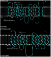
Conserved amino acid sequence motifs A and C of the major facilitator superfamily. The predicted 2D membrane topology structures of the multidrug efflux pumps (A) LmrS from S. aureus [111] and (B) EmrD-3 from V. cholerae [177] are shown. The consensus sequence of the highly conserved motif A [97,208], which resides in the loop between predicted transmembrane helices 2 and 3, is “G X X X (D/E) (R/K) X G X (R/K) (R/K).” Likewise, the consensus amino acid sequence of motif C is “G (X)8 G (X)3 G P (X)2 G G” and resides in the fifth predicted membrane spanning domains of most, if not all, transporters of the MFS [81]. Structures were generated using TMHMM and Tmpres2D servers.
Figure 5
Figure 5
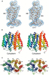
Crystal structure of the EmrD multidrug efflux pump from Escherichia coli. The general features of the three-dimensional structure of EmrD include 12 transmembrane α-helices that zig-zag through the inner membrane, a central channel for drug translocation, and a two-fold axis of rotational symmetry by which the antiporters are believed to mediate conformational changes that occur via an alternating access mechanism of the drug binding site. (A) An electron density map is shown as a stereo image indicating side-chain densities for α-helices 3 and 6 and the cytoplasmic-facing loop between α-helices 6 and 7; (B) The 12 membrane-spanning α-helices are shown in ribbon form as a stereo image; the N- and C-termini face the cytoplasmic side of the inner-membrane; (C) The EmrD structure is shown without loops and from a top perspective looking towards the cytoplasm. The EmrD structure is from Yin, et al. [217].

References

    1. Centers for Disease Control and Prevention Incidence and trends of infection with pathogens transmitted commonly through food—Foodborne diseases active surveillance network, 10 U.S. sites, 1996–2012. MMWR. 2013;62:283–287. - PMC - PubMed
    1. Kuchenmuller T., Hird S., Stein C., Kramarz P., Nanda A., Havelaar A.H. Estimating the global burden of foodborne diseases—A collaborative effort. Eur. Commun. Dis. Bull. 2009;14 doi: 10.1016/j.ijfoodmicro.2013.05.029. - DOI - PubMed
    1. Nyenje M.E., Odjadjare C.E., Tanih N.F., Green E., Ndip R.N. Foodborne pathogens recovered from ready-to-eat foods from roadside cafeterias and retail outlets in Alice, Eastern Cape Province, South Africa: Public health implications. Int. J. Environ. Res. Public Health. 2012;9:2608–2619. doi: 10.3390/ijerph9082608. - DOI - PMC - PubMed
    1. Ghosh M., Wahi S., Kumar M., Ganguli A. Prevalence of enterotoxigenic Staphylococcus aureus and Shigella spp. in some raw street vended Indian foods. Int. J. Environ. Res. Public Health. 2007;17:151–156. doi: 10.1080/09603120701219204. - DOI - PubMed
    1. Teplitski M., Wright A.C., Lorca G. Biological approaches for controlling shellfish-associated pathogens. Curr. Opin. Biotechnol. 2009;20:185–190. doi: 10.1016/j.copbio.2009.03.001. - DOI - PubMed

Publication types

MeSH terms

Substances

LinkOut - more resources