. 2015 Apr 17;10(4):1118-27.
doi: 10.1021/cb500820b. Epub 2015 Jan 30.

Structure-guided design of a high affinity inhibitor to human CtBP

Affiliations

Structure-guided design of a high affinity inhibitor to human CtBP

Brendan J Hilbert et al. ACS Chem Biol. .

Abstract

Oncogenic transcriptional coregulators C-terminal Binding Protein (CtBP) 1 and 2 possess regulatory d-isomer specific 2-hydroxyacid dehydrogenase (D2-HDH) domains that provide an attractive target for small molecule intervention. Findings that the CtBP substrate 4-methylthio 2-oxobutyric acid (MTOB) can interfere with CtBP oncogenic activity in cell culture and in mice confirm that such inhibitors could have therapeutic benefit. Recent crystal structures of CtBP 1 and 2 revealed that MTOB binds in an active site containing a dominant tryptophan and a hydrophilic cavity, neither of which are present in other D2-HDH family members. Here, we demonstrate the effectiveness of exploiting these active site features for the design of high affinity inhibitors. Crystal structures of two such compounds, phenylpyruvate (PPy) and 2-hydroxyimino-3-phenylpropanoic acid (HIPP), show binding with favorable ring stacking against the CtBP active site tryptophan and alternate modes of stabilizing the carboxylic acid moiety. Moreover, ITC experiments show that HIPP binds to CtBP with an affinity greater than 1000-fold over that of MTOB, and enzymatic assays confirm that HIPP substantially inhibits CtBP catalysis. These results, thus, provide an important step, and additional insights, for the development of highly selective antineoplastic CtBP inhibitors.


Figures

Figure 1
Figure 1
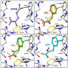
Ligand binding does not induce major conformational changes. (A) Ribbon diagram of the superposition of the PPy complex (orange) and HIPP complex (cyan) to the MTOB complex (grey). MTOB bound CtBP1 does not differ from apo CtBP1. Therefore, the position of the MTOB complex α-carbons were used for comparison with the HIPP (B) and PPy (C and D) structures. Small differences may be the result of different crystallization conditions relative to the MTOB structure. The phenylpyruvate structure contains a canonical (C) and alternate (D) conformation in the hinge conformation between the substrate binding and coenzyme binding domains. The alternate conformation (D) shows the change in position of the Cα of residue A123. The shift in position places A123 in the space normally occupied by the active site water network.
Figure 2
Figure 2
Two different ligand conformations both strongly interact with W318. (A) MTOB positioned in the CtBP1 active site. A single carboxylate oxygen and the carbonyl oxygen orient towards catalytic residue R266. The sulfur atom rests centered over the indole group of W318, positioned 3.8 Å from the ring at its closest distance (disks). (B) Phenylpyruvate assumes two distinct conformations in the crystal. In the substrate conformation (orange) the PPy positions analogous to MTOB for enzyme catalysis. The atoms of the phenyl group range from 3.2 – 4.4 Å in distance to the nearest atom of the W318 indole group (orange disks). For comparison, the non-canonical conformation (green) is semi-transparent. (C) The PPy non-canonical conformation (green) repositions the keto acid core of the molecule while maintaining phenyl ring stacking with W318. In this conformation, the phenyl ring positions at a similar distance to W318 of 3.2 – 4.2 Å (green disks). Both carboxylate oxygens orient towards R266, with the carbonyl positioned near S100 instead of catalytic residue H315. (D) HIPP assumes only the non-canonical conformation due to steric hindrance of the hydroxyimino group in the canonical conformation. The hydroxyimino group orients similar to the carbonyl in the noncanonical phenylpyruvate conformation. The phenyl ring stacks 3.0 – 4.1 Å from W318 (cyan disks).
Figure 3
Figure 3
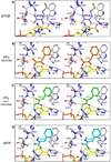
PPy and HIPP hydrogen bond networks and coulombic interactions shown in stereo. (A) The hydrogen bond network (dashes) of substrate MTOB as previously reported served as a comparison for the new structures. (B) The PPy substrate conformation (orange) possesses a similar hydrogen bond network to MTOB (orange dashes). This conformation has lost the hydrogen bond to R97, although PPy maintains proximity for coulombic interactions (yellow dashes). The substrate PPy has an additional hydrogen bond to the nicotinamide ribose. Conformational changes of NAD+ obscure this interaction in the MTOB structure. (C) The noncanonical phenylpyruvate conformation (green) has a distinct hydrogen bond network (green dashes). Orientation of the carboxylate towards R266 maximizes hydrogen bond potential as well as coulombic interactions (yellow dashes) with R97. (D) HIPP (cyan) forms similar hydrogen bonding network (cyan dashes) and coulombic interactions (yellow dashes) to the non-canonical PPy conformation, with the exception of losing the interaction with the nicotinamide ribose.
Figure 4
Figure 4
Calculated van der Waal’s contribution to binding energy by residue. PPy and HIPP exhibit more than a 2-fold increase for the calculated van der Waal’s contact energy with W318 compared to MTOB. The increased contact with W318 contributes to the increased potency of phenyl pyruvate and HIPP over MTOB. The two alternate PPy conformations have been calculated independently as shown in blue and purple.
Figure 5
Figure 5
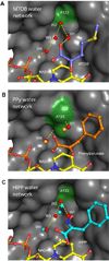
The effects of ligand binding on the CtBP1 water network. (A) MTOB possesses a water network (red spheres connected by yellow dashes) that connect the substrate to an NAD+ phosphate via four water molecules (W1–W4). The conformation of hinge residue A123 (dark green) helps create a cavity for the water molecules unique to CtBP. (B) The water network (orange spheres) in the PPy structure is disrupted by the novel conformation of A123. W2 and W3 are completely displaced as the cavity collapses. W1 is present with the substrate conformation of PPy (orange) and when no molecule is bound in the active site (~60% of the time). (C) The HIPP structure water network is altered by interactions with HIPP. W1 is completely displaced by the HIPP hydroxyl group. W2 has shifted position to interact with HIPP (Figure S4). Due to shifts in both W2 and W3 (now 1.9Å apart), their presence is mutually exclusive. Similar to PPy, HIPP has no effect of W4 position.
Figure 6
Figure 6
Example of single ITC experiments utilizing HIPP with (A) CtBP2 at pH 8.5, (B) CtBP1 at pH 8.5, and (C) CtBP1 at pH 7.5. The slope of the line at the midpoint is steeper at pH 7.5, indicating greater HIPP affinity at these conditions. (Full ITC results are provided in Table 1.)
Figure 7
Figure 7
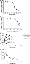
CtBP inhibition assays. IC50 measurements for HIPP (A) and PPy (B) after 15 minutes show that HIPP exhibits an IC50 value more than 100 fold lower than PPy. Data represent n=3 and n=2 triplicate experiments, respectively. Ki plots (C) demonstrate that HIPP inhibition results largely from a decrease in Vmax, which would not be expected for a purely competitive inhibitor. Points represent the average of n=7 reads. MTOB (D) exhibits substrate inhibition when in excess. n=2 independent triplicate experiments. All error bars represent standard deviation.

References

    1. Barroilhet L, Yang J, Hasselblatt K, Paranal RM, Ng SK, Rauh-Hain JA, Welch WR, Bradner JE, Berkowitz RS, Ng SW. C-terminal binding protein-2 regulates response of epithelial ovarian cancer cells to histone deacetylase inhibitors. Oncogene. 2013;32:3896–3903. - PubMed
    1. Birts CN, Harding R, Soosaipillai G, Halder T, Azim-Araghi A, Darley M, Cutress RI, Bateman AC, Blaydes JP. Expression of CtBP family protein isoforms in breast cancer and their role in chemoresistance. Biol Cell. 2010;103:1–19. - PMC - PubMed
    1. Di LJ, Byun JS, Wong MM, Wakano C, Taylor T, Bilke S, Baek S, Hunter K, Yang H, Lee M, Zvosec C, Khramtsova G, Cheng F, Perou CM, Ryan Miller C, Raab R, Olopade OI, Gardner K. Genome-wide profiles of CtBP link metabolism with genome stability and epithelial reprogramming in breast cancer. Nat Commun. 2013;4:1449. - PMC - PubMed
    1. Straza MW, Paliwal S, Kovi RC, Rajeshkumar B, Trenh P, Parker D, Whalen GF, Lyle S, Schiffer CA, Grossman SR. Therapeutic targeting of C-terminal binding protein in human cancer. Cell Cycle. 2010;9:3740–3750. - PMC - PubMed
    1. Wang R, Asangani IA, Chakravarthi BV, Ateeq B, Lonigro RJ, Cao Q, Mani RS, Camacho DF, McGregor N, Schumann TE, Jing X, Menawat R, Tomlins SA, Zheng H, Otte AP, Mehra R, Siddiqui J, Dhanasekaran SM, Nyati MK, Pienta KJ, Palanisamy N, Kunju LP, Rubin MA, Chinnaiyan AM, Varambally S. Role of transcriptional corepressor CtBP1 in prostate cancer progression. Neoplasia. 2012;14:905–914. - PMC - PubMed

Publication types

MeSH terms