. 2015 Apr;29(4):1456-66.
doi: 10.1096/fj.14-266742. Epub 2015 Jan 30.

Kinetic characterization of the sole nonmuscle myosin-2 from the model organism Drosophila melanogaster

Affiliations

Kinetic characterization of the sole nonmuscle myosin-2 from the model organism Drosophila melanogaster

Sarah M Heissler et al. FASEB J. 2015 Apr.

Abstract

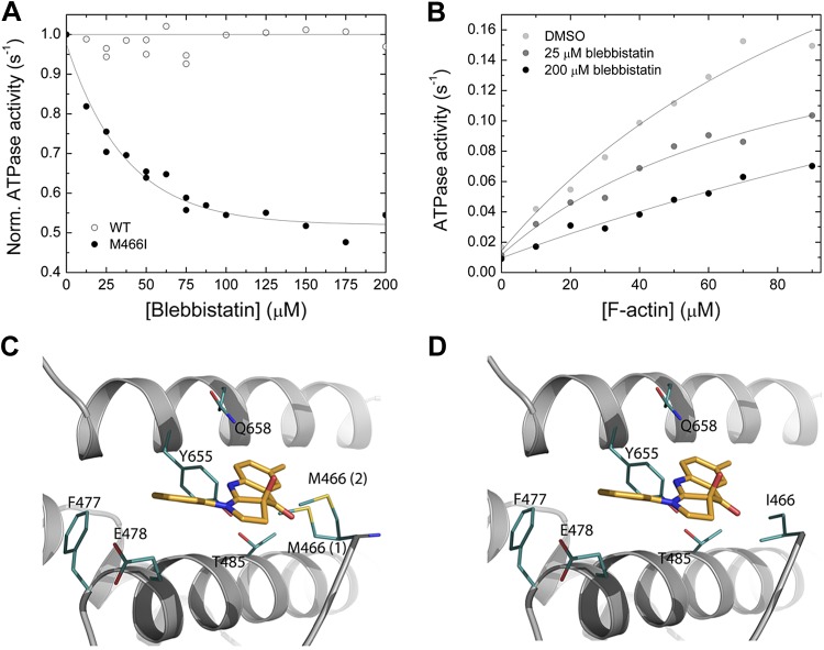
Nonmuscle myosin-2 is the primary enzyme complex powering contractility of the F-actin cytoskeleton in the model organism Drosophila. Despite myosin's essential function in fly development and homeostasis, its kinetic features remain elusive. The purpose of this in vitro study is a detailed steady-state and presteady-state kinetic characterization of the Drosophila nonmuscle myosin-2 motor domain. Kinetic features are a slow steady-state ATPase activity, high affinities for F-actin and ADP, and a low duty ratio. Comparative analysis of the overall enzymatic signatures across the nonmuscle myosin-2 complement from model organisms indicates that the Drosophila protein resembles nonmuscle myosin-2s from metazoa rather than protozoa, though modulatory aspects of myosin motor function are distinct. Drosophila nonmuscle myosin-2 is uniquely insensitive toward blebbistatin, a commonly used myosin-2 inhibitor. An in silico modeling approach together with kinetic studies indicate that the nonconsensus amino acid Met466 in the Drosophila nonmuscle myosin-2 active-site loop switch-2 acts as blebbistatin desensitizer. Introduction of the M466I mutation sensitized the protein for blebbistatin, resulting in a half-maximal inhibitory concentration of 36.3 ± 4.1 µM. Together, these data show that Drosophila nonmuscle myosin-2 is a bona fide molecular motor and establish an important link between switch-2 and blebbistatin sensitivity.

Keywords: actin; blebbistatin; cytoskeleton; rotamer; transient kinetics.


Figures

Figure 1.
Figure 1.
A) Minimum kinetic scheme of the myosin and actomyosin ATPase cycle. KA represents the affinity of myosin for F-actin, KD is the affinity of ADP for myosin, KAD is the affinity of ADP for the actomyosin complex, and KDA is the affinity of F-actin for myosin in the presence of saturating [ADP]. M refers to myosin. The asterisk represents different conformational states of the myosin motor domain detected by the intensity of the intrinsic tryptophan fluorescence. Kinetic parameters in the presence and absence of F-actin are in boldface (k+1 and K1) vs. normal (k+1 and K1) notation. Subscripts A and D refer to actin (KA) and ADP (KD), respectively. Dissociation equilibrium constants are calculated according to Kx = k-x/k+x. B) The overview figure shows the homology model of the Drosophila nonmuscle myosin-2 motor domain in cartoon representation. The nucleotide is in stick representation; switch-1, switch-2, and P-loop are in teal color. Sequence analysis is shown for the conserved structural elements P-loop, switch-1, and switch-2 in the nucleotide binding pocket. Dark gray highlights conserved amino acids. The nonconsensus amino acid Met466 in Drosophila nonmuscle myosin-2 is shown in teal. Dm, Drosophila melanogaster; Dd, Dictyostelium discoideum; Ac, Acanthamoeba castellanii; Sc, Saccharomyces cerevisiae; Ce, Caenorhabditis elegans; Rn, Rattus norvegicus; Mm, Mus musculus; Hs, Homo sapiens. C) Actin-activated steady-state ATPase activity. The dependence of the steady-state ATPase activity on [F-actin] was measured in the concentration range up to 200 µM. The data are corrected for the kbasal. Fitting of the data with the Michaelis-Menten equation yields the parameters kcat = 0.54 ± 0.06 s−1 and Kapp = 152 ± 39 µM.
Figure 2.
Figure 2.
Interaction between myosin and actomyosin and ATP. A) Increasing [ATP] was mixed under pseudo first-order conditions with 0.25 µM myosin in a stopped-flow spectrophotometer. The dependence of kobs on [ATP] is hyperbolic, yielding the parameter k+3 + k-3 of 11.65 ± 0.56 s−1 at saturation. 1/K1 is observed at [ATP] = 3.73 ± 1.32 µM. The inset shows a representative trace of the transient increase in tryptophan fluorescence after mixing 7.5 µM ATP with 0.25 µM myosin. Single-exponential fit to the data yields an amplitude of 14.2% and a kobs of 6.6 s−1. B) Comparison of the second-order rate constants for ATP and dmantATP binding to myosin at low [nucleotide]. The dependence of kobs on [nucleotide] is linear and results in a K1k+2 of 1.16 ± 0.06 µM−1 s−1 (ATP) and K1k+2 to 0.94 ± 0.02 µM−1 s−1 (d-mantATP). A fluorescence transient of the bimolecular interaction between 0.15 µM myosin and 3 µM d-mantATP is shown in the inset of (B). Single-exponential fit to the data set yields an amplitude of 2.8% and a kobs of 3.06 s−1. C) ATP-induced dissociation of the actomyosin complex. Fluorescence signals were obtained after mixing pyrene-actomyosin under pseudo first-order conditions with increasing [ATP] in a stopped-flow spectrophotometer. Hyperbolic fit to the data yields a k+2 of 482.8 ± 7.85 s−1 at saturating [ATP]. 1/K1 is observed at 1224.37 ± 88.39 µM [ATP]. Inset shows a representative transient of the interaction between 0.5 µM pyrene-actomyosin and 7.5 µM ATP. Single-exponential fit to the data set yields an amplitude of 69% and a kobs of 3.18 s−1. D) Comparison of the kobs upon the ATP-induced dissociation of the actomyosin complex. Monitored is either the postmixing pyrene or the light-scattering signal. Linear fits to the data sets up to 30 µM [ATP] define the second-order rate constants K1k+2 of 0.39 ± 0.01 µM−1 s−1 (pyrene), and K1k+2 of 0.34 ± 0.01 µM−1 s−1 (light-scattering).
Figure 3.
Figure 3.
Interaction between myosin and actomyosin with ADP. A) [d-mantADP] dependence of the kobs for myosin (closed circles) or actomyosin (open circles) binding to the fluorescent ADP analog. Linear fits to the data sets yield the second-order binding rate constants k+D of 0.52 ± 0.04 µM−1 s−1 and k+AD of 3.46 ± 0.1 s−1. Extrapolating the data to [d-mantADP] = 0 yields the ADP release rate constant k−D of 2.42 ± 0.13 s−1 and k−AD of 5.19 ± 0.55 s−1, in the absence and presence of F-actin, respectively. B) Representative transients for the ADP release from myosin and actomyosin (inset). Assay conditions were as follows in the absence of F-actin: 1.5 mM ATP versus 0.25 µM myosin and 15 µM ADP. The fluorescence was exited at a wavelength of 297 nm and the fluorescence emission detected after the passage of a 320 nm cutoff filter. The obtained transient was best described by a single exponential function yielding a kobs of 2.35 s−1. The inset shows a light-scattering signal upon the dissociation of a 0.25 µM pyrene-actomyosin complex in the presence of 10 µM ADP with 4 mM ATP. Single-exponential fit of the data yields a kobs of 4.27 s−1.
Figure 4.
Figure 4.
Interaction between myosin and F-actin. A) [F-actin] dependence of kobs for the reaction between myosin and filamentous actin. The second-order binding rate constants k+A of 1.14 ± 0.03 µM−1 s−1 (open circle) and k+DA of 0.26 ± 0.01 µM−1 s−1 (closed circle) in the absence or presence of 100 µM [ADP] were obtained from the linear fits to the data. B) Representative light-scattering signal of the interaction between 0.3 µM myosin and 1.5 µM F-actin. Single-exponential fit of the data yields an amplitude of 1.89% and a kobs of 2.65 s−1. The inset shows an observed F-actin release rate constant kobs of 0.07 s−1 that was obtained after fitting the transient, obtained from chasing 0.25 µM pyrene-actomyosin with 10 µM F-actin, to a single exponential function.
Figure 5.
Figure 5.
Interaction between myosin and blebbistatin. A) Normalized (Norm.) actin-activated steady-state ATPase activity of Drosophila nonmuscle myosin-2 (open circles) and M466I (closed circles) as a function of [blebbistatin]. [F-actin] was kept constant at 30 µM, and the data were normalized to the starting value of the ATPase activity in the absence of inhibitor. Single-exponential fit of the data set gives an IC50 of 36.3 ± 4.1 µM for M466I, whereas no inhibition is observed for the native protein [wild-type (WT)]. B) Blebbistatin-mediated decrease of the steady-state actin-activated ATPase activity. Increasing [blebbistatin] leads to a concentration-dependent decrease in the actin-activated ATP turnover. At 90 µM [F-actin], the ATPase activity is reduced >2-fold. For comparison, the uninhibited control was supplemented with 1.8% DMSO to account for the solvent effect. C, D) Close-up views of the blebbistatin binding site in the Drosophila nonmuscle myosin-2 and M466I motor domain homology models in prepower stroke state (ADP·Pi). Key residues are shown as sticks in teal, blebbistatin in mango color. There are 2 major side-chain rotamers [17% (1), and 13% (2)] of Met466 in the active-site loop switch-2 shown for nonmuscle myosin-2 (C). In silico substitution of switch-2 residue methionine 466 by “isoleucine” increases the conformational space available for blebbistatin binding in mutant M466I (D).

Similar articles

Cited by

References

    1. Young P. E., Richman A. M., Ketchum A. S., Kiehart D. P. (1993) Morphogenesis in Drosophila requires nonmuscle myosin heavy chain function. Genes Dev. 7, 29–41 - PubMed
    1. Sellers J. R. (2000) Myosins: a diverse superfamily. Biochim. Biophys. Acta 1496, 3–22 - PubMed
    1. Bloor J. W., Kiehart D. P. (2001) zipper Nonmuscle myosin-II functions downstream of PS2 integrin in Drosophila myogenesis and is necessary for myofibril formation. Dev. Biol. 239, 215–228 - PubMed
    1. Okumura T., Fujiwara H., Taniguchi K., Kuroda J., Nakazawa N., Nakamura M., Hatori R., Ishio A., Maeda R., Matsuno K. (2010) Left-right asymmetric morphogenesis of the anterior midgut depends on the activation of a non-muscle myosin II in Drosophila. Dev. Biol. 344, 693–706 - PubMed
    1. Aldaz S., Escudero L. M., Freeman M. (2013) Dual role of myosin II during Drosophila imaginal disc metamorphosis. Nat. Commun. 4, 1761. - PMC - PubMed

Publication types

MeSH terms

LinkOut - more resources