Review
. 2015 Mar;40(3):157-64.
doi: 10.1016/j.tibs.2015.01.003. Epub 2015 Jan 27.

Probing RNA-protein networks: biochemistry meets genomics

Affiliations
Review

Probing RNA-protein networks: biochemistry meets genomics

Zachary T Campbell et al. Trends Biochem Sci. 2015 Mar.

Abstract

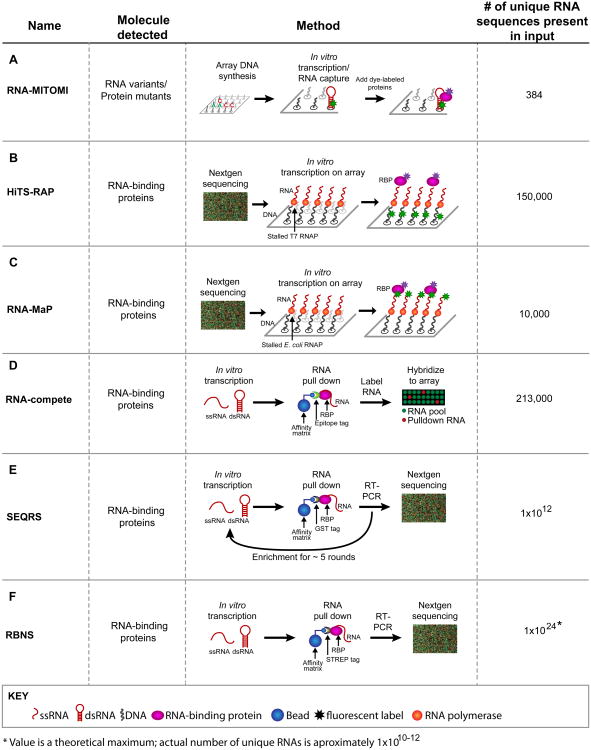
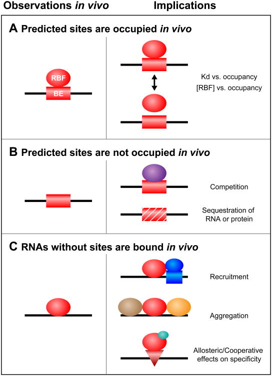
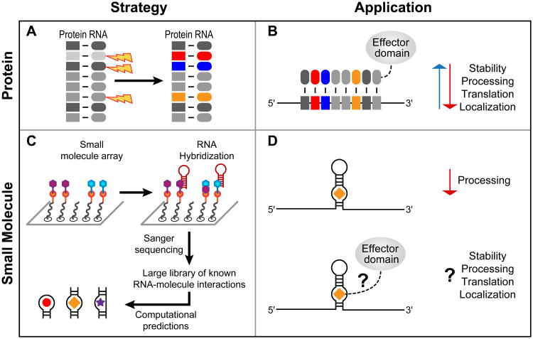
RNA-protein interactions are pervasive. The specificity of these interactions dictates which RNAs are controlled by what protein. Here we describe a class of revolutionary new methods that enable global views of RNA-binding specificity in vitro, for both single proteins and multiprotein complexes. These methods provide insight into central issues in RNA regulation in living cells, including understanding the balance between free and bound components, the basis for exclusion of binding sites, detection of binding events in the absence of discernible regulatory elements, and new approaches to targeting endogenous transcripts by design. Comparisons of in vitro and in vivo binding provide a foundation for comprehensive understanding of the biochemistry of protein-mediated RNA regulatory networks.


Figures

Figure 1
Figure 1
Methods to analyze RNA–protein and RNA–small molecule interactions. The diagrams for each method use the same color code. Red, RNA – structured (shown as a hairpin) or single stranded (squiggly lines); black, DNA; pink, RNA-binding proteins (RBPs); blue, affinity matrices; asterisks, fluorescent components. Use of arrays or deep sequencing is indicated in the diagrams. (A) In RNA mechanically induced trapping of molecular interactions (RNA-MITOMI), protein and RNA variants are analyzed by differential dye fluorescence for structure/function relationships. Both the RNA and the protein are fluorescent. (B) In high-throughput sequencing RNA affinity profiling (HiTS-RAP), RNA variants are synthesized on and tethered to an Illumina flow cell and the RBPs are detected by fluorescence at the site of DNA clusters. (C) Quantitative analysis of RNA on a massively parallel array (RNA-MaP) is similar to HiTS-RAP in that RNA variants are synthesized on a flow cell and incubated with fluorescently labeled proteins. (D) In RNA-compete, RNA–protein interactions are detected by retrieving RNAs bound to a protein of interest followed by detection of these RNAs via a microarray. Fluorescence intensity reveals binding preferences. (E) In in vitro selection, high-throughput sequencing of RNA, and sequence specificity landscapes (SEQRS), RNA–protein interactions are detected using iterative selection and high-throughput sequencing. The method is analogous to systematic evolution of ligands by exponential enrichment (SELEX) but with fewer rounds of selection and deep sequencing. (F) RNA Bind-n-Seq (RBNS) is similar to SEQRS in that RNA–protein complexes are retrieved from a complex library and detected via deep sequencing. However, unlike SEQRS only a single round of selection is used, allowing access to more RNA sequences.
Figure 2
Figure 2
Three classes of binding element (BE) in vivo and their potential implications. (A) A BE predicted to be functional in vitro is bound in vivo by an RNA-binding factor (RBF). (B) A BE predicted to be functional in vitro is not bound in vivo, due to either competition with other factors (purple) or physical sequestration. (C) RNAs without in vitro binding sites are associated with the protein in vivo. This situation might be due to recruitment by additional factors (dark blue circle and box), aggregation (other proteins are shown in brown and orange), or the consequences of allosteric effectors (light blue circle) that drive occupancy of latent sites (red triangle).
Figure 3
Figure 3
Approaches for tailored regulation of RNA in vivo. (A) Mutagenesis of the Pumilio/fem-3 mRNA-binding factor (PUF) scaffold provides a means to target cellular RNAs [17]. Repeat units (R1–R8) specify recognition of a single RNA base and can be manipulated to change RNA targeting. (B) Fusion of PUF scaffolds to effector domains results in desired regulatory outcomes. (C) Analysis of RNA–small molecule interactions using 2D combinatorial screening (2DCS) provides a starting place for the identification of bioactive compounds. (D) Addition of chemical effector domains to compounds should enable a diversity of outcomes for the targeted RNA, as in targeted PUF design.

References

    1. Licatalosi DD, et al. HiTS-CLIP yields genome-wide insights into brain alternative RNA processing. Nature. 2008;456:464–469. - PMC - PubMed
    1. Hafner M, et al. Transcriptome-wide identification of RNA-binding protein and microRNA target sites by PAR-CLIP. Cell. 2010;141:129–141. - PMC - PubMed
    1. Konig J, et al. iCLIP reveals the function of hnRNP particles in splicing at individual nucleotide resolution. Nat Struct Mol Biol. 2010;17:909–915. - PMC - PubMed
    1. Keene JD, et al. RIP-Chip: the isolation and identification of mRNAs, microRNAs and protein components of ribonucleoprotein complexes from cell extracts. Nat Protoc. 2006;1:302–307. - PubMed
    1. Guan L, Disney MD. Recent advances in developing small molecules targeting RNA. ACS Chem Biol. 2012;7:73–86. - PubMed

Publication types

LinkOut - more resources