. 2015 Mar 1;194(5):2390-8.
doi: 10.4049/jimmunol.1401064. Epub 2015 Jan 30.

Activation of human γδ T cells by cytosolic interactions of BTN3A1 with soluble phosphoantigens and the cytoskeletal adaptor periplakin

Affiliations

Activation of human γδ T cells by cytosolic interactions of BTN3A1 with soluble phosphoantigens and the cytoskeletal adaptor periplakin

David A Rhodes et al. J Immunol. .

Abstract

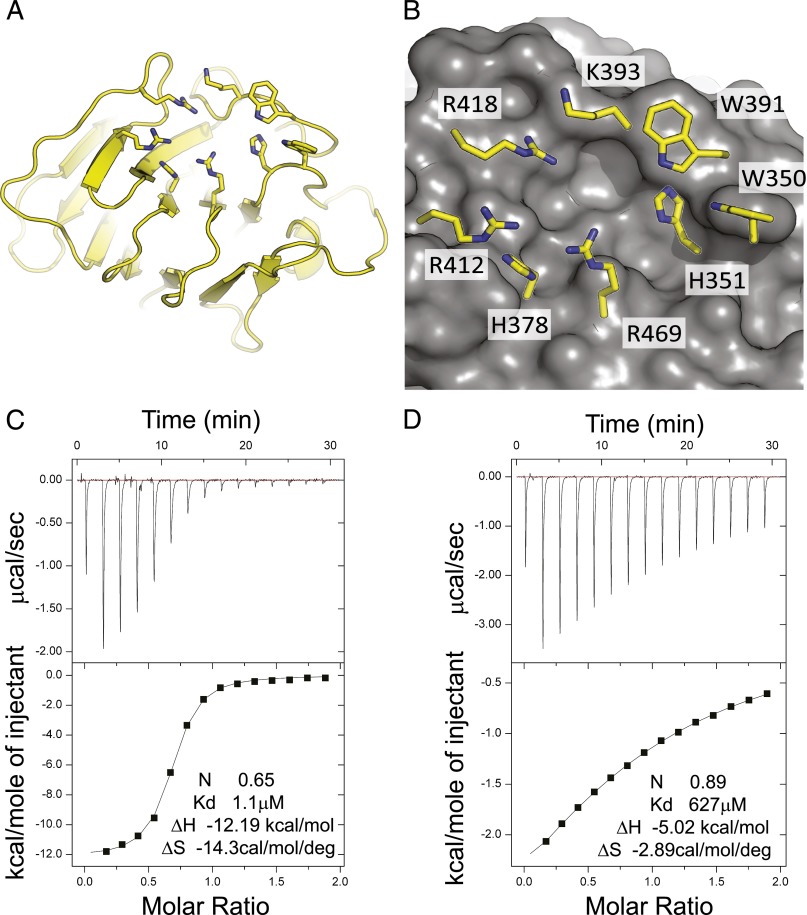
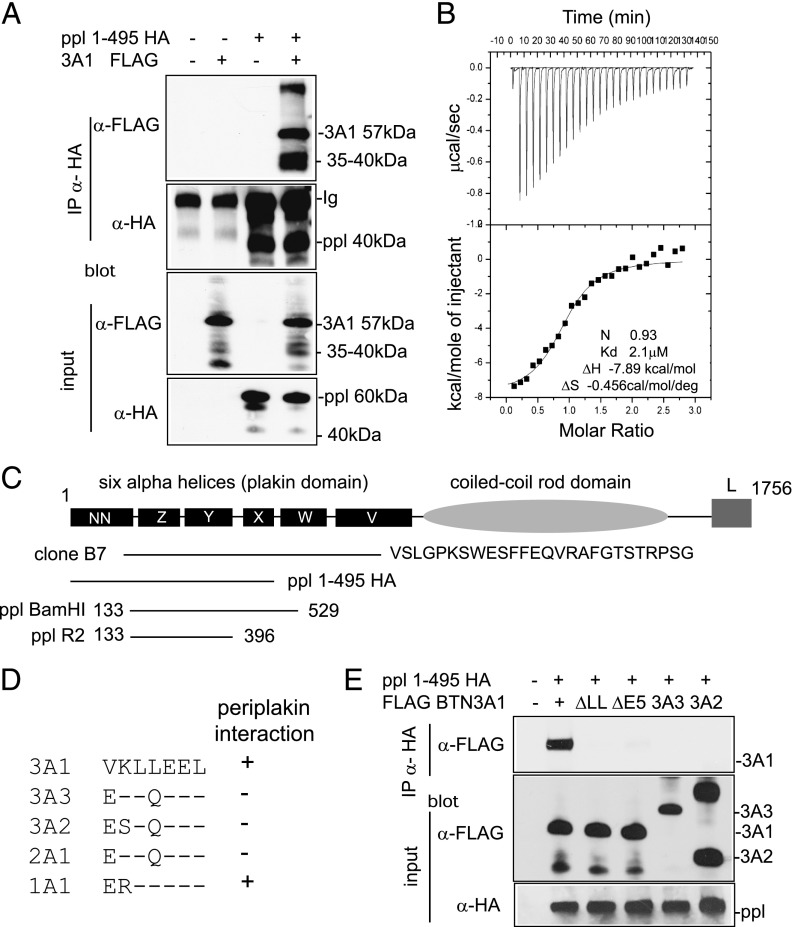
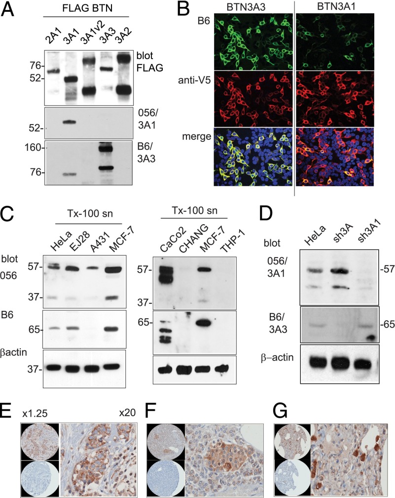
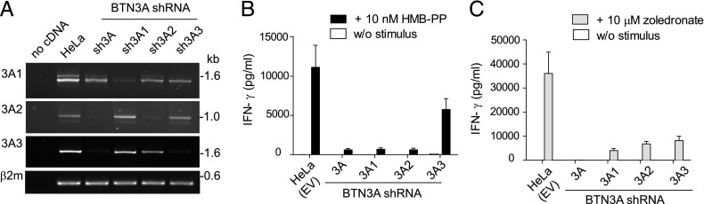
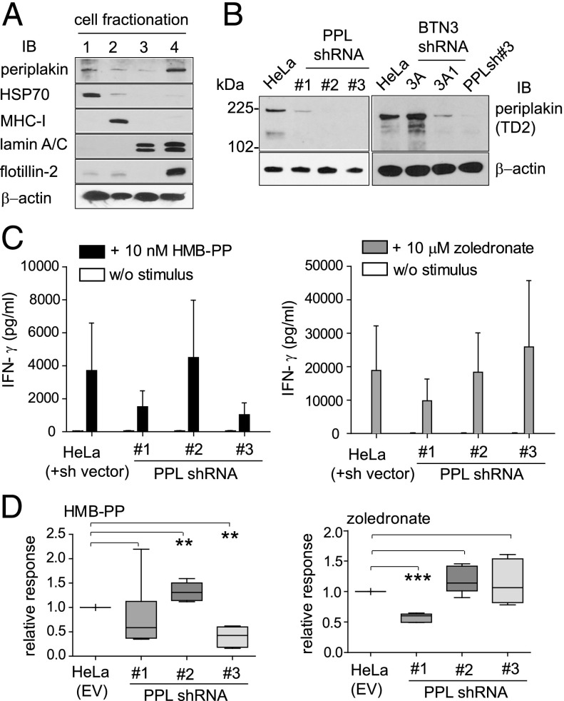
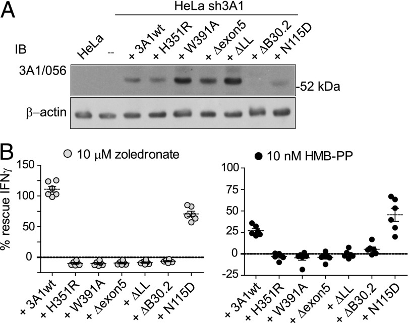
The three butyrophilin BTN3A molecules, BTN3A1, BTN3A2, and BTN3A3, are members of the B7/butyrophilin-like group of Ig superfamily receptors, which modulate the function of T cells. BTN3A1 controls activation of human Vγ9/Vδ2 T cells by direct or indirect presentation of self and nonself phosphoantigens (pAg). We show that the microbial metabolite (E)-4-hydroxy-3-methyl-but-2-enyl pyrophosphate binds to the intracellular B30.2 domain of BTN3A1 with an affinity of 1.1 μM, whereas the endogenous pAg isopentenyl pyrophosphate binds with an affinity of 627 μM. Coculture experiments using knockdown cell lines showed that in addition to BTN3A1, BTN3A2 and BTN3A3 transmit activation signals to human γδ T cells in response to (E)-4-hydroxy-3-methyl-but-2-enyl pyrophosphate and the aminobisphosphonate drug zoledronate that causes intracellular accumulation of isopentenyl pyrophosphate. The plakin family member periplakin, identified in yeast two-hybrid assays, interacted with a membrane-proximal di-leucine motif, located proximal to the B30.2 domain in the BTN3A1 cytoplasmic tail. Periplakin did not interact with BTN3A2 or BTN3A3, which do not contain the di-leucine motif. Re-expression into a BTN3A1 knockdown line of wild-type BTN3A1, but not of a variant lacking the periplakin binding motif, BTN3A1Δexon5, restored γδ T cell responses, demonstrating a functional role for periplakin interaction. These data, together with the widespread expression in epithelial cells, tumor tissues, and macrophages detected using BTN3A antiserum, are consistent with complex functions for BTN3A molecules in tissue immune surveillance and infection, linking the cell cytoskeleton to γδ T cell activation by indirectly presenting pAg to the Vγ9/Vδ2 TCR.


Figures

FIGURE 1.
FIGURE 1.
Structure of the BTN3A1 B30.2 domain. (A) Overview of the fold is shown with binding-site residues shown in stick representation. (B) A close-up view of the proposed pAg binding site showing the molecular surface and important interface residues (10). (C and D) ITC was used to show binding of pAg to BTN3A1 B30.2 domain. HMB-PP→BTN3A1 at 0.639 mM into HMB-PP at 0.064 mM; IPP→IPP at 6 mM into BTN3A1 at 0.597 mM.
FIGURE 2.
FIGURE 2.
Interaction of BTN3A1 with periplakin. (A) Interaction of periplakin and BTN3A1 in vivo by IP. 293T cells were transfected with expression constructs PPL1-495 HA and FLAG BTN3A1 either alone or together. Protein complexes were recovered by IP using anti-HA Ab and analyzed by IB using anti-FLAG (upper panel) or anti-HA (second panel) Abs. FLAG BTN3A1 was recovered from anti-HA IP only in the presence of PPL1-495 HA. Lower panels show input lysates analyzed by IB. Protein bands with molecular weights lower than the introduced expression constructs were detected for both BTN3A1 and periplakin in IP experiments, as shown, which likely represents postlysis proteolytic cleavage of proteins. (B) ITC was used to confirm thermodynamically favorable interaction between purified BTN3A1 and PPL BamH1. Binding affinity in the low-micromolar range (2.1 μM) was calculated. (C) Diagram of domain structure of periplakin (14) showing expression constructs used. Clone B7 represented periplakin aa 126–657 plus 26 additional amino acids, showing similarity to XP_001098863 periplakin-like sequence from Macaca mulatta. (D) Alignment of amino acids (306–312) encoded by exon 5 of BTN3A1 compared with other BTN proteins. (E) Periplakin interaction via the di-leucine motif in BTN3A1. IP of wild-type BTN3A1 and variants lacking either exon 5 or the di-leucine motif. 293T cells were transfected with the indicated BTN3A expression constructs together with PPL1-495 HA. Periplakin and associated proteins were recovered from cell lysates by IP using anti-HA Ab and analyzed by IB using anti-FLAG. Bottom panels show IB of input lysates using anti-FLAG and anti-HA acting as a loading control.
FIGURE 3.
FIGURE 3.
BTN3A protein expression in human cells and tissues. (A) 293T cells were transfected with pFLAG BTN expression constructs. Cell lysates were analyzed by nonreducing SDS-PAGE and IB using anti-FLAG and rabbit anti-B30.2 domain antisera 056 and B6 and goat anti-rabbit HRP secondary Abs. BTN3A1v2 is a truncated molecule lacking the B30.2 domain. Protein molecular mass in kDa. (B) Immunofluorescence microscopy of 293T cells transfected with expression constructs for BTN3A3 and BTN3A1. Fixed and Triton X-100 permeabilized cells (0.1%) were stained with antiserum B6/goat anti-rabbit Alexa 488 Abs (green) and carboxyl-terminal V5/goat anti-mouse Alexa 568 (red). Original magnification ×20. (C) BTN3A protein expression by IB of Triton X-100 detergent cell lysates from human epithelial carcinomas cell lines, as shown, probed with antisera 056 and B6 and goat anti-rabbit HRP secondary Abs. Loading control by β-actin Ab. (D) BTN3A expression in HeLa cells selected for expression of BTN3A shRNA knockdown vectors. Cell lysates were analyzed by IB with antisera 056 and B6 and by β-actin Ab. (EG) Immunohistochemistry on human tissue array probed with Ab B6. Original magnification ×20. (E) Breast tumor tissue. (F) Islets of Langerhans. (G) Macrophages in the lung. Control panels [lower panels in (E)–(G)] were serial tissue sections stained with preimmune serum.
FIGURE 4.
FIGURE 4.
BTN3A-dependent activation of γδ T cells. (A) BTN3A knockdown by expression of shRNA targeting each isoform. Full-length BTN3A transcripts were amplified by RT-PCR from cDNA derived from HeLa BTN3A knockdown cell lines. Amplification of β2microglobulin was used as a template control. RT-PCR products were analyzed by gel electrophoresis. Size in kb. (B and C) Coculture of γδ T cells with BTN3A knockdown lines and empty vector control (HeLa EV). T cell activation was induced by (B) 10 nM HMB-PP in coculture or (C) pretreatment of HeLa cells with 10 μM zoledronate. IFN-γ secretion into culture medium was detected by ELISA. Data shown are mean values obtained from four independent experiments using γδ T cells from PBMCs of three individual donors, with error bars (SEM). w/o, without pAg.
FIGURE 5.
FIGURE 5.
Role of periplakin in γδ T cell activation. (A) Cell fractionation based on detergent solubilization. Fractions from HeLa cells (2 × 10⁶) were analyzed by SDS-PAGE and IB using antiperiplakin antiserum TD2. Control Abs were used to show efficiency of fractionation into (1) cytosolic, (2) membrane, (3) nuclear, and (4) cytoskeletal/detergent-resistant membrane fractions. Anti–β-actin Ab acts as a loading control. (B) Periplakin knockdown by expression of shRNA in HeLa cells (left panel). Cytoskeletal/detergent-resistant membrane fractions were prepared and analyzed by IB using antiperiplakin antiserum. IB of detergent-resistant fractions in BTN3A and periplakin knockdown cells (right panel). Anti–β-actin Ab acts a loading control in each case. (C) Coculture of γδ T cells with periplakin shRNA knockdown lines PPLsh#1, PPLsh#2, and PPLsh#3. T cell activation was induced using 10 nM HMB-PP (left panel) or 10 μM zoledronate (right panel), and IFN-γ secretion into culture medium was detected by ELISA. HeLa EV empty vector control. Data shown are representative of five independent experiments, with error bars (SEM). (D) Analysis of data from (C) of γδ T cell stimulation induced from PPL knockdown cells. Box plots (depicting 25th percentile, median, 75th percentile, and highest/lowest data point whiskers) of relative change in IFN-γ responses compared with vector control (HeLa EV), from n = 5 determinations. Differences between groups were assessed by one-way ANOVA. A p value <0.05 was considered significant. **p < 0.01, ***p < 0.001. w/o, without pAg.
FIGURE 6.
FIGURE 6.
Activation of γδ T cells requires the B30.2 domain and periplakin interaction motif. (A) Analysis of re-expression lines using anti-BTN3A1 Ab 056. IB analysis of total protein (10 μg) from Triton-X100 detergent lysates from HeLa, HeLa sh3A1 knockdown line, and the parental sh3A1 cell line transduced additionally with IRES.GFP lentivirus–expressing wild-type BTN3A1, H351R, W391A, Δexon5, ΔLL, ΔB30.2, and N115D variants. Duplicate IB using β-actin acts as loading control. Endogenous BTN3A1 protein was not detectable by 056 antiserum in the untransfected HeLa cell lysate in these experimental conditions. BTN3A1 ΔB30.2 variant protein was also not detected using the 056 antiserum. (B) Analysis of re-expression lines in γδ T cell assays. Graph shows percent (%) IFN-γ recovery response from each of the re-expression lines compared with the vector control HeLa EV (positive) and sh3A1 knockdown (negative) lines. Data produced using γδ T cells from six individual donors.

Similar articles

Cited by

References

    1. Willcox C. R., Mohammed F., Willcox B. E. 2013. Resolving the mystery of pyrophosphate antigen presentation. Nat. Immunol. 14: 886–887. - PubMed
    1. Vantourout P., Hayday A. 2013. Six-of-the-best: unique contributions of γδ T cells to immunology. Nat. Rev. Immunol. 13: 88–100. - PMC - PubMed
    1. Boyden L. M., Lewis J. M., Barbee S. D., Bas A., Girardi M., Hayday A. C., Tigelaar R. E., Lifton R. P. 2008. Skint1, the prototype of a newly identified immunoglobulin superfamily gene cluster, positively selects epidermal gammadelta T cells. Nat. Genet. 40: 656–662. - PMC - PubMed
    1. Eberl M., Hintz M., Reichenberg A., Kollas A. K., Wiesner J., Jomaa H. 2003. Microbial isoprenoid biosynthesis and human gammadelta T cell activation. FEBS Lett. 544: 4–10. - PubMed
    1. Riganti C., Massaia M., Davey M. S., Eberl M. 2012. Human γδ T-cell responses in infection and immunotherapy: common mechanisms, common mediators? Eur. J. Immunol. 42: 1668–1676. - PubMed

Publication types

MeSH terms