. 2015 May;100(5):585-94.
doi: 10.3324/haematol.2014.118612. Epub 2015 Jan 30.

ANGPTL7 regulates the expansion and repopulation of human hematopoietic stem and progenitor cells

Affiliations

ANGPTL7 regulates the expansion and repopulation of human hematopoietic stem and progenitor cells

Yiren Xiao et al. Haematologica. 2015 May.

Abstract

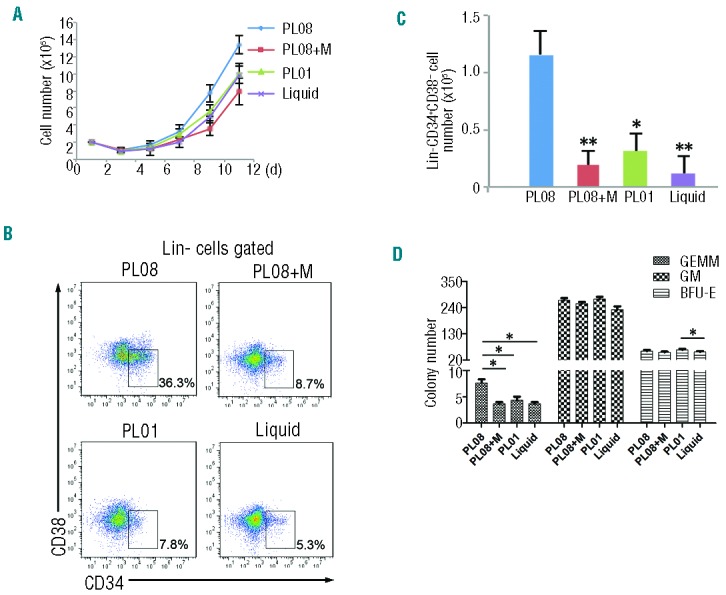
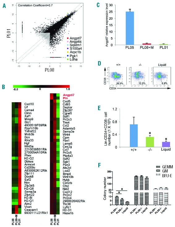
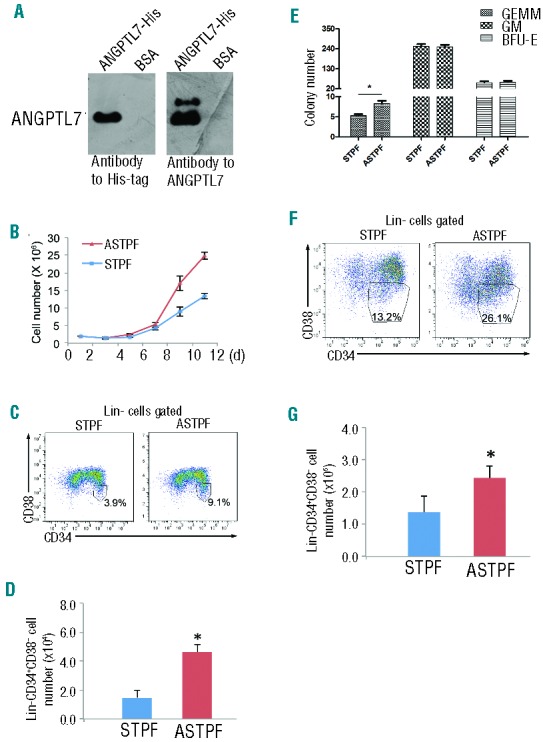
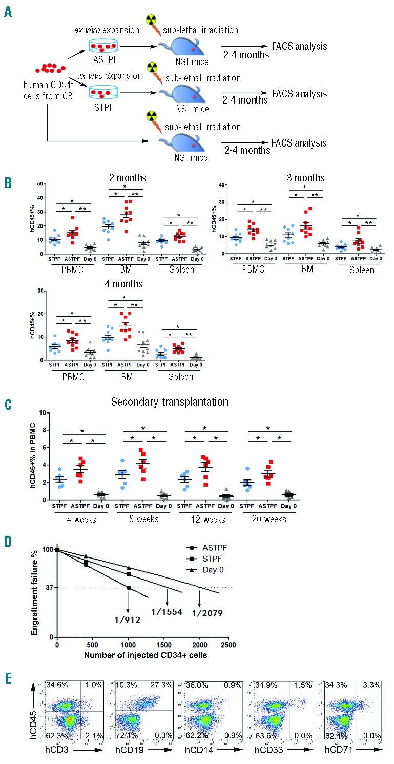
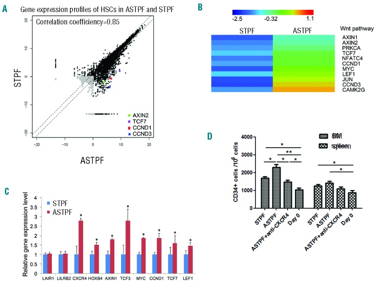
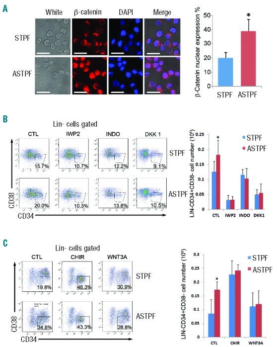
Successful expansion of hematopoietic stem cells would benefit the use of hematopoietic stem cell transplants in the clinic. Several angiopoietin-like proteins, including angiopoietin-like 7, can support the activity of hematopoietic stem cells. However, effects of ANGPTL7 on human hematopoietic stem cells and the downstream signaling cascade activated by ANGPTL7 are poorly understood. Here, we established a human hematopoietic stem and progenitor cell-supportive mouse fetal liver cell line that specifically expressed the Angptl7 protein. Furthermore, we found ANGPTL7 is capable of stimulating human hematopoietic stem and progenitor cell expansion and increasing the repopulation activities of human hematopoietic progenitors in xenografts. RNA-sequencing analysis showed that ANGPTL7 activated the expression of CXCR4, HOXB4 and Wnt downstream targets in human hematopoietic progenitors. In addition, chemical manipulation of Wnt signaling diminished the effects of ANGPTL7 on human hematopoietic stem and progenitor cells in culture. In summary, we identify the secreted growth factor ANGPTL7 as a regulator of both human hematopoietic stem and progenitor cell expansion and regeneration.


Figures

Figure 1.
Figure 1.
PL08 substantially stimulates the expansion of human hematopoietic stem cells (HSCs) in vitro. (A) A total of 2×10⁵ human umbilical cord blood nucleated cells (hUCBNCs) were isolated and co-cultured with PL08 cells, PL08 cells that were previously treated with Mito (PL08+M), or PL01 cells in StemSpan medium supplemented with hSCF, hTPO, PTN, and hFLT3L (STPF). In culture, hUCBNCs were separated from stromal cells using Transwell membranes. In another control group (Liquid), 2×10⁵ hUCBNCs were isolated and cultured without any stromal cells in StemSpan medium supplemented with STPF. Cultured hUCBNC were counted and analyzed every three days (n=4). (B) Representative FACS plots show the percentages of Lin-CD34+CD38− cells in PL08, PL08+M, PL01, and liquid culture conditions. (C) Summary of absolute numbers of Lin-CD34+CD38− cells cultured in PL08, PL08+M, PL01, or liquid conditions at day 12 from four independent experiments (n=4). Data represent mean+s.e.m. *P≤0.05 versus bar 3 for bar 1; **P≤0.01 versus bar 2 and bar 4 (for bar 1). (D) The colony numbers of CFU assays obtained from cultured cells as described in (A). Data represent mean+s.e.m. (n=4). *P≤0.05 versus bar 2, bar 3 and bar 4 (for bar 1). *P≤0.05 for bar 11 versus bar 12. BFU-E: erythroid colonies; GM: granulocyte-monocyte colonies; GEMM: granulocyte, erythrocyte, monocyte and megakaryocyte colonies.
Figure 2.
Figure 2.
Angptl7 is specifically expressed in the PL08 cell line. (A) Pairwise comparison of poly(A)+ RNA-Seq analysis of the global expression profiles in PL08 versus PL01 cells. Pearson correlation coefficient is shown. (B) Unsupervised hierarchical cluster analysis of expression levels of 77 secreted protein genes in PL08, PL08+M, and PL01 cells (red: increased expression; green: decreased expression). (C) qRT-PCR analysis of Angptl7 mRNA levels in PL08, PL08+M, and PL01 cells. The results were normalized to the β-actin mRNA levels and represent mean+s.e.m. (n=3). (D) A total of 2×10⁵ human umbilical cord blood nucleated cells (hUCBNCs) were isolated and co-cultured with PL08 cells, Angptl7-null PL08 cells in STPF conditions. In culture, hUCBNCs were separated from stromal cells using Transwell membranes. In another control group (Liquid), 2×10⁵ hUCBNCs were isolated and cultured without any stromal cells in StemSpan medium supplemented with STPF. Representative FACS plots show the percentages of Lin-CD34+CD38− cells in PL08, Angptl7-null PL08, and liquid culture conditions. (E) Summary of absolute numbers of Lin-CD34+CD38− cells in liquid culture condition (Liquid), or in co-culture with WT PL08 (+/−), or Angptl7−/− PL08 (−/−) cells at day 12 from four independent experiments (n=4). Data represent mean+s.e.m. *P≤0.05 versus bar 2 and bar 3 (for bar 1). (F) The colony numbers of CFU assays obtained cultured hUCBNCs as described in (D). Data represent mean+s.e.m. (n=4). *P≤0.05 versus bar 2 and bar 3 (for bar 1).
Figure 3.
Figure 3.
Effects of ANGPTL7 on human hematopoietic stem and progenitor cells (HSPCs). (A) Western blotting analysis of purified ANGPTL7 (left lanes) and control bovine serum albumin (BSA, right lanes) detected by antibodies as indicated. (B) Human umbilical cord blood nucleated cells (hUCBNC) cultures (2×10⁶) in ASTPF (red triangles) and STPF (blue squares) conditions. Viable cells were counted at the indicated time points. Data represent mean+s.e.m. (n=3). (C and D) Representative FACS plots (C) and summary of the absolute numbers (D) of Lin-CD34+CD38− cells in the ASTPF and STPF conditions. Data represent mean+s.e.m. (n=3). *P≤0.05 for bar 1 versus bar 2. (E) The colony numbers of CFU assays obtained from 1×10⁴ fresh hUCBNCs (day 0) or after culturing hUCBNCs in the ASTPF and STPF conditions. Data represent mean+s.e.m. (n=3). *P≤0.05 versus bar 1 for bar 2; **P≤0.01 for bar 2 versus bar 3. (F and G) Representative FACS profiles (F) and summary of absolute numbers (G) of purified CD34+ hUCBNCs cultured in the ASTPF and STPF conditions. Data represent mean+s.e.m. (n=3). *P≤0.05 for bar 1 versus bar 2.
Figure 4.
Figure 4.
ANGPTL7 stimulates expansion of human hematopoietic stem and progenitor cells (HSPCs) ex vivo. (A) Experimental design for assessing the repopulation capacities of ex vivo expanded human HSPCs. Total of 1×10⁴ fresh purified CD34+ human umbilical cord blood nucleated cells (hUCBNCs) and 1×10⁴ purified CD34+ hUCBNCs cultured in ASTPF or STPF conditions for seven days were injected into three groups of sub-lethally irradiated NSI mice (9 mice per group). Two, three, and four months post transplantation, peripheral blood (PB), BM, and spleen (SP) samples from NSI mice from each group were subjected to FACS analysis. (B) Summary of percentages of human CD45+ cells in the PB, BM, and SP of NSI mice at two, three, or four months after injection with 1×10⁴ purified CD34+ hUCBNCs or progeny of 1×10⁴ purified CD34+ hUCBNCs that had been cultured in ASTPF or STPF conditions. Bars represent the mean percentages of human CD45+ cells in the PB, BM and SP of mice from each group (n=9). The experiments were repeated three times. *P≤0.05 for ASTPF group versus STPF group; **P≤0.01 for ASTPF group versus day 0 group; *P≤0.05 for STPF group versus day 0 group. (C) Bone marrow aspirate from one hind leg from a primary recipient was transplanted into 2 secondary recipients (n=6); *P≤0.05 for ASTPF group and STPF group versus day 0 group; ASTPF group versus STPF group. (D) Limiting-dilution analysis of the repopulating ability of cells (400 or 1000 cells) before culture (day 0) and after culture for seven days in STPF medium and ASTPF medium. Plots show percentage of recipient mice containing less than 1% human hematopoietic populations in recipient mouse bone marrow eight weeks after transplantation versus the number of input or input-equivalent cells injected (n=10 mice transplanted at each dose per condition; n=60 mice total). (E) Representative FACS analysis of percentages of multiple hematopoietic lineages in the BM of NSI mice from the ASTPF group two months post transplantation, as described in (B).
Figure 5.
Figure 5.
Genes activated in human hematopoietic stem and progenitor cells (HSPCs) by ANGPTL7. (A) CD34+ human umbilical cord blood nucleated cells (hUCBNCs) were cultured in STPF and ASTPF conditions for seven days. Then human HSPCs (Lin-CD34+CD38) were purified from cultures and subjected for RNA-seq analysis. Pair-wise comparison of poly(A)+ RNA-Seq analysis of the global expression profiles of HSPCs (Lin-CD34+CD38) in STPF and ASTPF conditions. (B) Hierarchical clustering of representative Wnt signaling genes that were up-regulated with more than 2-fold change in human HSPCs upon ANGPTL7 stimulation. (C) Relative expression levels of selected genes in purified CD34+ hUCBNCs cultured in ASTPF and STPF conditions. The results were normalized to β-ACTIN mRNA levels and represent mean+s.e.m. (n=3). *P<0.05 for ASPTF group versus STPF group. (D) Cord blood CD34+ cells without culture, or cultured in the STPF medium, ASTPF medium or ASTPF medium plus anti-CXCR4 antibody were injected intravenously into NSI mice. Bone marrow (BM) and spleens (SP) of recipient mice were analyzed for the presence of human CD34+ cells by flow cytometry 16 h after transplantation (n=6); *P≤0.05 for bar 1, and bar 3 versus bar 4, bar 1 versus bar 2, bar 2 versus bar 3, bar 5 and bar 6 versus bar8; **P≤0.01 for bar 2 versus bar 4.
Figure 6.
Figure 6.
Wnt signaling participated in ANGPTL7-stimulation in hematopoietic stem and progenitor cells (HSPCs). (A) (Left) Immunofluorescence analysis of subcellular localization of β-catenin (red) in CD34+ human HSPCs treated with ANGPTL7 (ASTPF) or without ANGPTL7 (STPF) for 24 h. Scale bar: 100 μm. Nuclei were stained with DAPI (blue). In merged magnification images, the overlap of blue and red indicated the localization of β-catenin in nuclei. (Right) Chart depicting the percentage of β-catenin nuclear expression in CD34+ human HSPCs treated as indicated. Error bars show +/− s.e.m. of triplicates from three independent experiments; *P≤0.05 for bar 1 versus bar 2. (B) (Left) Representative FACS profiles of CD34+ purified human umbilical cord blood nucleated cells (hUCBNCs) cultured in ASTPF and STPF conditions with or without Wnt inhibitors (IWP2, INDO, or DKK1). (Right) Summary of absolute numbers of Lin-CD34+CD38− cells in purified CD34+ hUCBNCs cultured in ASTPF and STPF conditions with or without Wnt inhibitors (IWP2, INDO, or DKK1). Data represent mean+s.e.m. (n=3). *P≤0.05 for bar 1 versus bar 2. (C) (Left) Representative FACS profiles of CD34+ purified hUCBNCs cultured in ASTPF and STPF conditions with or without Wnt activators (CHIR or WNT3A). (Right) Summary of absolute numbers of Lin-CD34+CD38− cells in purified CD34+ hUCBNCs cultured in ASTPF and STPF conditions with or without Wnt activators (CHIR or WNT3A). Data represent mean+s.e.m. (n=3); *P≤0.05 versus bar 2 for bar 1.

Similar articles

Cited by

References

    1. Zon LI. Intrinsic and extrinsic control of haematopoietic stem-cell self-renewal. Nature. 2008;453(7193):306–313. - PubMed
    1. Kiel MJ, Morrison SJ. Uncertainty in the niches that maintain haematopoietic stem cells. Nat Rev Immunol. 2008;8(4):290–301. - PubMed
    1. Yilmaz OH, Valdez R, Theisen BK, et al. Pten dependence distinguishes haematopoietic stem cells from leukaemia-initiating cells. Nature. 2006;441(7092):475–482. - PubMed
    1. Antonchuk J, Sauvageau G, Humphries RK. HOXB4-induced expansion of adult hematopoietic stem cells ex vivo. Cell. 2002;109(1):39–45. - PubMed
    1. Will B, Vogler TO, Bartholdy B, et al. Satb1 regulates the self-renewal of hematopoietic stem cells by promoting quiescence and repressing differentiation commitment. Nat Immunol. 2013;14(5):437–445. - PMC - PubMed

Publication types

MeSH terms