. 2015 Jan 31:3:5.
doi: 10.1186/s40478-014-0181-z.

Early detection of structural abnormalities and cytoplasmic accumulation of TDP-43 in tissue-engineered skins derived from ALS patients

Early detection of structural abnormalities and cytoplasmic accumulation of TDP-43 in tissue-engineered skins derived from ALS patients

Bastien Paré et al. Acta Neuropathol Commun. .

Abstract

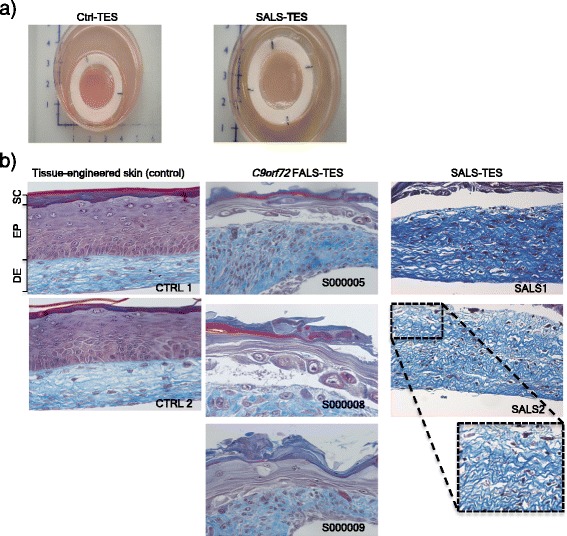
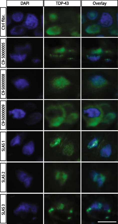
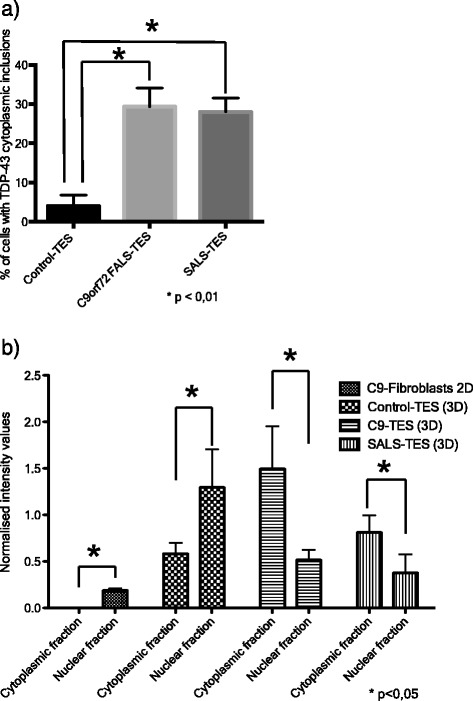
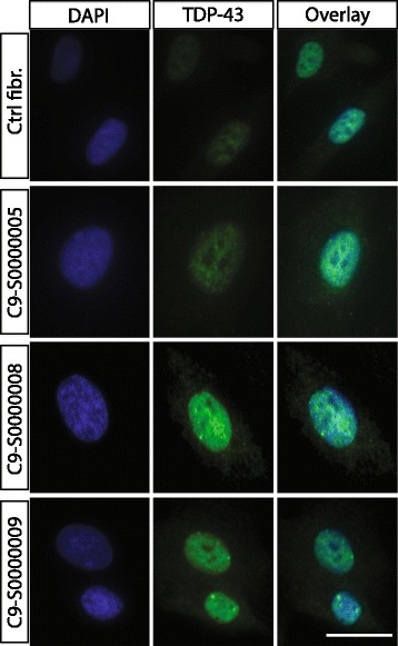
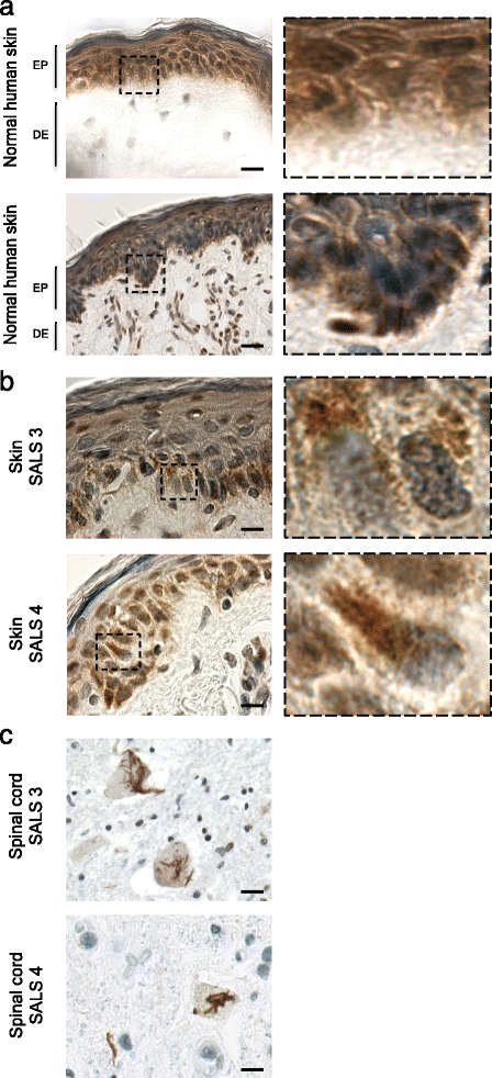
Amyotrophic lateral sclerosis (ALS) is an adult-onset disease characterized by the selective degeneration of motor neurons in the brain and spinal cord progressively leading to paralysis and death. Current diagnosis of ALS is based on clinical assessment of related symptoms. The clinical manifestations observed in ALS appear relatively late in the disease course after degeneration of a significant number of motor neurons. As a result, the identification and development of disease-modifying therapies is difficult. Therefore, novel strategies for early diagnosis of neurodegeneration, to monitor disease progression and to assess response to existing and future treatments are urgently needed. Factually, many neurological disorders, including ALS, are accompanied by skin changes that often precede the onset of neurological symptoms. Aiming to generate an innovative human-based model to facilitate the identification of predictive biomarkers associated with the disease, we developed a unique ALS tissue-engineered skin model (ALS-TES) derived from patient's own cells. The ALS-TES presents a number of striking features including altered epidermal differentiation, abnormal dermo-epidermal junction, delamination, keratinocyte infiltration, collagen disorganization and cytoplasmic TDP-43 inclusions. Remarkably, these abnormal skin defects, uniquely seen in the ALS-derived skins, were detected in pre-symtomatic C9orf72-linked ALS patients carrying the GGGGCC DNA repeat expansion. Consequently, our ALS skin model could represent a renewable source of human tissue, quickly and easily accessible to better understand the physiophatological mechanisms underlying this disease, to facilitate the identification of disease-specific biomarkers, and to develop innovative tools for early diagnosis and disease monitoring.


Figures

Figure 1
Figure 1
Structural abnormalities detected in ALS-derived tissue engineered skins. a) Macroscopic pictures of control-derived and ALS-derived tissue-engineered skins when cultured at the air-liquid interface. b) Masson’s trichrome colorations, specifically staining the dermis (DE) in blue and epidermis (EP) in purple, revealed a number of structural abnormalities including undifferentiated epidermis, abnormal dermo-epidermal junctions, delamination, abnormal collagen organization, keratinocyte infiltration and cohesive failure of the stratum corneum (SC) in both C9orf72 FALS- and SALS-derived skins. In contrast control-derived reconstructed skins showed a well-developed and differentiated epidermis and highly organized dermis.
Figure 2
Figure 2
Cytoplasmic TDP-43 accumulation detected in SALS- and C9orf72 FALS-derived tissue-engineered skins. Indirect immunofluorescence analysis using anti-TDP43 antibody (green) counterstained with DAPI (blue) revealed cytoplasmic TDP-43 accumulation in SALS-derived as well as in C9orf72 FALS-derived tissue-engineered skins. Note that representative pictures of 7-um thick tissue-sections were stained and visualized using a standard epifluorescent microscope. Each picture was taken using the same microscope, camera and exposure settings. Scale bar (white): 10 μm.
Figure 3
Figure 3
Cellular counts and Western blots quantification of TDP-43 cytoplasmic accumulation. a) Percentage of cell with positive cytoplasmic TDP-43 inclusions. 200 nuclei were counted for each of the generated tissue-engineered skins. *correspond to a P value < 0.01. b) Subcellular fractionation (cytoplasmic fraction vs nuclear fraction) of total protein extracted from ALS-fibroblast (2D culture), control-derived TES, C9ORF72-derived TES and SALS-derived TES (n = 5 for each group) was performed and loaded on a regular SDS-PAGE. TDP-43 expression in each fractionated sample was quantified using ImageJ after normalization against actin. Equal amount of proteins was used as shown on western blots after. *correspond to a P value < 0.05.
Figure 4
Figure 4
Nuclear TDP-43 expression in C9orf72 fibroblasts cultured cells. Indirect immunofluorescence using anti-TDP43 commercial antibody (green) conterstained with DAPI (nucleus) revealed no cytoplasmic TDP-43 accumulation in C9orf72 cultured fibroblasts collected from symptomatic and non-symptomatic C9orf72 FALS patients. These results indicate that the described 3D tissue-engineered skin model, allowing for cell-to-cell and cell-to-matrix interactions, is necessary to observe the pathological cytoplasmic accumulation of TDP-43. Scale bar (white): 10 μm.
Figure 5
Figure 5
Cytoplasmic TDP-43 detection in native skin biopsies and corresponding CNS tissues in SALS patients. Immunohistochemistry analysis, using anti-TDP-43 antibody, revealed the presence of TDP-43 cytoplasmic accumulation in both native skin biopsies (b) and post-mortem (c) spinal cord tissues collected from SALS patients. Such TDP-43 cytoplasmic accumulation was not detected in control individuals (a).

References

    1. Gros-Louis F, Gaspar C, Rouleau GA. Genetics of familial and sporadic amyotrophic lateral sclerosis. Biochim Biophys Acta. 2006;1762(11–12):956–972. doi: 10.1016/j.bbadis.2006.01.004. - DOI - PubMed
    1. Cellura E, Spataro R, Taiello AC, La Bella V. Factors affecting the diagnostic delay in amyotrophic lateral sclerosis. Clin Neurol Neurosurg. 2012;6:550–554. doi: 10.1016/j.clineuro.2011.11.026. - DOI - PubMed
    1. Phukan J, Pender NP, Hardiman O. Cognitive impairment in amyotrophic lateral sclerosis. Lancet Neurol. 2007;6((11):994–1003. doi: 10.1016/S1474-4422(07)70265-X. - DOI - PubMed
    1. Brettschneider J, Van Deerlin VM, Robinson JL, Kwong L, Lee EB, Ali YO, Safren N, Monteiro MJ, Toledo JB, Elman L, McCluskey L, Irwin DJ, Grossman M, Molina-Porcel L, Lee VM, Trojanowski JQ. Pattern of ubiquilin pathology in ALS and FTLD indicates presence of C9ORF72 hexanucleotide expansion. Acta Neuropathol. 2012;123(6):825–839. doi: 10.1007/s00401-012-0970-z. - DOI - PMC - PubMed
    1. Cooper-Knock J, Hewitt C, Highley JR, Brockington A, Milano A, Man S, Martindale J, Hartley J, Walsh T, Gelsthorpe C, Baxter L, Forster G, Fox M, Bury J, Mok K, McDermott CJ, Traynor BJ, Kirby J, Wharton SB, Ince PG, Hardy J, Shaw PJ. Clinico-pathological features in amyotrophic lateral sclerosis with expansions in C9ORF72. Brain. 2012;135(Pt 3):751–764. doi: 10.1093/brain/awr365. - DOI - PMC - PubMed

Publication types