

Clinical and radiological features and skeletal sequelae in childhood intra-/juxta-articular versus extra-articular osteoid osteoma
- PMID: 25637327
- PMCID: PMC4316764
- DOI: 10.1186/s12891-015-0456-y
Clinical and radiological features and skeletal sequelae in childhood intra-/juxta-articular versus extra-articular osteoid osteoma
Abstract
Background: To compare the clinical and radiological features of intra-/juxta-articular osteoid osteoma and extra-articular osteoid osteoma in skeletally immature patients, paying special attention to the skeletal complications.
Methods: Osteoid osteoma in 34 children (22 boys and 12 girls, mean age 10.4 years) was dichotomized according to the location of the nidus as intra-/juxta-articular (11 children) or extra-articular (23 children). The following features were compared: diagnostic delay, typical symptoms, synovitis and limited range of joint motion, response to treatment, typical radiographic findings, and skeletal complications.
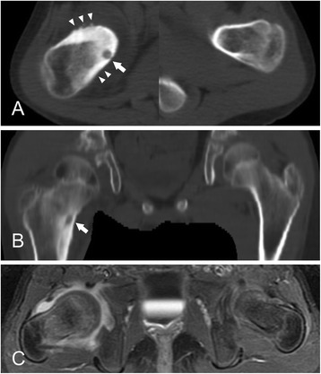
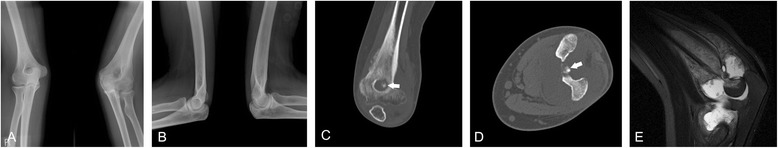
Results: Eight of the 11 children with intra-/juxta-articular osteoid osteoma presented with synovitis in the involved joint, which led to a delayed diagnosis for a median 9.5 months. Pain disappeared in all children with surgical or medical interventions, but at the mean 4.9-year follow-up evaluation, skeletal abnormalities around the joint were noted in 5 children (4 proximal femur and 1 distal humerus) with intra-/juxta-articular osteoid osteoma, 2 of whom required subsequent surgeries for limited hip motion caused by femoroacetabular impingement and limited range of elbow motion, respectively. In contrast, typical clinical and radiological features were observed more often in extra-articular osteoid osteoma, and only 1 child showed overgrowth of the tibia, which did not have clinical significance.
Conclusions: Intra-/juxta-articular osteoid osteomas in growing children exhibit different clinical and radiological features from extra-articular lesions. Skeletal abnormalities mainly develop in intra-/juxta-articular osteoid osteoma, and these may lead to permanent skeletal sequelae.
Figures
References
-
- Golding JS. The natural history of osteoid osteoma; with a report of twenty cases. J Bone Joint Surg Br. 1954;36-B(2):218–29. - PubMed
-
- Szendroi M, Kollo K, Antal I, Lakatos J, Szoke G. Intraarticular osteoid osteoma: clinical features, imaging results, and comparison with extraarticular localization. J Rheumatol. 2004;31(5):957–64. - PubMed
-
- Weber KL, Morrey BF. Osteoid osteoma of the elbow: a diagnostic challenge. J Bone Joint Surg Am. 1999;81(8):1111–9. - PubMed
MeSH terms
LinkOut - more resources
Full Text Sources
Other Literature Sources
Medical
