. 2015 Apr 1;308(7):H768-77.
doi: 10.1152/ajpheart.00779.2014. Epub 2015 Jan 30.

Mitochondrial N-formyl peptides induce cardiovascular collapse and sepsis-like syndrome

Affiliations

Mitochondrial N-formyl peptides induce cardiovascular collapse and sepsis-like syndrome

Camilla Ferreira Wenceslau et al. Am J Physiol Heart Circ Physiol. .

Abstract

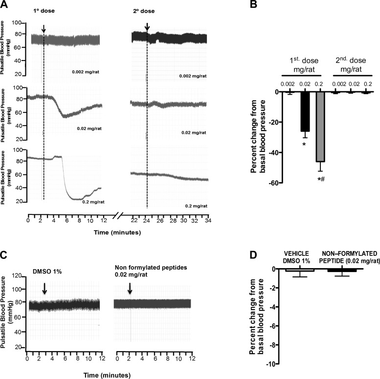
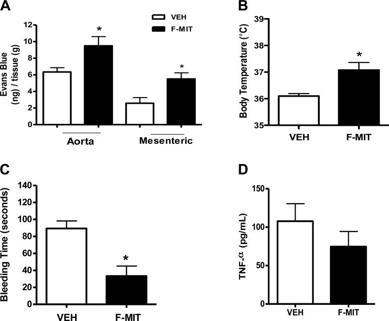
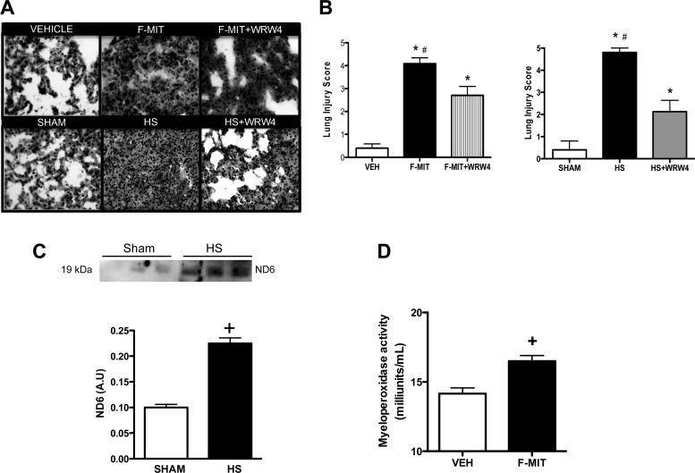
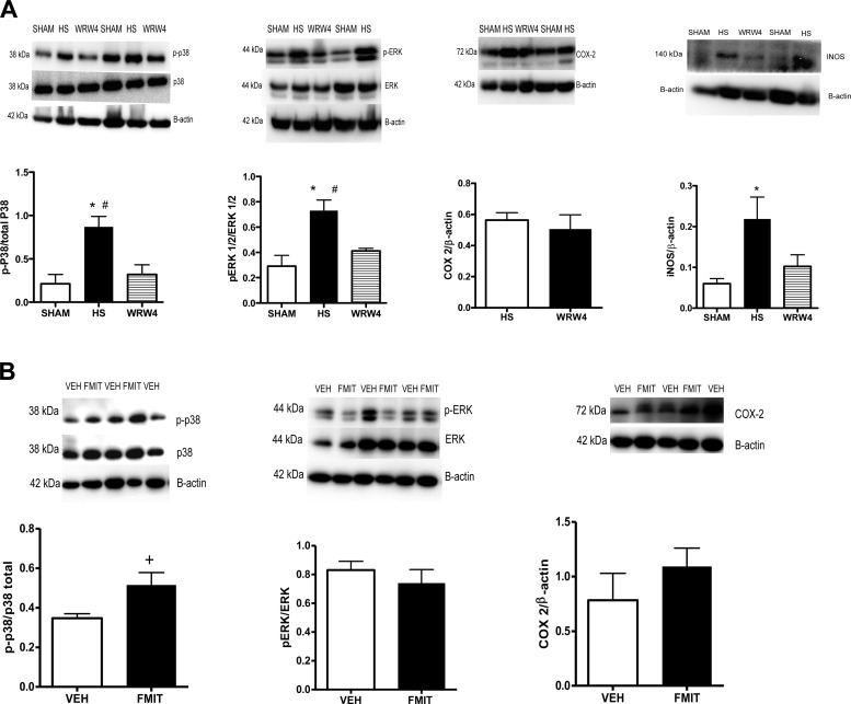
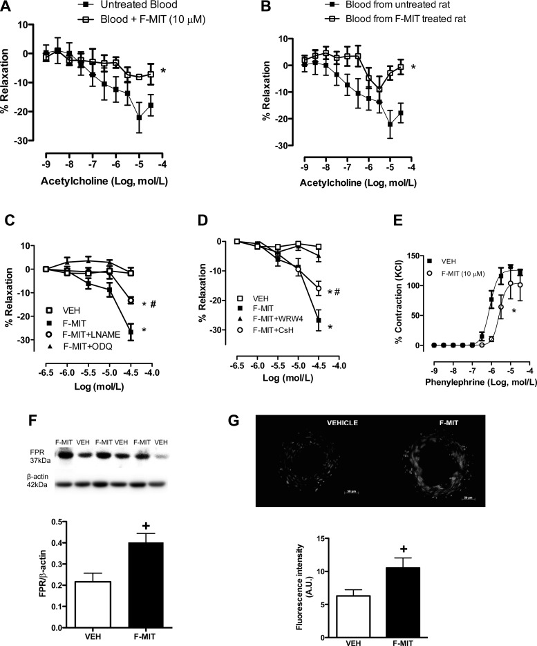
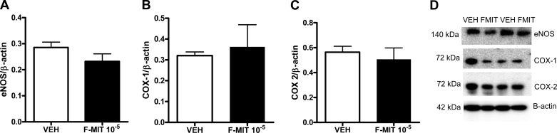
Fifty percent of trauma patients who present sepsis-like syndrome do not have bacterial infections. This condition is known as systemic inflammatory response syndrome (SIRS). A unifying factor of SIRS and sepsis is cardiovascular collapse. Trauma and severe blood loss cause the release of endogenous molecules known as damage-associated molecular patterns. Mitochondrial N-formyl peptides (F-MIT) are damage-associated molecular patterns that share similarities with bacterial N-formylated peptides and are potent immune system activators. The goal of this study was to investigate whether F-MIT trigger SIRS, including hypotension and vascular collapse via formyl peptide receptor (FPR) activation. We evaluated cardiovascular parameters in Wistar rats treated with FPR or histamine receptor antagonists and inhibitors of the nitric oxide pathway before and after F-MIT infusion. F-MIT, but not nonformylated peptides or mitochondrial DNA, induced severe hypotension via FPR activation and nitric oxide and histamine release. Moreover, F-MIT infusion induced hyperthermia, blood clotting, and increased vascular permeability. To evaluate the role of leukocytes in F-MIT-induced hypotension, neutrophil, basophil, or mast cells were depleted. Depletion of basophils, but not neutrophils or mast cells, abolished F-MIT-induced hypotension. Rats that underwent hemorrhagic shock increased plasma levels of mitochondrial formylated proteins associated with lung damage and antagonism of FPR ameliorated hemorrhagic shock-induced lung injury. Finally, F-MIT induced vasodilatation in isolated resistance arteries via FPR activation; however, F-MIT impaired endothelium-dependent relaxation in the presence of blood. These data suggest that F-MIT may be the link among trauma, SIRS, and cardiovascular collapse.

Keywords: cardiovascular collapse; mitochondrial N-formyl peptides; sepsis-like syndrome.


Figures

Fig. 1.
Fig. 1.
Effects of mitochondrial N-formyl peptides (F-MIT; A and B), nonformylated peptide (C and D), and DMSO (C and D) on blood pressure. A and B: subsequent infusion of the same dose of F-MIT (0.002, 0.02, or 0.2 mg/rat), administered 15 min after the first one, does not change blood pressure of Wistar rats. Arrows (A and C) indicate drug infusion. B and D: average values for percent change from basal blood pressure. Means ± SE; n = 4 to 5. One-way ANOVA: *P < 0.05 vs. 0.002 mg/rat; #P < 0.05 vs. 0.02 mg/rat.
Fig. 2.
Fig. 2.
Effects of F-MIT on vascular permeability (A), body temperature (B), bleeding time (C), and TNF-α production in plasma (D). Means ± SE; n = 4 to 5. t-test: *P < 0.05 vs. vehicle (Veh).
Fig. 3.
Fig. 3.
Representative hematoxylin-eosin staining of lung from rats treated with Veh, F-MIT, or F-MIT and formyl peptide receptor (FPR)-2 antagonist [Trp-Arg-Trp-Trp-Trp-Trp-NH2 (WRW4)] for 6 h (magnification, ×40) (A). Also, representative images of lung from rats that underwent hemorrhagic shock (HS) treated or not with FPR-2 antagonist (WRW4) (A). Graph represents lung injury score (B). C: protein expression (top) and densitometry analysis (bottom) from mitochondria NADPH dehydrogenase subunit 6 (ND6) in plasma from control rats (sham) and HS rats. AU, arbitrary units. Myeloperoxidase activity (D) in lung 6 h after Veh or F-MIT injection. Means ± SE; n = 4 to 5. One-way ANOVA: *P < 0.05 vs. Veh or Sham; #P < 0.05 vs. WRW4; t-test, +P < 0.05 vs. Veh.
Fig. 4.
Fig. 4.
Representative blots and densitometric analysis from protein expression for MAPK [phospho (p)- and total-p38 and -ERK 1/2], inducible nitric oxide synthase (iNOS) and cyclooxygenase-2 (COX-2) of lungs from HS rats treated or not with WRW4 (FPR-2 antagonist) (A), as well as animals treated with F-MIT for 6 h or Veh (B). Means ± SE; n = 4 to 5. One-way ANOVA: *P < 0.05 vs. sham; #P < 0.05 vs. WRW4; t-test, +P < 0.05 vs. Veh.
Fig. 5.
Fig. 5.
Pretreatment with cyclosporine H (CsH, FPR-1 antagonist), WRW4 (FPR-2 antagonist), NG-nitro-l-arginine methyl ester (l-NAME; NOS inhibitor), 1H-[1,2,4]oxadiazolo[4,3-a]quinoxalin-1-one (ODQ; guanylyl cyclase inhibitor), 1400W (iNOS inhibitor), or basophil depletion blocked F-MIT-induced hypotension. A: representative tracing and arrows show F-MIT infusion. B, C, and E: average of the values of blood pressure (in mmHg). D: guanylyl cyclase activity (GC) in plasma. Means ± SE; n = 4–8. One-way ANOVA: *P < 0.05 vs. basal; t-test, +P < 0.05 vs. Veh.
Fig. 6.
Fig. 6.
Acetylcholine-induced relaxation in blood incubated with F-MIT (A) or in blood from rats treated with F-MIT (B) for 6 h. Concentration-responses curves to F-MIT (C and D) or phenylephrine (E) in the presence or absence of l-NAME (C), ODQ (C), WRW4 (D), CsH (D), or F-MIT (E). Representative blots and densitometric analyses from protein expression for FPR of resistance arteries from Wistar rats treated with F-MIT or Veh for 15 min (F). Effect of F-MIT on reactive oxygen species generation (G). G, top: representative fluorescence photomicrographs. G, bottom: densitometric analysis. Means ± SE; n = 4 to 5. Two-way ANOVA: *P < 0.05 vs. blood from untreated rats, untreated blood, or Veh; #P < 0.05 vs. F-MIT; t-test, +P < 0.05vs. Veh.
Fig. 7.
Fig. 7.
Representative blots and densitometric analysis from protein expression for endothelial nitric oxide synthase (eNOS; A and D), COX-1 (B and D), COX-2 (C and D), and β-actin from mesenteric resistance arteries treated with F-MIT (10 μmol/l) or Veh for 15 min. Means ± SE; n = 4 to 5. t-test, P > 0.05.

References

    1. Angus DC, Wax RS. Epidemiology of sepsis: An update. Crit Care Med 29, Suppl: S109–S116, 2001. - PubMed
    1. Cohen J. The immunopsthogenesis of sepsis. Nature 420: 885–891, 2002. - PubMed
    1. de Paulis A, Prevete N, Fiorentino I, Walls AF, Curto M, Petraroli A, Castaldo V, Ceppa P, Fiocca R, Marone G. Basophils infiltrate human gastric mucosa at sites of Helicobacter pylori infection, and exhibit chemotaxis in response to H. pylori-derived peptide Hp(2–20). J Immunol 172: 7734–7743, 2004. - PubMed
    1. Hauser CJ, Sursal T, Rodriguez EK, Appleton PT, Zhang Q, Itagaki K. Mitochondrial DAMPs from femoral reamings activate neutrophils via formyl peptide receptors and P44/42 MAP kinase. J Orthop Trauma 24: 534–538, 2010. - PMC - PubMed
    1. Hotchkiss RS, Karl IE. The pathophysiology and treatment of sepsis. N Engl J Med 348: 138–150, 2003. - PubMed

Publication types

MeSH terms

Substances