. 2015 Jun;97(6):1011-22.
doi: 10.1189/jlb.3HI0614-303R. Epub 2015 Jan 30.

Chemokine receptor CCR7 regulates the intestinal TH1/TH17/Treg balance during Crohn's-like murine ileitis

Affiliations

Chemokine receptor CCR7 regulates the intestinal TH1/TH17/Treg balance during Crohn's-like murine ileitis

Eóin N McNamee et al. J Leukoc Biol. 2015 Jun.

Abstract

The regulation of T cell and DC retention and lymphatic egress within and from the intestine is critical for intestinal immunosurveillance; however, the cellular processes that orchestrate this balance during IBD remain poorly defined. With the use of a mouse model of TNF-driven Crohn's-like ileitis (TNF(Δ) (ARE)), we examined the role of CCR7 in the control of intestinal T cell and DC retention/egress during experimental CD. We observed that the frequency of CCR7-expressing TH1/TH17 effector lymphocytes increased during active disease in TNF(Δ) (ARE) mice and that ΔARE/CCR7(-/-) mice developed exacerbated ileitis and multiorgan inflammation, with a marked polarization and ileal retention of TH1 effector CD4(+) T cells. Furthermore, adoptive transfer of ΔARE/CCR7(-/-) effector CD4(+) into lymphopenic hosts resulted in ileo-colitis, whereas those transferred with ΔARE/CCR7(+/+) CD4(+) T cells developed ileitis. ΔARE/CCR7(-/-) mice had an acellular draining MLN, decreased CD103(+) DC, and decreased expression of RALDH enzymes and of CD4(+)CD25(+)FoxP3(+) Tregs. Lastly, a mAb against CCR7 exacerbated ileitis in TNF(Δ) (ARE) mice, phenocopying the effects of congenital CCR7 deficiency. Our data underscore a critical role for the lymphoid chemokine receptor CCR7 in orchestrating immune cell traffic and TH1 versus TH17 bias during chronic murine ileitis.

Keywords: CD103+ dendritic cells; Crohn’s disease; effector memory T cells; inflammatory bowel disease; lymphatics.


Figures

Figure 1.
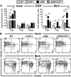
Figure 1.. The onset of ileitis in TNFARE mice is characterized by increased TH1/TH17 CD4+ T cells expressing CCR7.
(A and D) Cellular composition of WT and TNFARE (ARE) ileal infiltrate and MLN, with correlation of CD4+ and CD4+ CD44+ (TEM; CD44high CD62Lneg) lymphocytes by flow cytometry. (B and E) Flow cytometry analysis of CD4+ T cells from ilea and MLN of WT and TNFARE mice assessing the relative expression of IFN-γ and IL-17a. Gating was performed on live, CD45+MHCIIneg CD4+ T cells from indicated compartments at 8–10 wk of age. (C and F) Relative mRNA expression of CCR7 from sorted CD4+CD44+ from MLN and ileum of WT and TNFARE mice. Data expressed as mean ± sem; *P < 0.05; **P < 0.01 versus age-matched WT mice from 2 independent experiments (n = 5 mice/strain; A and D, ANOVA; C and F, t-test).
Figure 2.
Figure 2.. CCR7 deficiency exacerbates ileitis in TNFARE mice.
(A and B) Cellular composition of WT, CCR7−/−, TNFARE/CCR7+/+ (ARE) versus TNFARE/CCR7−/− (ARE/CCR7−/−) ileal infiltrate and MLN. (C) Histologic assessment of TNFARE/CCR7+/+ and TNFARE/CCR7−/− mice ilea at indicated ages, according to acute and chronic leukocytic infiltrates, villus distortion, and a combinatorial total inflammatory index. (D) Representative micrographs of ileum H&E from TNFARE/CCR7+/+ and TNFARE/CCR7−/− mice between 4 and 20 wk of age. Data expressed as mean ± sem; *P < 0.05; **P < 0.01 versus age-matched WT mice (n = 10–18 mice/strain, ANOVA). Original scale bars, 100 μm.
Figure 3.
Figure 3.. CCR7 deficiency increased ileal retention of CD4+ TEM and inhibits egress to draining MLN.
(A) Flow cytometric analyses characterizing the absolute cell numbers of TEM, TCM, and TNaive CD4+ T cells in the ilea and MLN of WT, CCR7−/−, ΔARE+/+ and ΔARE−/− mice. (B) Representative flow cytometry contour plots depicting frequency of TEM, TCM, and TNaive CD4+ T cells in the ilea and MLN of ΔARE/CCR7+/+ or ΔARE/CCR7−/− and WT mice. Data expressed as mean ± sem; **P < 0.01 versus its indicated counterpart from 3 independent experiments (n = 6 mice/strain, ANOVA).
Figure 4.
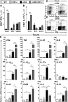
Figure 4.. Altered TH1/TH17 CD4+ ratio in ARE/CCR7−/− mice.
(A–D) Absolute numbers and representative flow cytometry plots of IFN-γ+ versus IL-17a+ from ileum and MLN CD4+ T cells. Gating was performed on live, CD45+ MHCIIneg CD4+ T cells at 8–10 wk of age. Data expressed as mean ± sem; **P < 0.01 versus age-matched indicated counterparts from 2 independent experiments (t-test). (E, I–XII) Cytokine mRNA analysis was performed by real-Time PCR on ileal tissues from CCR7-sufficient or -deficient WT and ARE/CCR7+/+ or ∆ARE/CCR7−/− mice at 10 wk of age. Data expressed as mean ± sem; *P < 0.05; **P < 0.01 versus indicated counterparts from 2 independent experiments (n = 6 mice/strain, ANOVA).
Figure 5.
Figure 5.. Changes in CD103+ versus CD11b+ DC subsets, loss of RALDH enzymes, and altered Treg profile in ARE/CCR7−/− mice.
(A) Frequency of ileal CD103+ and CD11b+ DC population and representative flow cytometry plots of ileal CD103+ and CD11b+ DC. SSC-A, Side-scatter-area. (B) Relative mRNA expression analysis of ileal RALDHs (RALDH1, RALDH2, RALDH3) from indicated genotypes. (C) Absolute numbers of CD4+CD25+FoxP3+ Tregs from the ileum of CCR7-sufficient or -deficient WT and ARE/CCR7+/+ or ∆ARE/CCR7−/− mice. (D) Frequency of MLN CD103+ and CD11b+ DC population. (E) Relative mRNA expression analysis of RALDHs (RALDH1, RALDH2, RALDH3) in the MLN from indicated genotypes. (F) Absolute numbers of CD4+CD25+FoxP3+ Tregs from the MLN of CCR7-sufficient or -deficient WT and ARE/CCR7+/+ or ∆ARE/CCR7−/− mice. Experiments were performed from indicated compartments and genotypes at 8–10 wk of age. Data expressed as mean ± sem from 3 independent experiments; *P < 0.05; **P < 0.01 (n = 4–6 mice/strain, ANOVA).
Figure 6.
Figure 6.. Antibody blockade of CCR7 exacerbates ileitis in TNFΔARE mice by inhibiting lymphatic egress and promoting retention of effector/memory CD4+ T cells.
Eight-wk-old TNFARE mice received 4 injections (i.p.) of anti-CCR7 (4B12; 500 μg) or IgG2a vehicle every 4 days for 2 wk. (A) Histology indices were assessed for ileal tissue post-treatment with representative micrographs. (B) Cytokine mRNA analysis was performed by real-time PCR on ileal tissues of isotype (IgG2a)- and anti-CCR7-treated mice. (C) Frequency of CD103+ and CD11b+ DC subsets from ilea post-treatment. Data expressed as mean ± sem; *P < 0.05; **P < 0.01 versus IgG2a from 2 independent experiments (n = 6–7 mice/treatment). (A and B) t-test versus IgG2a; (C) ANOVA). Original scale bars, 100 μm.
Figure 7.
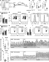
Figure 7.. CCR7-deficient CD4+ effector T cells from TNFΔARE mice drive a dysregulated ileo-colitis following adoptive transfer into RAG1−/− mice.
CD4+ TEM (CD44high CD62Lneg) from the spleen of 4- to 6-wk-old ΔARE/CCR7+/+ and ΔARE/CCR7−/− mice were isolated by FACS and adoptively transferred (1 × 10⁶; i.p.) into RAG1−/− recipients. (A) Representative assessment of CCR7 on adoptively transferred CD4+ T cells by flow cytometry. (B) Cell counts were assessed 8 wk post-transfer in ilea, colon, and MLN. (C and D) Percentage of Ki67+ in proliferating CD4+ T cells in the ilea and colon and representative flow cytometry plots from the ileum. (E) Representative flow cytometry histograms of ileal Tbet+ and RORγt+ CD4+. MFI, Mean fluorescence intensity. (F and G) Percentage of ileal IFN-γ+ CD4+ T cells and representative flow cytometry plots. (H and I) Percentage of colonic IL-17a+ CD4+ and representative flow cytometry plots. (J and K) Total inflammatory indices from adoptively transferred RAG1−/− recipient ilea and colons. Data expressed as mean ± sem; *P < 0.05; **P < 0.01 versus ΔARE/CCR7+/+ from 2 independent experiments (n = 6–7 mice/treatment). (B, J, and K, t-test; F and H, ANOVA). Original scale bars, 100 μm.
Figure 8.
Figure 8.. ∆ARE/CCR7−/− mice induce multiorgan inflammation and associated pathology.
(A) Histologic assessment and pathologic evaluation of ∆ARE/CCR7+/+ and ∆ARE/CCR7−/− mice at 10 wk of age. (B) Representative micrographs of tissue H&E from ∆ARE/CCR7+/+ and ∆ARE/CCR7−/− mice. (C) Representative macroscopic assessment at necropsy of ∆ARE/CCR7−/− mice displays a clear inability to thrive by 8 wk of age. Data expressed as mean ± sem; *P < 0.05; ***P < 0.001 versus age-matched ∆ARE/CCR7+/+ counterparts (n = 3–5 mice/strain, t-test). Original scale bars, 100 μm.

Comment in

References

    1. Maynard C. L., Weaver C. T. (2009) Intestinal effector T cells in health and disease. Immunity 31, 389–400. - PMC - PubMed
    1. Veny M., Esteller M., Ricart E., Piqué J. M., Panés J., Salas A. (2010) Late Crohn’s disease patients present an increase in peripheral Th17 cells and cytokine production compared with early patients. Aliment. Pharmacol. Ther. 31, 561–572. - PubMed
    1. Sakuraba A., Sato T., Kamada N., Kitazume M., Sugita A., Hibi T. (2009) Th1/Th17 immune response is induced by mesenteric lymph node dendritic cells in Crohn’s disease. Gastroenterology 137, 1736–1745. - PubMed
    1. Bromley S. K., Thomas S. Y., Luster A. D. (2005) Chemokine receptor CCR7 guides T cell exit from peripheral tissues and entry into afferent lymphatics. Nat. Immunol. 6, 895–901. - PubMed
    1. Debes G. F., Arnold C. N., Young A. J., Krautwald S., Lipp M., Hay J. B., Butcher E. C. (2005) Chemokine receptor CCR7 required for T lymphocyte exit from peripheral tissues. Nat. Immunol. 6, 889–894. - PMC - PubMed

Publication types

MeSH terms