Review
. 2015 Mar;117(2-3):157-165.
doi: 10.1016/j.pbiomolbio.2015.01.007. Epub 2015 Jan 29.

Molecular underpinnings of Aprataxin RNA/DNA deadenylase function and dysfunction in neurological disease

Affiliations
Review

Molecular underpinnings of Aprataxin RNA/DNA deadenylase function and dysfunction in neurological disease

Matthew J Schellenberg et al. Prog Biophys Mol Biol. 2015 Mar.

Abstract

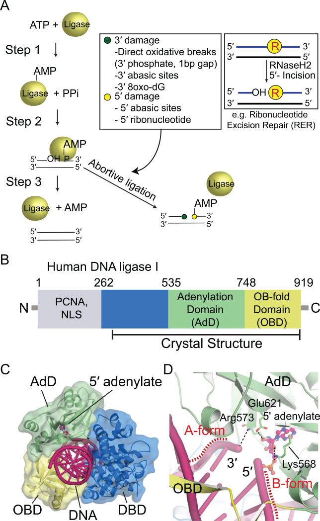
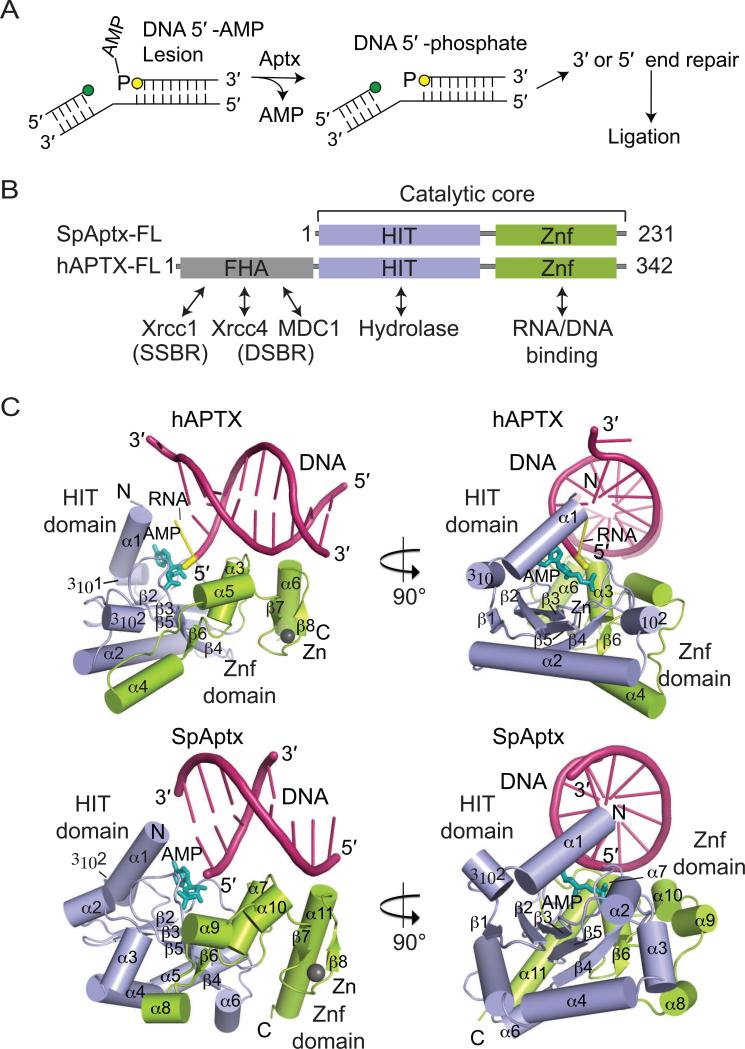
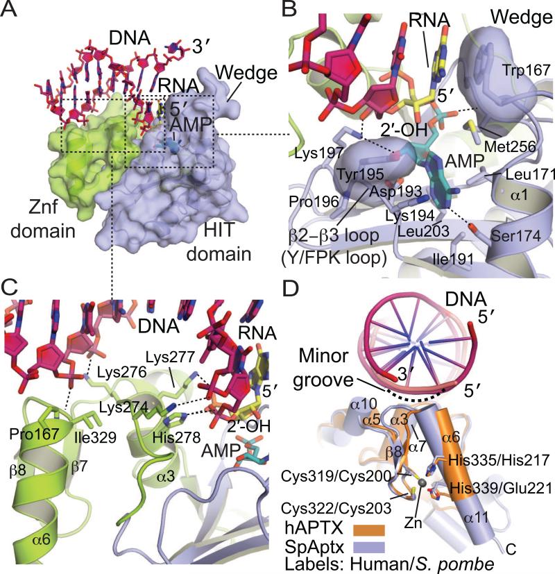
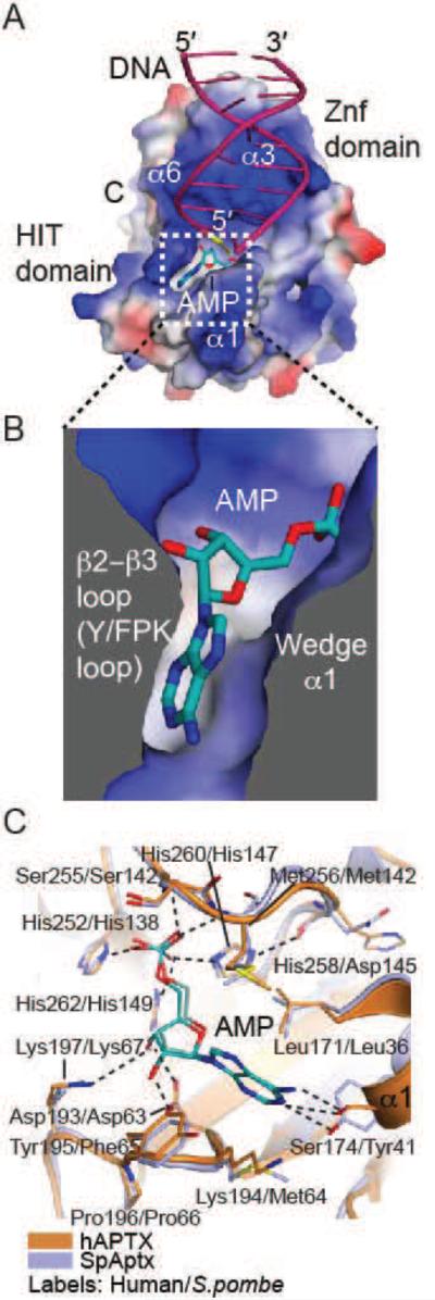
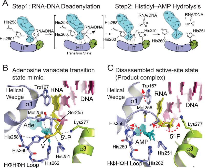
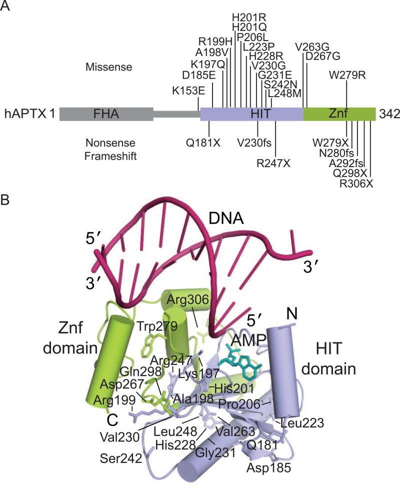
Eukaryotic DNA ligases seal DNA breaks in the final step of DNA replication and repair transactions via a three-step reaction mechanism that can abort if DNA ligases encounter modified DNA termini, such as the products and repair intermediates of DNA oxidation, alkylation, or the aberrant incorporation of ribonucleotides into genomic DNA. Such abortive DNA ligation reactions act as molecular checkpoint for DNA damage and create 5'-adenylated nucleic acid termini in the context of DNA and RNA-DNA substrates in DNA single strand break repair (SSBR) and ribonucleotide excision repair (RER). Aprataxin (APTX), a protein altered in the heritable neurological disorder Ataxia with Oculomotor Apraxia 1 (AOA1), acts as a DNA ligase "proofreader" to directly reverse AMP-modified nucleic acid termini in DNA- and RNA-DNA damage responses. Herein, we survey APTX function and the emerging cell biological, structural and biochemical data that has established a molecular foundation for understanding the APTX mediated deadenylation reaction, and is providing insights into the molecular bases of APTX deficiency in AOA1.

Keywords: AOA1; Aprataxin; Aptx; DNA damage response; DNA ligase; Neurodegenerative disease.


Figures

Fig. 1
Fig. 1
DNA ligation and sources of abortive ligation. (A) Three-step ligation reaction utilized by ATP-dependent DNA ligases. Repair of the DNA backbone is coupled to ATP hydrolysis. Inset (left): Lesions found at the 3' (green circle) and 5' (yellow circle) terminal ends of the nicked strands that can trigger abortive ligation are listed in the box. Inset (right): an example of generation of non-canonical DNA termini during ribonucleotide excision repair that triggers abortive ligation is diagrammed. (B) Human DNA ligase I (LigI) domain architecture. The N-terminal domain (gray) mediates nuclear localization and protein interactions with PCNA. The DBD (blue), AdD (green), and OBD (yellow) contain key catalytic residues that comprise the catalytic core of eukaryotic DNA ligases. (C) X-ray structure of LigI/DNA complex (PDB 1X9N) showing three domains (DBD, AdD, OBD) encircle the 5’-adenylated DNA intermediate. DNA is shown in pink and 5’-adenylate in magenta. (D) Structure of the active site of LigI showing key catalytic residues from AdD and OBD that align the nicked strands for the final strand–sealing reaction.
Fig. 2
Fig. 2
APTX and deadenylation of 5’-adenylated intermediates. (A) Lesions at the 3’ (green circle) or 5’ (yellow circle) terminal ends of the nicked strands can trigger abortive ligation. APTX hydrolyses 5’-adenylates to restore the 5’-phosphate of the nicked strand. Other DNA repair pathways can remove the lesions before DNA ligase reseals the nick. (B) Domain architectures of hAPTX and SpAptx. The N-terminal FHA domain (gray) mediates protein interactions and cellular localization in humans. The Histidine Triad domain (HIT) (blue) and the Zn finger motif-containing C-term domain (Znf, orange) comprise the catalytic core of APTX. (C) Orthogonal views of the hAPTX (PBD 4NDF) and SpAptx(PDB 3SZQ) X–ray structures. The HIT (blue) and Znf (green) domains are represented as cylindrical α–helices and β-strands. DNA is colored in pink, 5'-ribonucleotide in yellow, AMP in teal, and Zn in gray.
Fig. 3
Fig. 3
APTX–DNA interactions. (A) Molecular details of the APTX Znf-DNA interactions. The HIT (blue) and Znf (green) domains are represented as cylindrical α–helices and β-strands. DNA is colored in pink, 5’-ribonucleotide in yellow, and AMP in teal. (B) Molecular details of the APTX HIT-DNA interactions, colored as in panel A (C) Surface representation of the APTX HIT-Znf DNA interaction interface. An extended DNA binding surface comprised of the HIT (blue) and Znf (green) mediates DNA (pink) contacts. (D) Superposition of the Znf domains of APTX (orange, PDB 4NDF) and SpAptx (blue, PDB 3SZQ). DNA is colored in pink and Zn in gray. C2H2 (hAPTX) and C2HE (SpAptx) Znf motifs adopt a similar fold and the Zn is coordinated by four zinc binding residues (Cys319, Cys322, His335 and His339 of hAPTX and Cys200, Cys203, His217, Glu221 of SpAptx).
Fig. 4
Fig. 4
APTX AMP interactions. (A) Surface potential charge representation (red – negatively charged, blue – positively charged, gray – neutral/hydrophobic) of APTX with the bound AMP (teal), 5’ terminal RNA (yellow), and DNA (pink). The AMP binding pocket is outlined in a dotted-line box. (B) Helix α1 (Wedge) and β2-β3 loop (Y/FPK loop) elements comprise the AMP binding pocket. (C) Structure of the AMP binding pockets of APTX (orange, PDB 4NDF) and SpAptx (blue, PDB 3SZQ), showing conserved active–site architecture. AMP is colored in teal.
Fig. 5
Fig. 5
APTX deadenylation reaction mechanism. (A) Steps of the proposed two–step APTX reaction mechanism. APTX HIT domain is colored in blue, Znf in green, AMP in teal. (B) Molecular details of active site environment during the transition state shown in the structure of APTX/AMP/RNA-DNA/Vanadate transition state mimic complex (PDB 4NDG). (C) Molecular details of the active-site disassembled state shown by the structure of APTX bound to product RNA/DNA (PDB 4NDF).
Fig. 6
Fig. 6
Ataxia Oculomotor Apraxia 1 (AOA1) mutations. (A) Residues mutated in AOA1 are located in the HIT (blue) and Znf (green) domains of APTX. (B) The location of residues mutated in AOA1 mapped onto the X-ray structure of human APTX (PDB 4NDF). DNA is colored in pink, AMP in teal.

Similar articles

Cited by

References

    1. Ahel I, Rass U, El-Khamisy SF, Katyal S, Clements PM, McKinnon PJ, Caldecott KW, West SC. The neurodegenerative disease protein aprataxin resolves abortive DNA ligation intermediates. Nature. 2006;443:713–716. - PubMed
    1. Aicardi J, Barbosa C, Andermann E, Andermann F, Morcos R, Ghanem Q, Fukuyama Y, Awaya Y, Moe P. Ataxia-ocular motor apraxia: a syndrome mimicking ataxia-telangiectasia. Ann. Neurol. 1988;24:497–502. - PubMed
    1. Andres SN, Schellenberg MJ, Wallace BD, Tumbale P, Williams RS. Recognition and repair of chemically heterogeneous structures at DNA ends. Environ. Mol. Mutagen. 2014 doi: 10.1002/em.21892. [Epub ahead of print] - PMC - PubMed
    1. Baba Y, Uitti RJ, Boylan KB, Uehara Y, Yamada T, Farrer MJ, Couchon E, Batish SD, Wszolek ZK. Aprataxin (APTX) gene mutations resembling multiple system atrophy. Parkinsonism Relat. Disord. 2007;13:139–142. - PubMed
    1. Becherel OJ, Gueven N, Birrell GW, Schreiber V, Suraweera A, Jakob B, Taucher-Scholz G, Lavin MF. Nucleolar localization of aprataxin is dependent on interaction with nucleolin and on active ribosomal DNA transcription. Hum Mol Genet. 2006;15:2239–2249. - PubMed

Publication types

MeSH terms

Supplementary concepts

LinkOut - more resources