. 2015 Mar:321:52-64.
doi: 10.1016/j.heares.2015.01.008. Epub 2015 Jan 28.

Deletion of Shank1 has minimal effects on the molecular composition and function of glutamatergic afferent postsynapses in the mouse inner ear

Affiliations

Deletion of Shank1 has minimal effects on the molecular composition and function of glutamatergic afferent postsynapses in the mouse inner ear

Jeremy P Braude et al. Hear Res. 2015 Mar.

Abstract

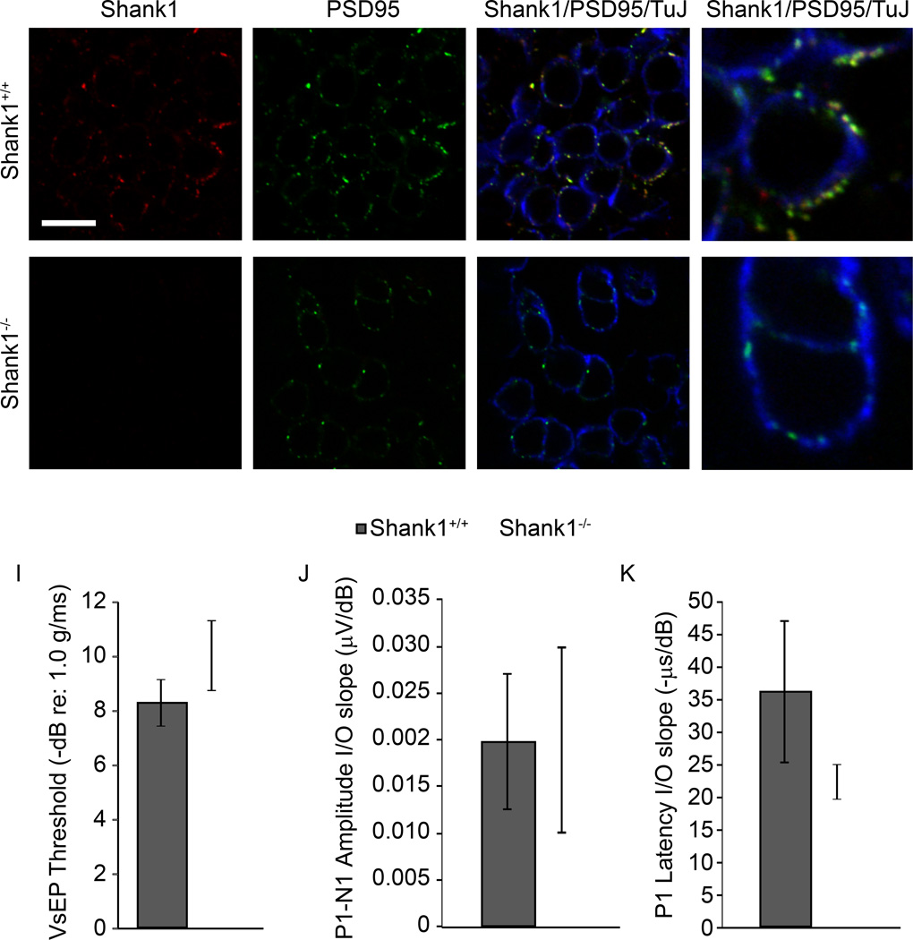
Shank proteins (1-3) are considered the master organizers of glutamatergic postsynaptic densities in the central nervous system, and the genetic deletion of either Shank1, 2, or 3 results in altered composition, form, and strength of glutamatergic postsynapses. To investigate the contribution of Shank proteins to glutamatergic afferent synapses of the inner ear and especially cochlea, we used immunofluorescence and quantitative real time PCR to determine the expression of Shank1, 2, and 3 in the cochlea. Because we found evidence for expression of Shank1 but not 2 and 3, we investigated the morphology, composition, and function of afferent postsynaptic densities from defined tonotopic regions in the cochlea of Shank1(-/-) mice. Using immunofluorescence, we identified subtle changes in the morphology and composition (but not number and localization) of cochlear afferent postsynaptic densities at the lower frequency region (8 kHz) in Shank1(-/-) mice compared to Shank1(+/+) littermates. However, we detected no differences in auditory brainstem responses at matching or higher frequencies. We also identified Shank1 in the vestibular afferent postsynaptic densities, but detected no differences in vestibular sensory evoked potentials in Shank1(-/-) mice compared to Shank1(+/+) littermates. This work suggests that Shank proteins play a different role in the development and maintenance of glutamatergic afferent synapses in the inner ear compared to the central nervous system.


Figures

Figure 1
Figure 1. Shank1 is a component of inner hair cell (IHC) afferent postsynaptic densities (PSD)
Organs of Corti from 4 week old mice were immunostained with either a mouse monoclonal IgG1 antibody against CTBP2 (green, A) or rabbit polyclonal antibody against Shank1 (green, E) and a mouse monoclonal IgG2A antibody against PSD95 (red, B,F). Observations of individual samples revealed that almost every presynaptic CTBP2-positive ribbon was juxtaposed to a PSD95-positive PSD and vice versa (C) and that almost every PSD95-positive PSD also expressed Shank1 immunoreactivity and vice versa (G). Images are presented as Z-projections of a stack of confocal micrographs from the 32 kHz region. Mean values (± SEM) of CTBP2-positive presynaptic ribbons (D) or Shank1-positive PSDs (H) and PSD95-positive PSDs across samples are compared at two tonotopic regions (8 and 32 kHz). Statistically significant differences are indicated with an asterisk.
Figure 2
Figure 2. Shank proteins are not detected in the cochleae of inner hair cell (IHC) afferent postsynaptic densities (PSD) from Shank1−/− mice nor is there compensatory upregulation of Shank2 or Shank3 in cochlea from Shank1−/− mice
Organs of Corti from 4 week old Shank1+/+ (A–D) and Shank1−/− (E–H) littermate mice were immunostained with a mouse monoclonal IgG1 antibody against CTBP2 (blue, A and E), a rabbit polyclonal antibody against Shank1 (red, B and F) and a goat polyclonal antibody that recognizes all three Shank isoforms (panShank, green, C and G). Colocalized Shank1 and panShank immunoreactivity was observed juxtaposed to CTBP2-positive presynaptic ribbons in Shank1+/+ mice (D). Although CTBP2-positive presynaptic ribbons were observed, neither Shank1 nor panShank immunoreactivity was observed in Shank1−/− mice (H). Images are presented as Z-projections of a stack of confocal micrographs from the 16 kHz region. Shank1–3 transcript expression was investigated in the cochleae of Shank1+/+ mice (and presented normalized to control gene expression in Shank1+/+ mice, I). Shank2–3 transcript expression was investigated in Shank1−/− mice (and presented relative to Shank2 and Shank3 expression in Shank1+/+ mice, J). Two-fold or more greater changes in expression are indicated with asterisks.
Figure 3
Figure 3. Cochleae from Shank1−/− mice show minimal changes in transcript abundances for genes encoding GluA1–4 and GKAP
Gria1–4 and Dlgap1 transcript expression was investigated in the cochleae of Shank1+/+ mice (normalized to control gene expression in Shank1+/+ mice, A) and Shank1−/− mice (relative to Gria1–4 and Dlgap1 expression in Shank1+/+ mice, B). Two-fold or more greater changes in expression are indicated with asterisks.
Figure 4
Figure 4. Inner hair cell (IHC) afferent postsynaptic densities (PSDs) from Shank1−/− mice show subtle changes in their morphology and composition
Organs of Corti from 4 week old Shank1+/+ (A-D,I,K) and Shank1−/− (E-H,J,L) littermate mice were immunostained with a mouse monoclonal IgG1 antibody against CTBP2 (green, A and E), a rabbit polyclonal antibody against GluA2/3 (red, B and F,I-L), and a mouse monoclonal IgG2B antibody against GKAP (blue, C and G,I-L). Colocalized GluA2/3 and GKAP immunoreactivity was observed juxtaposed to CTBP2-positive presynaptic ribbons in both Shank1+/+ (D) and Shank1−/− mice (H). Images are presented as Z-projections through a stack of confocal micrographs from the 32 kHz region (AH) or as 3D reconstructions comparing the 8 and 32 kHz region (I–L) from Shank1+/+ and Shank1−/− mice. Mean values (± SEM) of GluA2/3-positive and GKAP-positive immunopuncta per hair cell (M) and mean values (± SEM) of GluA2/3-positive and GKAP-positive immunopuncta volumes (N) for each genotype are compared at two tonotopic regions (8 and 32 kHz). Statistically significant differences are indicated with an asterisk.
Figure 5
Figure 5. Auditory brainstem responses are comparable in Shank1+/+ and Shank1−/− mice
ABR raw traces (A), thresholds (B), P1-N1 amplitude (I/O) slopes (C), and P1 latency I/O slopes (D) are compared between genotypes across frequencies.
Figure 6
Figure 6. Vestibular function is comparable in Shank1+/+ and Shank1−/− mice
The vestibular sensory epithelia of the utricular maculae was isolated from 4 week old Shank1+/+ (A–D) and Shank1−/− (E–H) mice and immunostained with a rabbit polyclonal antibody against Shank1 (red, A and E), a mouse monoclonal IgG2A antibody against PSD95 (green, B and F), and a mouse monoclonal antibody against tubulin J (TuJ, blue, C and G). Shank1 immunoreactivity was colocalized to PSD95-positive PSDs observed in TuJ-positive calyx and bouton afferent terminals in Shank1+/+ mice (D). In contrast, no Shank1 immunoreactivity was observed in PSD95-positive PSDs in Shank1−/− mice (H). Images are presented as a single optical section from the striolar region. Vestibular evoked potentials (VsEPs) showed comparable thresholds (I), P1-N1 amplitude (I/O) slopes (J) and P1 latency I/O slopes (K) between genotypes.

References

    1. Beurg M, Michalski N, Safieddine S, Bouleau Y, Schneggenburger R, Chapman ER, Petit C, Dulon D. Control of exocytosis by synaptotagmins and otoferlin in auditory hair cells. J Neurosci. 2010;30:13281–13290. - PMC - PubMed
    1. Boeckers TM, Winter C, Smalla KH, Kreutz MR, Bockmann J, Seidenbecher C, Garner CC, Gundelfinger ED. Proline-rich synapse-associated proteins ProSAP1 and ProSAP2 interact with synaptic proteins of the SAPAP/GKAP family. Biochem Biophys Res Commun. 1999;264:247–252. - PubMed
    1. Boeckers TM, Segger-Junius M, Iglauer P, Bockmann J, Gundelfinger ED, Kreutz MR, Richter D, Kindler S, Kreienkamp HJ. Differential expression and dendritic transcript localization of Shank family members: identification of a dendritic targeting element in the 3’ untranslated region of Shank1 mRNA. Mol Cell Neurosci. 2004;26:182–190. - PubMed
    1. Burkard R, Feldman M, Voigt HF. Brainstem auditory-evoked response in the rat. Normative studies, with observations concerning the effects of ossicular disruption. Audiology. 1990;29:146–162. - PubMed
    1. Chen Z, Kujawa SG, Sewell WF. Auditory sensitivity regulation via rapid changes in expression of surface AMPA receptors. Nat Neurosci. 2007;10:1238–1240. - PubMed

Publication types

MeSH terms