. 2015 Apr 2:290:227-42.
doi: 10.1016/j.neuroscience.2015.01.028. Epub 2015 Jan 28.

GABAergic somatostatin-immunoreactive neurons in the amygdala project to the entorhinal cortex

Affiliations

GABAergic somatostatin-immunoreactive neurons in the amygdala project to the entorhinal cortex

A J McDonald et al. Neuroscience. .

Abstract

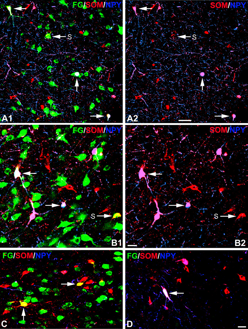
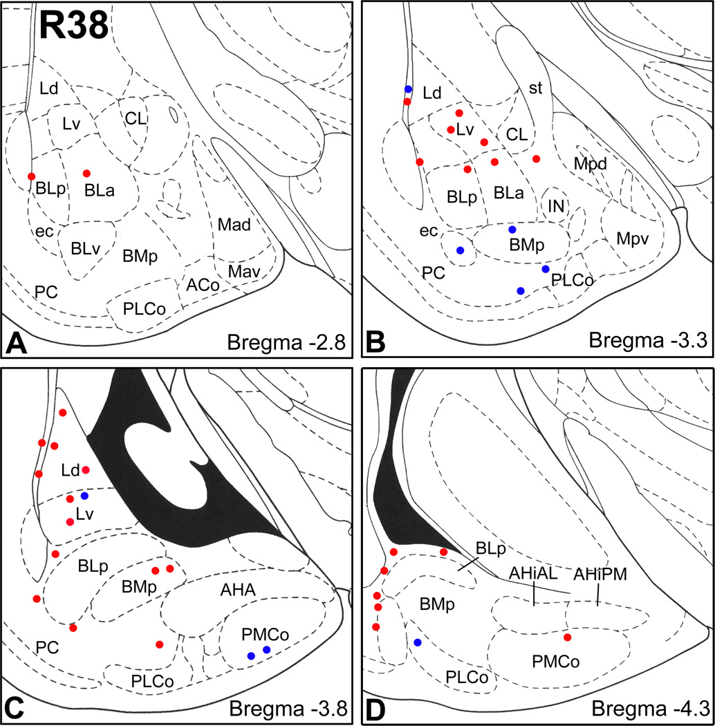
The entorhinal cortex and other hippocampal and parahippocampal cortices are interconnected by a small number of GABAergic nonpyramidal neurons in addition to glutamatergic pyramidal cells. Since the cortical and basolateral amygdalar nuclei have cortex-like cell types and have robust projections to the entorhinal cortex, we hypothesized that a small number of amygdalar GABAergic nonpyramidal neurons might participate in amygdalo-entorhinal projections. To test this hypothesis we combined Fluorogold (FG) retrograde tract tracing with immunohistochemistry for the amygdalar nonpyramidal cell markers glutamic acid decarboxylase (GAD), parvalbumin (PV), somatostatin (SOM), neuropeptide Y (NPY), vasoactive intestinal peptide (VIP), and the m2 muscarinic cholinergic receptor (M2R). Injections of FG into the rat entorhinal cortex labeled numerous neurons that were mainly located in the cortical and basolateral nuclei of the amygdala. Although most of these amygdalar FG+ neurons labeled by entorhinal injections were large pyramidal cells, 1-5% were smaller long-range nonpyramidal neurons (LRNP neurons) that expressed SOM, or both SOM and NPY. No amygdalar FG+ neurons in these cases were PV+ or VIP+. Cell counts revealed that LRNP neurons labeled by injections into the entorhinal cortex constituted about 10-20% of the total SOM+ population, and 20-40% of the total NPY population in portions of the lateral amygdalar nucleus that exhibited a high density of FG+ neurons. Sixty-two percent of amygdalar FG+/SOM+ neurons were GAD+, and 51% were M2R+. Since GABAergic projection neurons typically have low perikaryal levels of GABAergic markers, it is actually possible that most or all of the amygdalar LRNP neurons are GABAergic. Like GABAergic LRNP neurons in hippocampal/parahippocampal regions, amygdalar LRNP neurons that project to the entorhinal cortex are most likely involved in synchronizing oscillatory activity between the two regions. These oscillations could entrain synchronous firing of amygdalar and entorhinal pyramidal neurons, thus facilitating functional interactions between them, including synaptic plasticity.

Keywords: entorhinal cortex; long-range GABAergic neurons; nonpyramidal neurons; parasubiculum; prefrontal cortex; retrograde tract tracing.


Figures

Figure 1
Figure 1
Drawings showing the locations of the FG injection sites in the brains used for this study. The letters “L” (left) and “R” (right) in two of the brains (R40 and R41) indicate whether the injection site in these two bilaterally-injected rats was on the left or right side. Drawings of brain sections are from the atlas by Paxinos and Watson (1997). Abbreviations: AID, dorsal agranular insular cortex; AIP, posterior agranular insular cortex; AIV, ventral agranular insular cortex; DI, dysgranular insular cortex; DLEA, dorsolateral entorhinal area, GI, granular insular cortex; IL, infralimbic cortex; PaS, parasubiculum; PL, prelimbic cortex; S2, secondary somatosensory cortex; VLEA, ventrolateral entorhinal area; VMEA, ventromedial entorhinal area; VS, ventral subiculum. Scale bar = 1 mm.
Figure 2
Figure 2
A) Photomicrograph of the FG injection site in case R13 in an immunoperoxidasestained section that was counterstained with pyronin Y. The injection site mainly involved the DLEA. Arrows indicate the pipette track traversing the lateral part of the ventral subiculum (VS). B) Photomicrograph of FG retrogradely-labeled neurons in the amygdala (black) at the bregma −3.1 level in case R13 in an immunoperoxidase-stained section. Abbreviations: BLa, anterior basolateral nucleus; BLp, posterior basolateral nucleus; BLv, ventral basolateral nucleus; BMp, posterior basomedial nucleus; CL, lateral central nucleus; DLEA, dorsolateral entorhinal area; Ld, dorsolateral lateral nucleus; Lv, ventromedial lateral nucleus; Mpd, posterodorsal medial nucleus; Mpv, posteroventral medial nucleus; PC, piriform cortex; PLCo, posterolateral cortical nucleus; VLEA, ventrolateral entorhinal area. Scale bars = 500 µm in A and B.
Figure 3
Figure 3
Plots of FG+ retrogradely-labeled neurons at three rostrocaudal levels (Bregma −1.8, −2.8, and −3.8) in cases R13, R41L and R38. Each dot represents one cell. Sections were stained with a Nissl stain (pyronin Y) to determine nuclear borders. See Fig. 2 for most abbreviations. Additional abreveviations: ACo, anterior cortical nucleus; AHA, amygdalohippocampal area; BAOT, bed nucleus of the accessory olfactory tract; BMa, anterior basomedial nucleus; CLC, lateral capsular subdivision of the central nucleus; CM, medial central nucleus; ec, external capsule; IN, intercalated nucleus; L, lateral nucleus; Mad, anterodorsal medial nucleus; Mav, anteroventral medial nucleus; ot, optic tract; PMCo, posteromedial cortical nucleus; s, commissural portion of the stria terminalis.
Figure 4
Figure 4
Photomicrographs of FG/SOM/NPY triple labeling in the amygdala. FG is green, SOM is red, NPY is blue, FG+/SOM+ structures are yellow, FG+/SOM+/NPY+ structures are white, and FG−/SOM+/NPY+ structures in triple-labeled sections are magenta. A1) FG/SOM/NPY triple labeling in the lateral nucleus with a FG injection into the caudal DLEA (case R38). This field contains three FG+/SOM+/NPY+ triple-labeled neurons (unlabeled arrows) and one FG+/SOM+ double-labeled neuron (S). A2) The same field as A1, but with the green channel omitted. Note that the three triple-labeled neurons in A1 (unlabeled arrows) are SOM+/NPY+ (magenta) and constitute a small subpopulation of the SOM+ neurons. B1) FG/SOM/NPY triple labeling in the lateral nucleus with a FG injection into the rostral DLEA (case R13). This field contains two FG+/SOM+/NPY+ triple-labeled neurons (unlabeled arrows) and one FG+/SOM+ double-labeled neuron (S). B2) The same field as B1, but with the green channel omitted. The two triple-labeled neurons in B1 (unlabeled arrows) are SOM+/NPY+ (magenta). C) FG/SOM/NPY triple labeling in the posteromedial cortical nucleus in a case with a FG injection into the VMEA (case R40L). There are two FG+/SOM+ double-labeled neurons (arrows), but no triple-labeled neurons, in this field. D) FG/SOM/NPY triple-labeled neuron (arrow) in the external capsule bordering the posterior basolateral nucleus in a case with a FG injection into the parasubiculum (R40R). Note that this triple-labeled neuron is isolated (i.e., it is not surrounded by other green FG+ retrogradely-labeled neurons). Scale bars = 50 µm in A, 20 µm in B and D (C is the same magnification as D).
Figure 5
Figure 5
Sections arranged from rostral (A) to caudal (F) depicting the locations of FG+ neurons in the amygdala co-expressing SOM and NPY (red dots), expressing SOM but not NPY (blue dots), or expressing NPY but not SOM (green dot) in case R13 (injection: rostral DLEA). Each bregma level shows the locations of neurons plotted from two non-adjacent 50 µm-thick sections at this level; each dot represents one neuron. Templates are modified from the atlas by Paxinos and Watson (1997).
Figure 6
Figure 6
Sections arranged from rostral (A) to caudal (F) depicting the locations of FG+ neurons in the amygdala co-expressing SOM and NPY (red dots) or expressing SOM but not NPY (blue dots) in case R39 (injection: DLEA/VSub). Each bregma level shows the locations of neurons plotted from two non-adjacent 50 µm-thick sections at this level with the exception of bregma levels −1.8 and −2.3 where only one section was available for analysis; each dot represents one neuron. Templates are modified from the atlas by Paxinos and Watson (1997).
Figure 7
Figure 7
Sections arranged from rostral (A) to caudal (D) depicting the locations of FG+ neurons in the amygdala co-expressing SOM and NPY (red dots) or expressing SOM but not NPY (blue dots) in case R38 (injection: caudal DLEA). Since peroxidase preparations revealed relatively few FG+ neurons rostral to bregma −2.8 in this case, sections at these rostral levels were not processed for FG/SOM/NPY immunohistochemistry. Each bregma level shows the locations of neurons plotted from two non-adjacent 50 µm-thick sections at this level; each dot represents one neuron. Templates are modified from the atlas by Paxinos and Watson (1997).
Figure 8
Figure 8
Sections arranged from rostral (A) to caudal (C) depicting the locations of FG+ neurons in the amygdala co-expressing SOM and NPY (red dots) or expressing SOM but not NPY (blue dots) in cases R41R (injection: VLEA), R40L (injection: VMEA) and R40R (injection: parasubiculum). Since peroxidase preparations revealed relatively few FG+ neurons rostral to bregma −3.3 in these cases, sections at these rostral levels were not processed for FG/SOM/NPY immunohistochemistry. Each bregma level shows the locations of neurons plotted from two non-adjacent 50 µm-thick sections at this level; each dot represents one neuron. Templates are modified from the atlas by Paxinos and Watson (1997).
Figure 9
Figure 9
Photomicrographs showing M2R-ir in a subpopulation of amygdalar SOM+ neurons that are retrogradely labeled by FG in a case with a large injection that involved VLEA, VMEA and the ventral subiculum (R54). A1) FG/SOM/M2R triple labeling in the lateral nucleus (FG is green, SOM is red, M2R is blue). A FG+/SOM+/M2R+ triple-labeled neuron (arrow) is white. A2) The same field as A1 showing only SOM-ir with the red channel. A3) The same field as A1 showing only M2R-ir with the blue channel. B1) FG/SOM/M2R triple labeling in the posteromedial cortical nucleus (FG is green, SOM is red, M2R is blue). A FG+/SOM+/M2R+ triple-labeled neuron (unlabeled arrow) is white. Two M2R-negative FG+/SOM+ neurons (S) are yellow. B2) The same field as B1 showing only SOM-ir with the red channel. B3) The same field as B1 showing only M2R-ir with the blue channel. Scale bar = 20 µm in B (A is at the same magnification).
Figure 10
Figure 10
Photomicrographs of FG-labeled neurons in the amygdala that are SOM+/GAD+. A1) FG/SOM/GAD triple labeling in case R51 (FG is green, SOM is red, GAD is blue). A FG+/SOM+/GAD+ triple-labeled neuron in the external capsule (arrow) is white. A FG-negative SOM+/GAD+ double-labeled neuron in the adjacent posterior basolateral nucleus is magenta (arrowhead). A2) The same field as A1 showing only SOM-ir with the red channel. A3) The same field as A1 showing only GAD-ir with the blue channel. B1) FG/SOM/GAD triple labeling in case R54 (FG is green, SOM is red, GAD is blue). Two FG+/SOM+/GAD+ triple-labeled neurons in the lateral nucleus (arrows) are white. This field also contains a GAD+ neuron with very low levels of FG (arrowhead). B2) The same field as B1 showing only the SOM-ir with the red channel. B3) The same field as B1 showing only the GAD-ir with the blue channel. Scale bar = 20 µm in A (B is at the same magnification).

References

    1. Apergis-Schoute J, Pinto A, Paré D. Muscarinic control of long-range GABAergic inhibition within the rhinal cortices. J Neurosci. 2007;27:4061–4071. - PMC - PubMed
    1. Bauer EP, Paz R, Paré D. Gamma oscillations coordinate amygdalo-rhinal interactions during learning. J Neurosci. 2007;27:9369–9379. - PMC - PubMed
    1. Beckstead RM. Afferent connections of the entorhinal area in the rat as demonstrated by retrograde cell-labeling with horseradish peroxidase. Brain Res. 1978;152:249–264. - PubMed
    1. Buzsáki G, Chrobak JJ. Temporal structure in spatially organized neuronal ensembles: a role for interneuronal networks. Curr Opin Neurobiol. 1995;5:504–510. - PubMed
    1. Canteras NS, Simerly RB, Swanson LW. Connections of the posterior nucleus of the amygdala. J Comp Neurol. 1992;324:143–179. - PubMed

Publication types

MeSH terms