. 2015 Feb 1:12:7.
doi: 10.1186/s12985-014-0220-1.

Agnoprotein of polyomavirus BK interacts with proliferating cell nuclear antigen and inhibits DNA replication

Affiliations

Agnoprotein of polyomavirus BK interacts with proliferating cell nuclear antigen and inhibits DNA replication

Nancy Gerits et al. Virol J. .

Abstract

Background: The human polyomavirus BK expresses a 66 amino-acid peptide referred to as agnoprotein. Though mutants lacking agnoprotein are severely reduced in producing infectious virions, the exact function of this peptide remains incompletely understood. To elucidate the function of agnoprotein, we searched for novel cellular interaction partners.

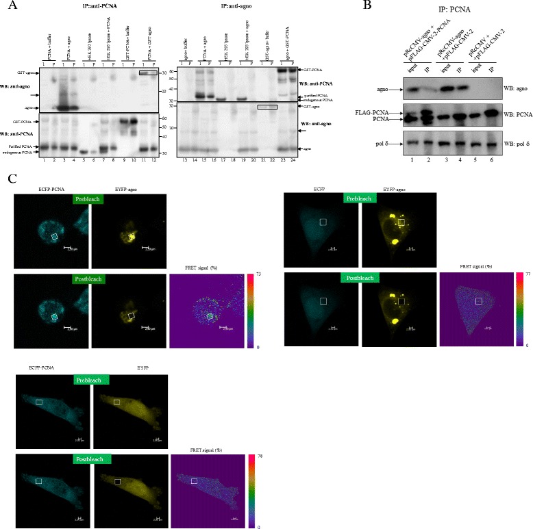
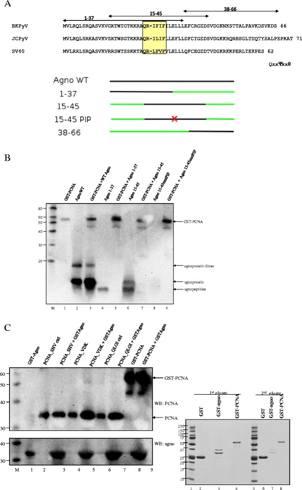
Methods: Yeast-two hybrid assay was performed with agnoprotein as bait against human kidney and thymus libraries. The interaction between agnoprotein and putative partners was further examined by GST pull down, co-immunoprecipitation, and fluorescence resonance energy transfer studies. Biochemical and biological studies were performed to examine the functional implication of the interaction of agnoprotein with cellular target proteins.

Results: Proliferating cell nuclear antigen (PCNA), which acts as a processivity factor for DNA polymerase δ, was identified as an interaction partner. The interaction between agnoprotein and PCNA is direct and occurs also in human cells. Agnoprotein exerts an inhibitory effect on PCNA-dependent DNA synthesis in vitro and reduces cell proliferation when ectopically expressed. Overexpression of PCNA restores agnoprotein-mediated inhibition of cell proliferation.

Conclusion: Our data suggest that PCNA is a genuine interaction partner of agnoprotein and the inhibitory effect on PCNA-dependent DNA synthesis by the agnoprotein may play a role in switching off (viral) DNA replication late in the viral replication cycle when assembly of replicated genomes and synthesized viral capsid proteins occurs.


Figures

Figure 1
Figure 1
BKPyV agnoprotein and PCNA interact in vitro and in vivo. (A) In vitro pull down experiment shows direct interaction between PCNA and agnoprotein. Purified agnoprotein, PCNA or GST-PCNA fusion protein were incubated alone or together in PBS (buffer) or with lysate of HEK293 cells (cell lysate) at 4°C for 1 h. Complexes were immunoprecipitated with PCNA antibodies (IP:anti-PCNA; lanes 1–12) or with antibodies against agnoprotein (IP:anti-agno; lanes 13–24). Samples were run on gel and western blot was performed with antibodies against PCNA (WB:anti-PCNA) or against agnoprotein (WB: anti-agno) as indicated. I = input, P = precipitate. The position of GST-agno, agnoprotein, GST-PCNA, purified PCNA and endogenous PCNA are indicated by arrows. The arrow with dashed line probably represents agnoprotein dimers (see also Figure 2B). (B) Co-immunoprecipitation of agnoprotein and PCNA. HEK293 cells were transfected with following plasmids: lanes 1 and 2: pRcCMV-agno plus pFLAG-CMV-2-PCNA, lanes 3 and 4: pRcCMV-agno plus pFLAG-CMV-2, lanes 5 and 6: pRcCMV plus pFLAG-CMV-2. Lanes 1, 3, and 5: input; lanes 2, 4, and 6: immunoprecipitates (IP). Protein complexes were precipitated with antibodies against PCNA and the presence of polδ, PCNA and agnoprotein was examined with antibodies against these proteins. (C) FRET measurements of the interaction between ECFP-PCNA (donor) and EYFP-agnoprotein (acceptor) fusion proteins in A375 cells by acceptor photo bleaching. FRET efficiency (FRET signal %) is calculated by fluorescence before (prebleach) and after bleaching (postbleach) and shown by the colour code bar. Control experiments with ECFP plus EYFP-agno and ECFP-PCNA plus EYFP were included.
Figure 2
Figure 2
Mapping of the agnoprotein and PCNA regions required for interaction. (A) Amino acid sequences of the agnoproteins of BKPyV, JCPyV, and SV40 (top part) and schematic representation of BKPyV wild-type agnoprotein (agno WT) and truncated peptides (bottom part). The conserved “PIP box like sequence” is indicated. The regions encompassing peptides 1–37, 15–45, and 8–66 are indicated by arrows. The number at the end of the amino acid sequence refers to the number of residues in the protein. The name of the peptides corresponds to the residue numbers. PIP signifies the PCNA Interaction Protein box and the mutation QRIFIF into ARAFIA is symbolized by an X. (B) GST-pull down of purified GST-PCNA fusion protein and purified wild-type agnoprotein or agnopeptides. GST-PCNA (lanes 1, 3, 5, 7, 9) was mixed with full-length agnoprotein (lane 3) or agnoprotein peptide fragments comprising residues 1–37 (lane 5), residues 15–45 (lane 7) or residues 15–45 with mutated PIP motif (lane 9), respectively. The presence of full-length agnoprotein or peptides and PCNA was monitored by immunoblotting using antibodies against agnoprotein and PCNA simultaneously. The band representing agnoprotein dimer (lanes 2 and 3) is indicated. The additional weaker bands in lanes 3, 5, 7 and 9 probably represent degradation products. (C) GST-pull down experiments using purified GST-agnoprotein. Left panel: GST-agnoprotein was incubated with different mutants of PCNA (lanes 3, 5, 7) or GST-PCNA (lane 9) and complexes were pulled down. The presence of agnoprotein and PCNA was investigated by immunoblotting using antibodies against agnoprotein and PCNA, respectively. M = molecular mass marker in kDa. Right panel: Coomassie blue staining of purified GST (lanes 2 and 6), GST-agno (lanes 3 and 7) and GST-PCNA (lanes 4 and 8). Lanes 1 and 5: Precision Plus Protein Dual Color Standards (BioRad) marker.
Figure 3
Figure 3
Agnoprotein inhibits in vitro DNA replication mediated by PCNA:DNApolymerase δ. (A) Left panel: In vitro DNA synthesis by PCNA plus pol δ was monitored in the presence of increasing amounts of agnoprotein. DNA synthesis in the presence of pol δ alone was arbitrary set as 1 and the increase by adding PCNA is shown as fold PCNA stimulation. Right panel: Pol λ-mediated DNA synthesis was monitored in the presence of variable nM ratios of agnoprotein:PCNA. Incorporation of radioactivity was measured by scintillation counting. (B) DNA synthesis was assayed by monitoring elongation of a partially double-stranded 39:72 oligonucleotide dimer (=template DNA). Lanes 1 and 16: 32P-labelled template DNA; lanes 2–5: PCNA and Pol δ were allowed to interact before template DNA and increasing amounts of agnoprotein were added; lanes 6–9: agnoprotein and pol δ were pre-incubated before supplementing PCNA and template DNA; lanes 10–14: agnoprotein and PCNA were mixed prior to the addition of pol δ and template DNA; lane 15: PCNA and template DNA; lane 17: template DNA and pol δ. The upper symbol (=) represents the elongated DNA template, while the lower symbol (− ) is the template. (C) Pol λ-mediated DNA synthesis in the presence of PCNA or agnoprotein. The upper band is ssDNA, the middle band is dsDNA, while the lower band represents incomplete dsDNA. Lanes 2–6: increasing amounts of PCNA were added; lanes 9–14: increasing amounts of agnoprotein were added; lane 1: template DNA; lane 8: DNA was incubated with only pol λ. DNA synthesis was visualized by autoradiography as described in 3B. The upper symbol (=) represents the elongated DNA template, while the lower symbol (− ) is the template.
Figure 4
Figure 4
Agnoprotein inhibits cell proliferation. A375 cells were transfected with empty expression plasmid pRcCMV (CTRL), expression plasmid for agnoprotein (Agno), expression plasmid for PCNA, or expression plasmids for agnoprotein and PCNA, respectively. (A) Cell proliferation was measured by the MTT method 48 hours after transfection. The value obtained for pRcCMV-transfected cells was arbitrary set as 100% and the values for the other cells were related to this. The values are the average of six independent parallels measured in triplicate. The standard deviation is shown and a student’s t-test was performed. Similar results were obtained in a separate experiment. (B) Ectopically expressed agnoprotein and FLAG-tagged PCNA were detected by western blot on lysates of transfected cells using anti-agnoprotein and anti-FLAG-specific antibodies. Lane 1: lysates of cells transfected with empty expression plasmid pRcCMV; lane 2: lysates of cells transfected with pRcCMV-agnoprotein expression plasmid; lane 3: lysates of cells transfected with an expression plasmid encoding FLAG-tagged PCNA; lane 4 lysates of cells co-transfected with expression plasmids for agnoprotein and FLAG-tagged PCNA. To assure equal loading, expression levels of ERK2 in each lysate were determined (bottom panel).

References

    1. Imperiale MJ, Major EO. Polyomaviruses. In: Knipe DM, Howley PM, Griffin DE, Lamb RA, Martin MA, Roizman B, Straus SE, editors. Fields Virology. Philadelphia: Lippincott Williams and Wilkins; 2007. pp. 2263–2298.
    1. DeCaprio JA, Garcea RL. A cornucopia of human polyomaviruses. Nat Rev Microbiol. 2013;11(4):264–276. doi: 10.1038/nrmicro2992. - DOI - PMC - PubMed
    1. Ehlers B, Moens U. Genome Analysis of Non-human primate polyomaviruses. Infect Genet Evol. 2014;26:283–294. doi: 10.1016/j.meegid.2014.05.030. - DOI - PubMed
    1. Schowalter RM, Buck CB. The Merkel cell polyomavirus minor capsid protein. PLoS Pathog. 2013;9(8):e1003558. doi: 10.1371/journal.ppat.1003558. - DOI - PMC - PubMed
    1. Khalili K, Whie MK, Sawa H, Nagashima K, Safak M. The agnoprotein of polyomaviruses: a multifunctional auxiliary protein. J Cell Physiol. 2005;204(1):1–7. doi: 10.1002/jcp.20266. - DOI - PubMed

Publication types

MeSH terms

Substances