. 2015 Apr;157(2):103-13.
doi: 10.1016/j.clim.2015.01.007. Epub 2015 Jan 28.

Involvement of interleukin-18 in the pathogenesis of human eosinophilic esophagitis

Affiliations

Involvement of interleukin-18 in the pathogenesis of human eosinophilic esophagitis

Rituraj Niranjan et al. Clin Immunol. 2015 Apr.

Abstract

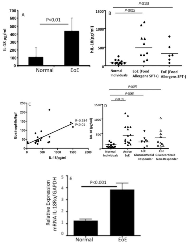
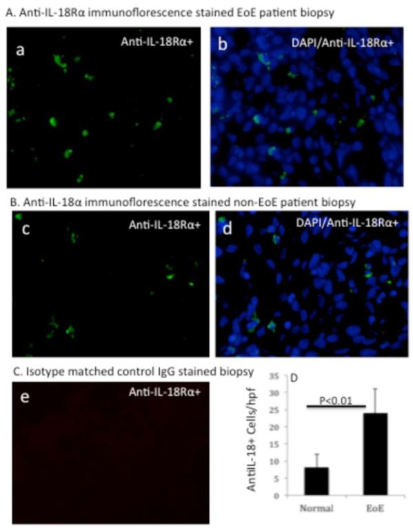
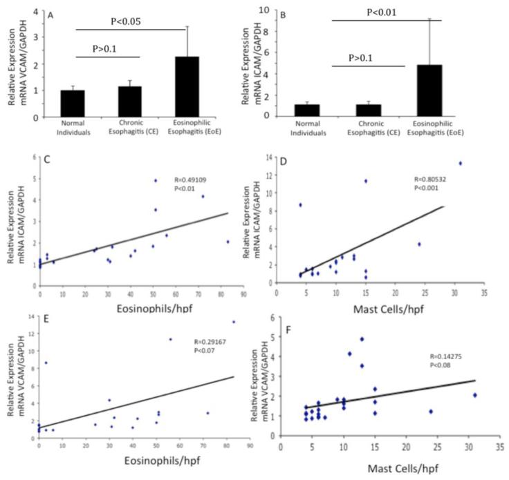
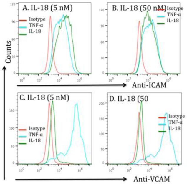
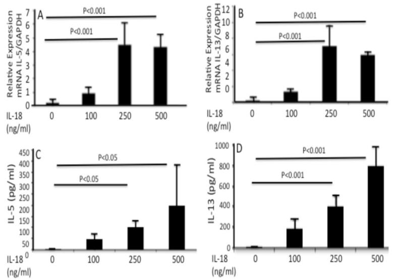
IL-18 is induced in food allergy and EoE is food allergen-induced disease. Therefore, we tested the hypothesis whether IL-18 is involved in food allergen-induced EoE pathogenesis. Accordingly, we examined normal SPT+ and SPT- EoE patient blood and biopsy samples for IL-18, IL-18Rα, ICAM and VCAM expression. Herein, we show increased IL-18 level is highly significant in food allergen SPT+ compared to SPT- EoE patients. We also report that IL-18Rα+ cells and mRNA levels are induced in the esophageal biopsies of EoE patients and blood IL-18 levels correlate with esophageal eosinophilia (P<0.01). Additionally, we report that the levels of esophageal eosinophil and mast cells correlate with ICAM expression in human EoE. Mechanistically, we show that IL-18 in vitro stimulates iNKT cells and endothelial cells and induce eosinophil active cytokines IL-5 and IL-13. We provide the evidence that IL-18 is critical cytokine involved in activation of iNKT cells and ICAM in promoting human EoE.

Keywords: EoE; ICAM; IL-18Rα; Interleukin-IL-18; iNKT cells.


Figures

Figure 1
Figure 1. Induced expression of blood IL-18 and esophageal IL-18Rα+ cells in non-EoE, and active EoE patients
Blood IL-18 was measured in non-EoE (normal) individuals and EoE patient samples by performing ELISA analysis. Additionally the patients are divided to skin test positive (SPT+) and (SPT-) for food allergens. Their blood IL-18 levels are also analyzed and shown (A, B). A correlation between the maximum eosinophil number/hpf of EoE patients with blood IL-18 levels is statistically analyzed in human EoE and is found r=0.58 and p<0.01 (C). The blood IL-18 levels in non-EoE (normal) individuals, active EoE patients (> 24 eosinophils/hpf); patients responsive to glucocorticoid treatment or non-responsive to the treatment groups are analyzed and are shown (D). The relative expression of IL-18Rα mRNA of normal (non-EoE) and EoE patients biopsies are analyzed by real time PCR analysis and is shown (E). Each data point represents an individual patient The data is expressed as mean ± SD. p-values were in between each groups are calculated using Kreskas–Wallis test followed by Dunn’s multiple comparison tests.
Figure 2
Figure 2. Immunofluorescence analysis of esophageal IL-18Rα+ cells in active EoE patients and non-EoE individual
A representative photomicrograph of anti-IL-18Rα FITC conjugated and DAPI mounted esophageal biopsy section of EoE patient and non-EoE individual is shown (A, B). The analysis detected IL-18Rα+ cells (stained green) in EoE patient esophageal biopsy. [A (a)], the overlapped merged figure of DAPI/IL-18Rα+ cells is shown [A, (b)]. A similar analysis was performed in the biopsy of non-EoE individual and shown [B, (c-d)]. The isotype matched control antibody has not detected any staining of IL-18Rα+ cells in EoE patient biopsy [C, (e)]. All photomicrograph shown has original magnification in 400×. The quantitation of IL-18Rα+ cells in normal (comparison control) individuals and EoE patients are shown in [D]. The level of significance was calculated by using the Mann-Whitney test, P <0.01, n= 7-8.
Figure 3
Figure 3. Analysis of IL-18-induced ICAM-1 and VCAM-1 genes expression in human esophageal biopsies
The mRNA levels of ICAM-1 and VCAM-1 were examined by performing quantitative real-time PCR analysis on the biopsy samples of normal individuals, CE and EoE patients. The mRNA expression was normalized to GAPDH mRNA and expressed as relative expression. Statistical significance was calculated using the Mann-Whitney test. p values for each experiment are provided in the figure (A-B). Correlations between the peak eosinophil and mast cell numbers/hpf vs. ICAM-1 and VCAM-1 mRNA expression normalized with GADPH mRNA in human EoE is shown (C and F). The r-value was calculated using the Spearman correlation test (n=17-20). Statistical significance was calculated using both the Mann–Whitney and Kruskal–Wallis test.
Figure 4
Figure 4. IL-18 induces ICAM-1 and VCAM-1 on endothelial cells; but VCAM-1-deficient mice are not protected from allergen-induced EoE
A flowcytometric analysis was performed to examine ICAM-1 and VCAM-1 expression on IL-18 treated (5 and 50 nM) and a positive control TNF-α (10 nM) on endothelial cells. The histogram showed that both doses of IL-18 treatment of endothelial cells show a significant induction in the expression of ICAM-1 (A, B) and also induces some expression of VCAM-1 (C, D). The positive control TNF-α also confirms the induced expression of both adhesion molecules on endothelial cells. The histogram is representative of three independent experiments.
Figure 5
Figure 5. iNKT cells express IL-18Rα in experimental EoE and generate Th2 cytokine from iNKT cell line following exposure with IL-18
iNKT cell line was exposed to IL-18 (0, 50 and 100, 250 and 500 ng/ml) for 48 hrs. IL-18 activates iNKT cells and induces eosinophil active cytokines IL-5 and IL-13 mRNA (A, C), and protein (B, D). The cytokines mRNA expression was normalized with GAPDH and shown as a relative expression of each cytokine.

Similar articles

Cited by

References

    1. Rothenberg ME, Mishra A, Collins MH, Putnam PE. Pathogenesis and clinical features of eosinophilic esophagitis. The Journal of allergy and clinical immunology. 2001;108:891–894. - PubMed
    1. Blanchard C, Wang N, Stringer KF, Mishra A, Fulkerson PC, Abonia JP, Jameson SC, Kirby C, Konikoff MR, Collins MH, Cohen MB, Akers R, Hogan SP, Assa’ad AH, Putnam PE, Aronow BJ, Rothenberg ME. Eotaxin-3 and a uniquely conserved gene-expression profile in eosinophilic esophagitis. The Journal of clinical investigation. 2006;116:536–547. - PMC - PubMed
    1. Abonia JP, Blanchard C, Butz BB, Rainey HF, Collins MH, Stringer K, Putnam PE, Rothenberg ME. Involvement of mast cells in eosinophilic esophagitis. The Journal of allergy and clinical immunology. 2010;126:140–149. - PMC - PubMed
    1. Aceves SS, Newbury RO, Dohil R, Bastian JF, Broide DH. Esophageal remodeling in pediatric eosinophilic esophagitis. The Journal of allergy and clinical immunology. 2007;119:206–212. - PubMed
    1. Mishra A, Wang M, Pemmaraju VR, Collins MH, Fulkerson PC, Abonia JP, Blanchard C, Putnam PE, Rothenberg ME. Esophageal Remodeling Develops as a Consequence of Tissue Specific IL-5-Induced Eosinophilia. Gastroenterology. 2008;134:204–214. - PMC - PubMed

Publication types

MeSH terms