. 2015 Feb;6(1):17-32.
doi: 10.1007/s13244-014-0380-y. Epub 2015 Feb 1.

Aortic emergencies-diagnosis and treatment: a pictorial review

Affiliations

Aortic emergencies-diagnosis and treatment: a pictorial review

Esther Voitle et al. Insights Imaging. 2015 Feb.

Abstract

Objectives: To demonstrate the various presentations of acute aortic pathology and to present diagnostic and therapeutic approaches.

Methods: Diagnostic imaging is the key to the reliable diagnosis of acute aortic pathology with multi-slice computed tomography angiography (CTA) as the fastest and most robust modality. Endovascular aortic repair (EVAR) with stent grafts and open surgical repair are therapeutic approaches for aortic pathology.

Results: CTA is reliable in diagnosing and grading aortic trauma, measuring aortic diameter in aortic aneurysms and detecting vascular wall pathology in acute aortic syndrome and aortic inflammation. CTA enables planning the optimal therapeutic approach. Stent graft implantation and/or an open surgical approach can address vascular wall pathology and exclude aortic aneurysms.

Conclusion: Aortic emergencies have to be detected quickly. CTA is the imaging method of choice and helps to decide whether elective, urgent or emergent treatment is necessary with EVAR and open surgical repair as the main treatment approaches.

Teaching points: • To present aortic pathology caused by trauma • To present acute aortic syndrome (aortic dissection, intramural haematoma and penetrating ulcers) • To present symptomatic and ruptured aortic aneurysm • To present infection (mycotic aneurysms/aorto-duodenal fistulae) or iatrogenic injury of the aorta • To understand different presentations for treatment planning (EVAR and open surgery).


Figures

Fig. 1
Fig. 1
Grading of traumatic aortic injury in para-sagittal CT reformations with additional schematic presentation: (a) Grade 1 injury in a 38-year-old patient after a fall from 10 m. Intimal flaps are demonstrated at the level of the upper and lower curvature of the proximal descending aorta and 10 cm distal to the subclavian artery (arrowheads) and accompanying mediastinal haematoma. (b) Grade 3 injury in a 35-year-old female patient after a motor vehicle accident. A large pseudoaneurysm formation is seen in the typical position. (c) and (d) Traumatic aortic transection (grade 4) in a 79-year-old female patient after a fall from 4 m height. There is also a massive para-aortic haematoma. The patient died immediately after the CT examination
Fig. 2
Fig. 2
Traumatic aortic injury with stent graft implantation. A 49-year-old male patient presented with aortic trauma after a motor vehicle accident. An aortic transection with a grade 3 lesion of the proximal descending aorta (a) and accompanying mediastinal haematoma (b) is seen on a para-sagittal reformation. CTA and DSA demonstrate patency of both vertebral arteries. More pronounced pseudoaneurysm formation prior to stent graft placement and exclusion of the pseudoaneurysm after stent graft placement (e) over the left subclavian artery (arrowhead) that is still filling during control angiography
Fig. 3
Fig. 3
Type A dissection. A 56-year-old patient presented with chest pain in the emergency department. CTA demonstrated a type A dissection. The tear starts at the level of the sinotubular junction and extends into the supraaortic arteries (arrowhead) and the abdominal aorta. The false lumen opacifies to a lesser extent (and later) than the true lumen (ac). The patient was treated with a modified Bentall procedure (composite graft replacement of the aortic valve, aortic root and ascending aorta, with re-implantation of the coronary arteries) (ce). Ruptured type A dissection in a 71-year-old female patient (after implantation of a mechanical aortic valve). She presented with acute chest pain. CTA demonstrated a type A dissection restricted to the ascending aorta starting at the sinotubular junction [coronal (g) and sagittal (h) CT reformations]. The axial CT scans (Ij) demonstrate extravasation (arrowhead) and haematopericardium (asterisk)
Fig. 3
Fig. 3
Type A dissection. A 56-year-old patient presented with chest pain in the emergency department. CTA demonstrated a type A dissection. The tear starts at the level of the sinotubular junction and extends into the supraaortic arteries (arrowhead) and the abdominal aorta. The false lumen opacifies to a lesser extent (and later) than the true lumen (ac). The patient was treated with a modified Bentall procedure (composite graft replacement of the aortic valve, aortic root and ascending aorta, with re-implantation of the coronary arteries) (ce). Ruptured type A dissection in a 71-year-old female patient (after implantation of a mechanical aortic valve). She presented with acute chest pain. CTA demonstrated a type A dissection restricted to the ascending aorta starting at the sinotubular junction [coronal (g) and sagittal (h) CT reformations]. The axial CT scans (Ij) demonstrate extravasation (arrowhead) and haematopericardium (asterisk)
Fig. 4
Fig. 4
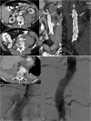
Ruptured type B dissection with stent graft implantation. An 83-year-old patient with ruptured type B dissection in para-sagittal reformations demonstrating intimal tear (a) and haematothorax (a, b). Stent graft implantation (c) (partially covering the left subclavian artery) and exclusion of the dissection after expansion of the stent graft (d)
Fig. 5
Fig. 5
Traumatic type B dissection in the infrarenal abdominal aorta in a 55-year-old female patient who fell from 3 m height. The patient presented with abdominal pain and acute bilateral limb ischaemia caused by the dissection reaching into the common iliac artery on both sides (a-e). She was treated by a surgical approach with implantation of a bifurcated prosthesis (f)
Fig. 6
Fig. 6
Intramural haematoma in a 79-year-old female patient. The patient presented with thoracic pain. Initial CTA with coronal reformations (a) and axial section (c) CTA demonstrated a small ulcer-like configuration (arrowhead) in the intramural haematoma that ascended up to the left subclavian artery (a). One-week follow-up CTA demonstrated enlargement of the ulcer-like configuration (arrow) (b). Native CT demonstrates the hyperdense intramural haematoma (d) and contrast-enhanced CT the smooth circumferential configuration of the intramural haematoma (e)
Fig. 7
Fig. 7
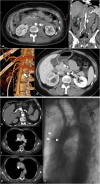
Ruptured penetrating aortic ulcer in a 57-year-old male patient. Chronic renal failure led to haemodialysis. The patient presented with hypotension and abdominal pain. CTA demonstrated a large retroperitoneal haematoma (a) and an aortic ulcer projecting beyond the aortic contour (arrowhead) (b). Coronal reformation demonstrated the mushroom-like appearance of the ulcer (arrow) (c). The patient was treated with implantation of a uni-iliac stent graft (d). Three years later he presented with hypotension and thoracic pain. CTA demonstrated a ruptured aortic ulcer in the distal descending aorta (e) that was again treated with implantation of a thoracic stent graft (f-g)
Fig. 8
Fig. 8
Signs of impending rupture. Focal “blebbing” (arrow) of the aortic wall (a, c). “Crescent sign” with hyperdense thrombus (b). Draped aorta signs (b, e) with flattening of the posterior aspect of the abdominal aorta. Focal “discontinuation” of the aortic contour (here with discontinuation of the wall calcification and blebbing) (c) in a 6-cm abdominal aortic aneurysm. The patient refused further treatment. He presented with hypotension 3 months later. CTA demonstrated a ruptured aneurysm (d)
Fig. 9
Fig. 9
Ruptured abdominal aortic aneurysm with stent graft treatment. The 72-year-old male patient presented with left-sided abdominal pain and hypotension. CTA demonstrated a ruptured infrarenal aortic aneurysm on coronal and axial sections (ab). The patient was treated with aorto-uni-iliac stent graft implantation (d) to the left, a crossover bypass and placement of an occluder (arrow) in the right common iliac artery. Follow-up CTA (c) demonstrated complete exclusion of the large aneurysm
Fig. 10
Fig. 10
Mycotic abdominal aortic aneurysm. The 51-year-old female patient presented with abdominal/lumbar discomfort and sub-febrile temperatures. A lumbar MRI demonstrated no spondylodiscitis and a small abdominal aortic aneurysm with 35 mm diameter. One month later the abdominal aortic diameter had grown to 52 mm (b–c) and PET-CT (ce) demonstrated wall thickening and PET uptake in the aortic wall and an atypical eccentric aneurysm presentation in coronal reformations (e). This was the only spot of infectious arthritis. Endocarditis of the mitral valve was demonstrated to be the focus. Additionally infectious arthritis of the left AC joint was treated by resection. Surgical reconstruction with deep vein (f) was performed; surgery confirmed a contained rupture and an aorto-duodenal fistulae
Fig. 11
Fig. 11
Aorto-duodenal fistulae in a 75-year-old male patient presenting with melena and haematemesis. He had an aortic repair with a surgical tube graft 10 years ago. CTA (axial and coronal reformations) demonstrates contrast extravasation of the infrarenal aorta directly into the adjacent duodenum (ab). The patient died immediately after the CT scan before stent graft implantation could be performed. In a 63-year-old male patient with severe haematemesis ultrasound detected an aortic aneurysm. He had previous aortic repair (15 years ago) and had also been suffering from intermittent haematemesis for 3 months. CTA (c) and DSA (d) demonstrated an aneurysm of the distal anastomosis in close proximity to the duodenum. A bifurcated stent graft was implanted (e). The aneurysm was completely excluded. A follow-up CT on the next day (after oral contrast) nicely demonstrated the aorto-duodenal fistulae (arrow) (f)
Fig. 12
Fig. 12
Iatrogenic aortic injury. A 36-year-old female patient underwent laparoscopic removal of an ovarian cyst; 6 h later she presented with hypotension and severe abdominal pain. CTA [(a) axial section, arrowhead; (b) coronal reformation; (c) volume-rendered image, arrow] demonstrated a left lateral laceration of the aorta, confirmed to be a trocar injury during vascular surgical reconstruction. A 6-month follow-up CT demonstrates a rather smooth aortic contour at the level of the original injury (d arrowhead). A 66-year-old patient presented with an aneurysm of the coeliac trunk/common hepatic artery (e). During an endovascular approach for stent graft implantation, when the Simmons type II catheter was configured in the aortic arch, the patient reported severe thoracic pain; DSA (f) and CTA [(gh) on a single slice CT scanner] confirmed the suspicion of a type A aortic dissection with a true lumen (arrowhead) and false lumen (arrows) on unsubtracted angiographic images

References

    1. Knaut AL, Cleveland JC., Jr Aortic emergencies. Emerg Med Clin North Am. 2003;21(4):817–845. doi: 10.1016/S0733-8627(03)00063-4. - DOI - PubMed
    1. Go AS, Mozaffarian D, Roger VL, et al. Heart disease and stroke statistics–2013 update: a report from the American Heart Association. Circulation. 2013;127(1):e6–e245. doi: 10.1161/CIR.0b013e31828124ad. - DOI - PMC - PubMed
    1. Authors/Task Force m. Erbel R, Aboyans V, et al. 2014 ESC guidelines on the diagnosis and treatment of aortic diseases: document covering acute and chronic aortic diseases of the thoracic and abdominal aorta of the adult. The task force for the diagnosis and treatment of aortic diseases of the European Society of Cardiology (ESC) Eur Heart J. 2014;35(41):2873–2926. doi: 10.1093/eurheartj/ehu281. - DOI - PubMed
    1. Hiratzka LF, Bakris GL, Beckman JA, et al. 2010 ACCF/AHA/AATS/ACR/ASA/SCA/SCAI/SIR/STS/SVM guidelines for the diagnosis and management of patients with thoracic aortic disease: a report of the American College of Cardiology Foundation/American Heart Association Task Force on Practice Guidelines, American Association for Thoracic Surgery, American College of Radiology, American Stroke Association, Society of Cardiovascular Anesthesiologists, Society for Cardiovascular Angiography and Interventions, Society of Interventional Radiology, Society of Thoracic Surgeons, and Society for Vascular Medicine. Circulation. 2010;121(13):e266–369. doi: 10.1161/CIR.0b013e3181d4739e. - DOI - PubMed
    1. Fattori R, Cao P, De Rango P, et al. Interdisciplinary expert consensus document on management of type B aortic dissection. J Am Coll Cardiol. 2013;61(16):1661–1678. doi: 10.1016/j.jacc.2012.11.072. - DOI - PubMed

LinkOut - more resources