. 2015:4:328-39.
doi: 10.1016/j.redox.2015.01.012. Epub 2015 Jan 20.

Induction of heme oxygenase-1 contributes to survival of Mycobacterium abscessus in human macrophages-like THP-1 cells

Affiliations

Induction of heme oxygenase-1 contributes to survival of Mycobacterium abscessus in human macrophages-like THP-1 cells

Maher Y Abdalla et al. Redox Biol. 2015.

Abstract

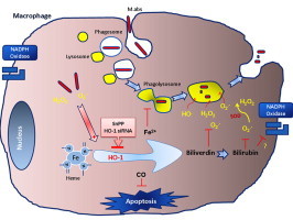
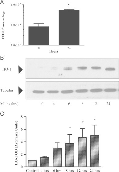
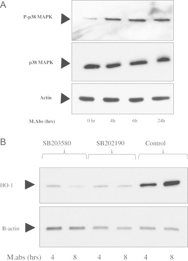
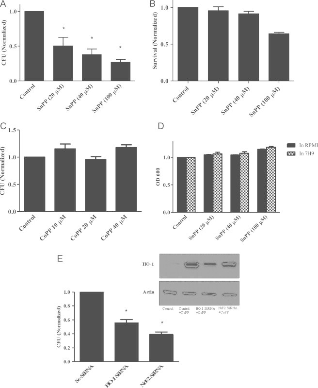
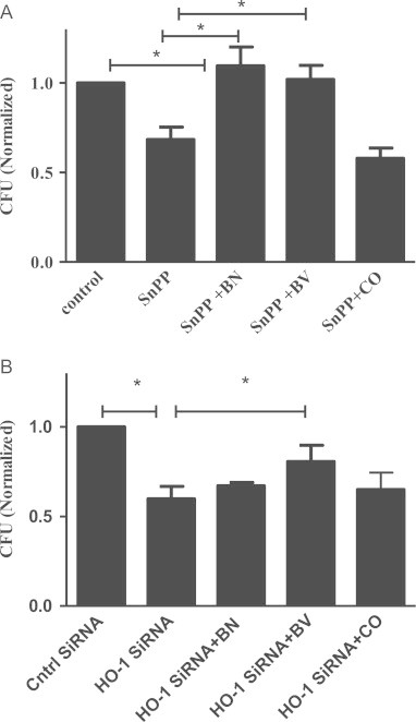
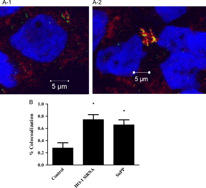
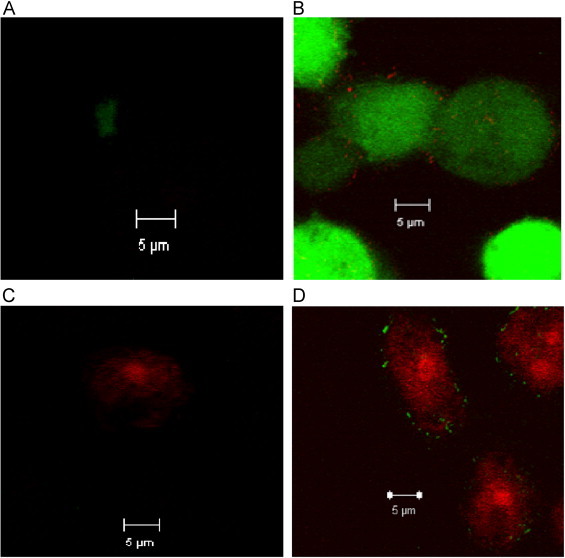
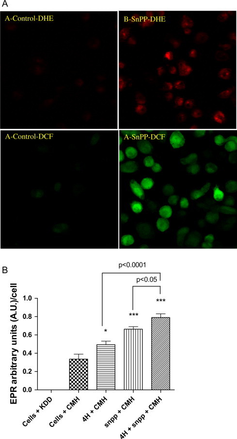
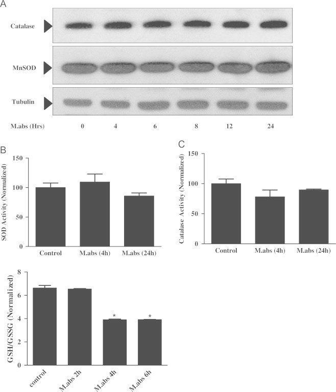
Mycobacterium abscessus (M.abs) is a rapidly growing mycobacterial species that infects macrophages, and is an important pathogen in patients with cystic fibrosis. We studied the early stages of M.abs infection of macrophages, with emphasis on the role of heme-oxygenase-1 (HO-1) in this infection. THP-1 cells were activated using TPA into macrophage-like cells and infected with M.abs for different time points. M.abs infection robustly induced HO-1 expression in the THP-1 cells. Production of HO-1 was p38 MAPK-dependent, as p38 inhibitors suppressed HO-1 induction. Pretreatment with HO-1 inhibitors tin-protoporphyrin (SnPP) significantly inhibited M.abs growth inside macrophages. Furthermore, inhibiting HO-1 using HO-1 siRNA or the HO-1 upstream signaling molecule; Nrf2 using Nrf2 siRNA resulted in similar inhibition of M.abs. In contrast, inducing HO-1 did not increase M.abs intracellular growth above control. Products of HO-1 metabolism of heme are bilirubin, biliverdin, carbon monoxide (CO) and iron. The addition of either bilirubin or biliverdin, but not CO, completely restored the SnPP inhibitory effect and partially that with HO-1 siRNA. To understand the mechanisms, we used Syto-62 labeled M.abs to infect macrophages. Interestingly, HO-1 inhibition promoted M.abs-containing phagosome fusion with lysosomes, which should enhance M.abs killing. M.abs infection enhanced THP-1 ROS production as demonstrated by increased DHE, DCF fluorescence, and EPR signal. HO-1 inhibition further increased ROS production in infected macrophages. Our results indicate that HO-1 induction is important for M.abs growth during the early stages of infection, and that the HO-1 products bilirubin and biliverdin, perhaps through modulation of intracellular ROS levels, may be involved.

Keywords: HO-1; Mycobacterium abscessus; Oxidative stress; THP-1 cells.


Figures

None
Graphical abstract
Fig. 1
Fig. 1
M.abs increased HO-1 expression. TPA-differentiated THP-1 cells were infected with M.abs for 1 h, washed and incubated in media containing gentamicin until harvested at different time points. M.abs cfu were determined and cells were prepared for immunoblotting as described in Materials and methods. (A) M.abs showed robust intracellular growth, as shown by the increase in CFU at days 0, and 1 post-infection. (B) Proteins were run and HO-1 visualized by immunoblotting. Results showed induction of HO-1 after infection. (C) Densitometric analysis of HO-1 bands in (B). M.abs infection resulted in a significant increase in expression of HO-1 (n=4, *indicates p<0.001).
Fig. 2
Fig. 2
p38 is involved in M.abs-mediated HO-1 induction signal transduction pathway. (A) TPA-differentiated THP-1 cells were infected with M.abs for 1 h, washed and incubated in media as shown in materials and methods. Cells were collected at different time points. Phosphorylated and total forms of p38MAPK were determined by immunoblotting. (B) Addition of the p38 inhibitors; SB203580 and SB202190 reduced HO-1 induction by M.abs.
Fig. 3
Fig. 3
HO-1 inhibition suppresses M.abs growth in macrophages. (A) TPA-differentiated THP-1 cells were cultured in the presence of the HO-1 inhibitor SnPP for 24 h, infected with M.abs for 1 h, and collected 24 h post-infection for CFU determination, as described in materials and methods. SnPP significantly inhibited M.abs growth as detected by CFU (p<0.05, n=5). Results obtained were normalized to vehicle-treated controls. B) TPA-differentiated THP-1 cells were cultured in the presence of different concentrations of SnPP for 48 h and assayed for toxicity by MTT assay. No toxicity was seen using SnPP concentrations up to 40 µM for 48 h. Results were normalized to vehicle-treated controls. (C) No growth inhibition of M.abs was detected when bacteria were cultured in RPMI or 7H9 in the presence of different concentrations of SnPP. (D) TPA-differentiated THP-1 cells were cultured in the presence of different concentrations of CoPP for 24 h, and then infected with M.abs for 1 h. Cells were washed, and cultured in the presence of CoPP for 24 h. M.abs cfu were counted as described in materials and methods. There was no significant difference in M.abs growth in control versus CoPP treated THP-1 cells (n=3, p>0.05). CFU counts were normalized to vehicle-treated controls. (E) TPA-differentiated THP-1 cells were cultured in the presence of siRNA for 24 h and then infected with M.abs 1 hr. Cells were washed and cultured for 24 h before collected for CFU assay as described in materials and methods. Significant inhibition of M.abs growth was observed with treatment of THP-1 cells by HO-1 siRNA (p<0.05, n=3) or Nrf2 siRNA (p<0.05, n=2). CFU counts were normalized to Sc-siRNA-treated controls infected with M.abs. The inset shows decreased HO-1 protein level in the presence of HO-1 siRNA or Nrf2 siRNA. Cells were collected after 24 h of culture in the presence of HO-1 siRNA, Nrf2 siRNA, or CoPP (HO-1 inducer). Control cells treated with HO-1 siRNA shows no detectable protein by immunoblot (not shown). CoPP slightly induced HO-1 in the presence of siRNAs as determined by immunoblot (p<0.05, n=3).
Fig. 4
Fig. 4
HO-1 enzymatic products restore M.abs growth reduced by HO-1 inhibition. TPA-differentiated THP-1 cells were cultured in the presence of SnPP (30 µM) or HO-1 siRNA for 24 h. Cells were then infected with M.abs and cultured in media with the addition of either bilirubin (10 µM), biliverdin (10 µM), or CO (10 µM) for 24 h. Cells were collected and M.abs CFU assayed. CFU counts were normalized to vehicle-treated controls. (A) Both bilirubin and biliverdin significantly increased CFU in SnPP-treated cells (n=7, p<0.05). (B) Biliverdin significantly increased CFU HO-1 siRNA-treated cells (n=5, p<0.05).
Fig. 5
Fig. 5
HO-1 inhibition increases M.abs–lysosomal fusion in macrophages. TPA-differentiated THP-1 cells were cultured in the presence of SnPP or HO-1 siRNA for 24 h. Macrophages were infected with Syto-62 labeled M.abs for 1 h, and incubated for 4 h. Lysotracker was added 2 h before preparation for confocal microscopy. (A-1) control cells infected with M.abs (shown in green), lysotracker is shown in red, and nucleus in blue. (A-2) M.abs infection in the presence of HO-1 siRNA. M.abs–lysosomal co-localization is shown in yellow shows (n=5). (B) Percent co-localization in SnPP and HO-1 siRNA treated macrophages as compared to control. HO-1 inhibition resulted in a significant increase in the percentage of M.abs showing co-localization with lysosomes (n=6, p<0.05).
Fig. 6
Fig. 6
M.abs infection increases THP-1 cell ROS production. TPA-differentiated THP-1 cells were infected with Syto-62 labeled M.abs for 4 h, and loaded with 10 µM DCFH2 or DHE for the 30 min, washed and prepared for confocal microscopy study. Panels A and C are control cells with no infection showing minimal DCF or DHE fluorescence. Panel B shows increased DCF fluorescence and Panel D shows increased DHE fluorescence after 4 h of infection. Syto-62 labeled M.abs bacteria are shown inside the cells in red or green (n=4).
Fig. 8
Fig. 8
HO-1 inhibition increase ROS production. TPA-differentiated THP-1 cells were infected with M.abs for 1 h, washed and cultured with media for 4 h. Cells were then loaded with DCFH2 or DHE for the 30 min, washed and prepared for confocal microscopy study. (A) HO-1 inhibition using SnPP increased ROS production as shown by DHE (A and B) and DCF fluorescence (C and D). (B) TPA-differentiated THP-1 cells were infected with M.abs for 4 h. Cells were then collected and studied for O2•− production by EPR as described in materials and methods. CMH is the spin probe 1-hydroxy-3-methoxycarbonyl-2,2,5,5-tetramethylpyrrolidine hydrochloride, KDD is the EPR buffer. A significant increase in O2•− production was observed at 4 h post-M.abs infection (p<0.05), and when infection is combined with HO-1 inhibition (p<0.0001). Inhibiting HO-1 also increased ROS production (p<0.0001) (n=5).
Fig. 7
Fig. 7
M.abs infection increases THP-1 cell GSH turnover, but not MnSOD or catalase protein expression or activity. TPA-differentiated THP-1 cells were infected with M.abs for 1 h, washed and cultured in media until collected at different time points. Immunoblotting and enzyme activity were determined as described in materials and methods. (A) No difference in catalase or MnSOD protein (Panel A), SOD activity (Panel B, n=3, p>0.05) or catalase activity (Panel C, n=3, p>0.05) was observed between M.abs-infected and non-infected THP-1 cells at different points. (Panel D) Cells were then collected and studied for reduced glutathione (GSH), and oxidized glutathione (GSSG) levels as described in materials and methods. M.abs significantly decreased the ratio of GSH/GSSG, which reflects glutathione turnover (n=3, p<0.05). Data counts in B–D were normalized to uninfected controls.

References

    1. Petrini B. Mycobacterium abscessus: an emerging rapid-growing potential pathogen. Acta Pathologica, Microbiologica et Immunologica Scandinavica. 2006;114(5):319–328. doi: 10.1111/j.1600-0463.2006.apm_390.x. 16725007 - DOI - PubMed
    1. Griffith D.E., Girard W.M., Wallace R.J. Clinical features of pulmonary disease caused by rapidly growing mycobacteria. An analysis of 154 patients. American Review of Respiratory Disease. 1993;147(5):1271–1278. doi: 10.1164/ajrccm/147.5.1271. 8484642 - DOI - PubMed
    1. Chan E.D., Bai X., Kartalija M., Orme I.M., Ordway D.J. Host immune response to rapidly growing mycobacteria, an emerging cause of chronic lung disease. American Journal of Respiratory Cell and Molecular Biology. 2010;43(4):387–393. doi: 10.1165/rcmb.2009-0276TR. 20081053 - DOI - PubMed
    1. Román A., Ussetti P., Solé A., Zurbano F., Borro J.M., Vaquero J.M., de Pablo A., Morales P., Blanco M., Bravo C., Cifrian J., de la Torre M., Gámez P., Laporta R., Monforte V., Mons R., Salvatierra A., Santos F., Solé J., Varela A. Guidelines for the selection of lung transplantation candidates. Archivos de BronconeumologÃa. 2011;47(6):303–309. doi: 10.1016/j.arbres.2011.03.007. English Version. - DOI - PubMed
    1. Bryant J.M., Grogono D.M., Greaves D., Foweraker J., Roddick I., Inns T., Reacher M., Haworth C.S., Curran M.D., Harris S.R., Peacock S.J., Parkhill J., Floto R.A. Whole-genome sequencing to identify transmission of Mycobacterium abscessus between patients with cystic fibrosis: a retrospective cohort study. Lancet. 2013;381(9877):1551–1560. doi: 10.1016/S0140-6736(13)60632-7. 23541540 - DOI - PMC - PubMed

Publication types

MeSH terms

LinkOut - more resources