. 2015 Apr;44(2):66-75.
doi: 10.1111/jmp.12162. Epub 2015 Jan 29.

Initial gene vector dosing for studying symptomatology of amyotrophic lateral sclerosis in non-human primates

Affiliations

Initial gene vector dosing for studying symptomatology of amyotrophic lateral sclerosis in non-human primates

Kasey L Jackson et al. J Med Primatol. 2015 Apr.

Abstract

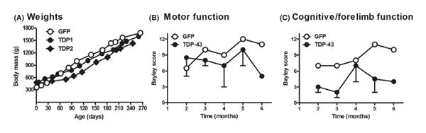
Background: Most amyotrophic lateral sclerosis (ALS) research has focused on mice, but there are distinct differences in the functional neuroanatomy of the corticospinal pathway in primates vs. rodents. A non-human primate model may be more sensitive and more predictive for therapeutic efficacy.

Methods: Rhesus macaques received recombinant adeno-associated virus (AAV9) encoding either the ALS-related pathological protein TDP-43 or a green fluorescent protein (GFP) control by intravenous administration. Motor function and electromyography were assessed over a nine-month expression interval followed by post-mortem analyses.

Results: Recombinant TDP-43 or GFP was stably expressed long term. Although the TDP-43 subjects did not manifest severe paralysis and atrophy, there were trends of a partial disease state in the TDP-43 subjects relative to the control.

Conclusions: These data indicate that a higher gene vector dose will likely be necessary for more robust effects, yet augur that a relevant primate model is feasible.

Keywords: TDP-43; adeno-associated virus; amyotrophic lateral sclerosis; frontotemporal lobar degeneration; gene therapy; gene transfer.


Figures

Fig. 1
Fig. 1
Green fluorescent protein (GFP) expression in the macaque, 9 months after gene transfer. Detection of GFP used an immunofluorescent method. (A) Cross-section of the lumbar spinal cord. (B) Ventral horn of the lumbar spinal cord. Cells with neuronal (arrow) and non-neuronal (arrowhead) morphologies were expressing GFP. (C) GFP fibers in the dorsal columns of the spinal cord. (D) Motor cortex, cells with neuronal and non-neuronal morphologies. (E) Brainstem, non-neuronal morphologies. (F) Hippocampus, neuronal (arrow) and non-neuronal morphologies (arrowhead). (G) Sagittal section of the cerebellum, efficient transduction of Purkinje neurons. (H) The gray matter of the cerebellum is counterstained in red for the neuronal marker, NeuN. (I) Efficient transduction of cells in the heart. (J–M) Confocal images from cerebellum showing GFP-positive Purkinje neurons, brainstem showing GFP in cells with non-neuronal morphologies, and gastrocnemius muscle showing GFP-positive muscle fibers. Counterstains: J, L: TO-PRO3 DNA in blue; K: TDP-43 in red. Bar in A = 536 μm, same magnification in G, H. Bar in B = 134 μm, same magnification in C–E. Bar in F = 67 μm. Bar in I = 268 μm. Bar in J = 150 μm. Bar in K = 38 μm. Bar in L = 75 μm, same magnification in M.
Fig. 2
Fig. 2
Functional trends in the TDP-43 subjects consistent with a disease state. Neonatal macaques were injected with AAV9 vectors i.v. (A) Weight gain curves: lower weights in the two TDP-43 subjects between 90 and 240 days. (B) Bayley composite ethogram score of motor function (GFP, N = 1; TDP-43, N = 2 from 2 to 5 months and N = 1 at 6 months due to technical issues; bars = SEM shown in the minus direction). (C) Bayley scale of cognitive functions, which also involves forelimb motor function: trend of impairment in the TDP-43 subjects.
Fig. 3
Fig. 3
Lumbar spinal cord and gastrocnemius muscle: no loss of motor neurons or muscle atrophy in the TDP-43 subjects relative to control. (A–C) Spinal cord was stained for Nissl substance to visualize large motor neurons in the anterior horn. (D–F) Gastrocnemius was stained with hematoxylin and eosin (H & E). Bar in C = 134 μm, same magnification in A–C. Bar in F = 67 μm, same magnification in D–F.
Fig. 4
Fig. 4
(A) Detection of recombinant human TDP-43 mRNA or recombinant GFP mRNA in primates. The cerebellum was analyzed because of relatively efficient gene transfer in this area. TDP primer set 1 amplified a portion of human TDP-43 with mismatches between the human and macaque TDP-43 sequences, which demonstrated upregulated expression in the two TDP-43 subjects compared to the GFP control. TDP primer set 2 amplified recombinant TDP-43 only by specifically recognizing a downstream sequence in the expression cassette. GFP mRNA was specifically present in only the GFP subject as expected. (B) TDP-43 protein levels in primates. Western blots of dorsal root ganglia, lumbar spinal cord, and cerebellum at 9–10 months post-injection. There was evidence of elevated expression in the TDP-43 subjects in the dorsal root ganglia and spinal cord but not the cerebellum. The density of the TDP-43 bands (T) was normalized to glyceraldehyde-3-phosphate dehydrogenase (GAPDH) expression (G).
Fig. 5
Fig. 5
TDP-43 immunoreactivity in the lumbar spinal cord and cerebellum. (A, B) Anterior horn and motor neurons in the control GFP subject. Arrows, TDP-43 staining in cell nuclei, found in both GFP and TDP-43 subjects. (C–F) Anterior horn and motor neurons in the TDP-43 subjects with examples of dense cytoplasmic immunoreactivity and staining in neuronal processes (arrowheads). (G, H) Purkinje cell layer in cerebellum. Cells with cytoplasmic immunoreactivity were relatively more frequent in the TDP-43 subjects. Bar in A = 134 μm, same magnification in C. Bar in B = 13 μm, same magnification in D–F. Bar in G = 21 μm, same magnification in H.

References

    1. Watson C, Paxinos G, Kayalioglu G. Chapter 7: Localization of motoneurons in the spinal cord. In: McHanwell S, Watson C, editors. The Spinal Cord: A Christopher and Dana Reeve Foundation Text and Atlas. 1st. Sydney: Academic Press; 2009. pp. 94–114.
    1. Chen-Plotkin AS, Lee VM, Trojanowski JQ. TAR DNA-binding protein 43 in neurodegenerative disease. Nat Rev Neurol. 2010;6:211–20. - PMC - PubMed
    1. Kiernan MC, Vucic S, Cheah BC, Turner MR, Eisen A, Hardiman O, Burrell JR, Zoing MC. Amyotrophic lateral sclerosis. Lancet. 2011;377:942–55. - PubMed
    1. Mackenzie IR, Bigio EH, Ince PG, Geser F, Neumann M, Cairns NJ, Kwong LK, Forman MS, Ravits J, Stewart H, Eisen A, McClusky L, Kretzschmar HA, Monoranu CM, Highley JR, Kirby J, Siddique T, Shaw PJ, Lee VM, Trojanowski JQ. Pathological TDP-43 distinguishes sporadic amyotrophic lateral sclerosis from amyotrophic lateral sclerosis with SOD1 mutations. Ann Neurol. 2007;61:427–34. - PubMed
    1. Neumann M, Sampathu DM, Kwong LK, Truax AC, Micsenyi MC, Chou TT, Bruce J, Schuck T, Grossman M, Clark CM, McClus-key LF, Miller BL, Masliah E, Mackenzie IR, Feldman H, Feiden W, Kretzschmar HA, Trojanowski JQ, Lee VM. Ubiquitinated TDP-43 in frontotemporal lobar degeneration and amyotrophic lateral sclerosis. Science. 2006;314:130–3. - PubMed

Publication types

MeSH terms

Substances