. 2015 Mar 2;25(5):606-12.
doi: 10.1016/j.cub.2015.01.009. Epub 2015 Jan 29.

A non-canonical pathway from cochlea to brain signals tissue-damaging noise

Affiliations

A non-canonical pathway from cochlea to brain signals tissue-damaging noise

Emma N Flores et al. Curr Biol. .

Abstract

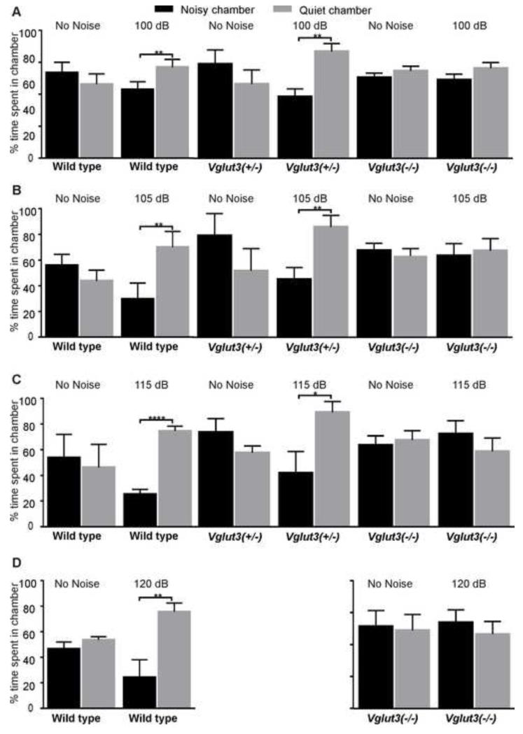
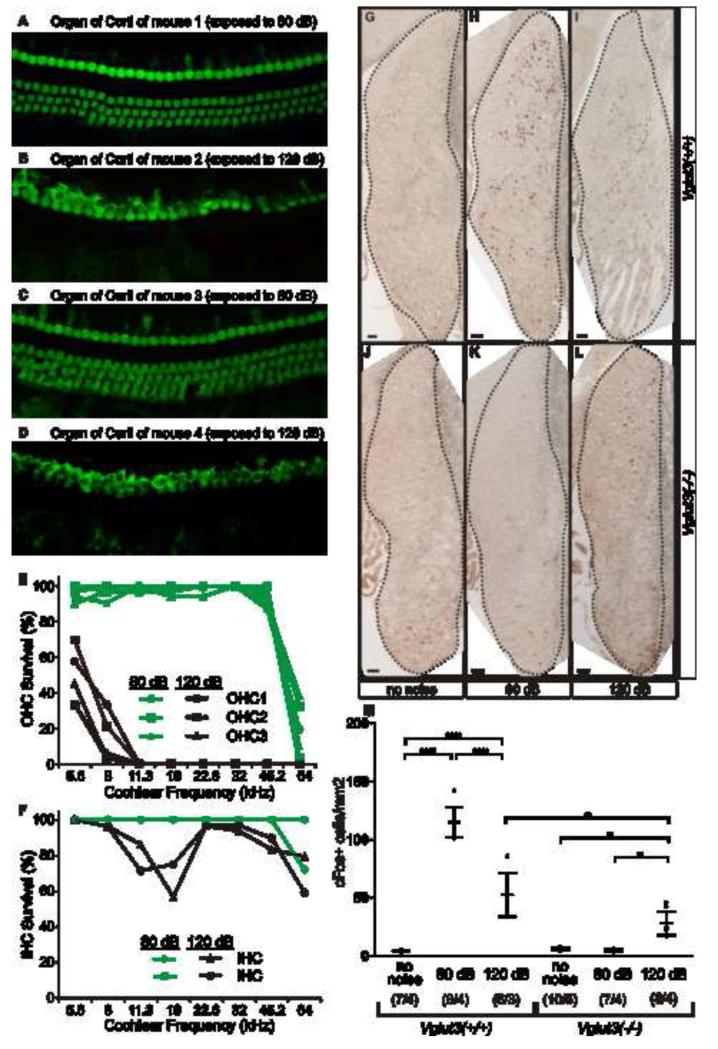
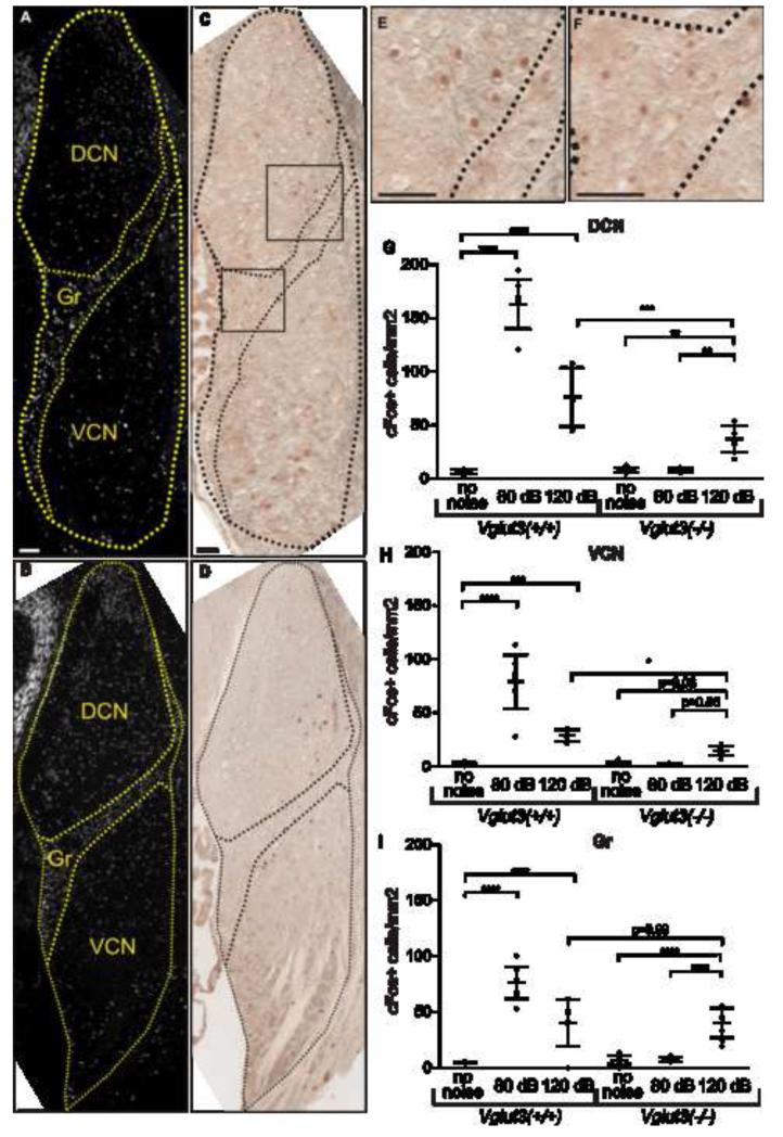
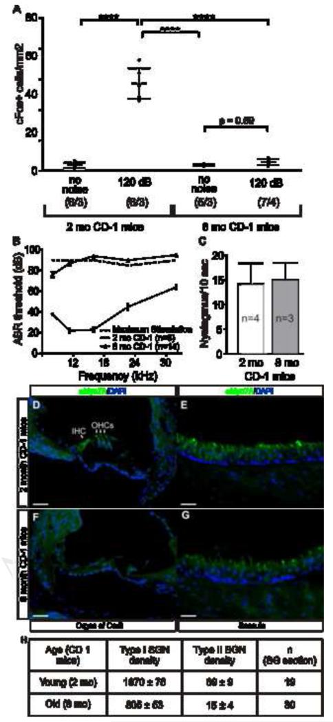
Intense noise damages the cochlear organ of Corti, particularly the outer hair cells (OHCs) [1]; however, this epithelium is not innervated by nociceptors of somatosensory ganglia, which detect damage elsewhere in the body. The only sensory neurons innervating the organ of Corti originate from the spiral ganglion, roughly 95% of which innervate exclusively inner hair cells (IHCs) [2-4]. Upon sound stimulation, IHCs release glutamate to activate AMPA-type receptors on these myelinated type-I neurons, which carry the neuronal signals to the cochlear nucleus. The remaining spiral ganglion cells (type IIs) are unmyelinated and contact OHCs [2-4]. Their function is unknown. Using immunoreactivity to cFos, we documented neuronal activation in the brainstem of Vglut3(-/-) mice, in which the canonical auditory pathway (activation of type-I afferents by glutamate released from inner hair cells) is silenced [5, 6]. In these deaf mice, we found responses to noxious noise, which damages hair cells, but not to innocuous noise, in neurons of the cochlear nucleus, but not in the vestibular or trigeminal nuclei. This response originates in the cochlea and not in other areas also stimulated by intense noise (middle ear and vestibule) as it was absent in CD1 mice with selective cochlear degeneration but normal vestibular and somatosensory function. These data imply the existence of an alternative neuronal pathway from cochlea to brainstem that is activated by tissue-damaging noise and does not require glutamate release from IHCs. This detection of noise-induced tissue damage, possibly by type-II cochlear afferents, represents a novel form of sensation that we term auditory nociception.


Figures

Figure 1
Figure 1. VGLUT3 is required for noise avoidance behavior
(A-D) Behavioral noise avoidance assays measuring the preference of an animal for a noisy vs. quiet environment. We place animals in a chamber with two interconnected compartments, and every 4 min present octave-band noise (8-16 kHz) lasting 2 min to one compartment and measure the time spent in the noisy (100, 105, 115, or 120 dB SPL) vs. quiet (attenuated by ~25 dB) environments. To prevent spatial bias, we alternate the source of noise between the two compartments. Each pair of columns represents the average time spent in the noisy compartment (black columns) and the quiet compartment (grey columns). We also measure the % time spent in each chamber during the intercalating 2 min silent periods, which demonstrates no compartment preference in the absence of noise (columns under “No Noise” labels). Mice expressing VGLUT3 (wild types and/or Vglut3+/− mice), but not mice lacking VGLUT3 (Vglut3−/−), display avoidance to noise at 100 dB SPL (A), 105 dB SPL (B), 115 dB SPL (C), and 120 dB SPL (D) (***p<l0.0001,**p<l0.001; Student’s t-test, n=4 for each genotype and sound exposure level). All error bars represent the standard deviation.
Figure 2
Figure 2. Tissue-damaging noise activates neurons in the cochlear nucleus (CN) via a VGLUT3-independent pathway
(A-D) Noise-induced hair cell loss is documented with myosin VIIa labeling. Here, we show the middle of the cochlear spiral (22.6 kHz), from 2 wild type mice 2 wks after 1 hr exposure to octave-band noise (8-16 kHz) at (A,C) 80 dB SPL or (B,D) 120 dB SPL. (E,F) The 120 dB exposure destroys OHCs throughout much of the cochlea (E) and destroys a significant number of IHCs in the region of the noise band (F). By contrast, hair cell loss is minimal after 80 dB: the loss at the extreme base is likely the age-related degeneration characteristic of C57BL/6 [40]. (G-M) Immunohistochemistry with an antibody to c-Fos on coronal sections of the cochlear nucleus (CN) following (G,J) no noise, (H,K) 80 dB SPL or (I,L) 120 dB SPL exposures to same noise band shown in A-F. In wild type mice, 80 or 120 dB SPL triggers c-Fos expression in neurons throughout the CN. InVglut3−/− mice, stimulation with innocuous (80 dB SPL) noise triggers no c-Fos immunoreactivity (density of c-Fos+ neurons is indistinguishable from that in unexposed controls), while the noxious (120 dB SPL) noise triggers cFos expression. (M) Average densities of c-Fos expressing cells reveal significant increases in CN activity (compared to baseline) in wild types following 80 dB SPL and, to a lesser extent 120 dB SPL. However, in Vglut3−/− mice only the noxious (120 dB SPL) noise triggered CN activity. Error bars represent the standard deviation. The sample size (# of CN / # of animals) is indicated for each group in parentheses. Pair wise comparisons were calculated with Type III F-test [F (2,22) = 72.56] where P values are <l0.0001 (****), and <l0.01 (**). Scale bars are 50 μm.
Figure 3
Figure 3. Tissue-damaging noise activates neurons in CN granule cell region primarily through a VGLUT3-independent pathway
(A,B) DAPI fluorescence of coronal sections of Vglut3−/− CN (A) and Vglut3+/+CN (B) exposed to 120 dB SPL reveals its three subdivisions: dorsal cochlear nucleus (DCN), ventral cochlear nucleus (VCN), and granule cell region (Gr). (C, D) c-Fos immunoreactivity within the same coronal section reveals positive cells throughout the three CN subdivisions. Areas of panel (C) magnified in (E,F) demonstrate positive neurons within the DCN and Gr areas of Vglut3−/− CN. (G-I) Densities of c-Fos-expressing neurons in each CN subdivision: DCN (G), VCN (H), and Gr (I). Wild type mice display significant increases of neuronal activity in all three subdivisions (G-I) following exposure to 80 dB SPL and 120 dB SPL. Vglut3−/− mice display significant increases of neuronal activity following exposure to 120 dB SPL in the DCN (G) and Gr (I), while increases in VCN approached significance (p=0.06; H). However, in the granule cell region, which is not innervated by type-I afferents, neuronal activity induced by noxious (120 dB SPL) noise is VGLUT3 independent (indistinguishable between wild type and Vglut3−/− mice). Error bars represent the standard deviation. The sample size in G-I is the same as in Fig 2M. Pair wise comparisons were calculated for each CN area with Type III F-test [DCN (F) (2, 22) = 76.95]; [VCN (F) (2, 22) = 40.01]; [Gr (F) (2, 22) = 44.85]. P values are <l0.0001 (****), <l0.001 (***), and <l0.01 (**). Scale bars are 50 μm.
Figure 4
Figure 4. CN activation following tissue-damaging noise requires an intact cochlea
(A) Tissue-damaging noise activates CN neurons in young (2 months) but not middle-age (8 months) CD1 mice, which have (B) severe cochlear dysfunction due to (D,F) a massive loss of hair cells but (c) normal vestibular function and (E,G) appearance as well as normal somatosensory pain thresholds [21]. Pair wise comparisons were calculated with Type III F-test [F (1,11) = 90.72] where P values are <l0.0001 (****); the sample size (# of CN/ # of animals) is indicated for each group in parentheses. (B) Average ABR thresholds of young (n = 6) and middle-age (n = 14) CD1 mice. The dotted line represents the maximum sound tested at each frequency, so values shown above it represent non-responding ears. (C) Nystagmus, assessed as the number of saccades during 10 seconds after rotation at 250 r.p.m., reveals no difference (p = 0.68; Student’s t-test) in vestibulo-ocular reflex between young and middle-age CD1 mice. (D-G) Immunohistochemistry for Myosin VIIA and nuclear counterstain with DAPI to (D, E) young and (F, G) middle-age CD1 inner ear reveals loss of (F) cochlear but not (G) saccular hair cells in aged CD1 mice. Scale bars are 50 μm. (H) Neuronal counts show that middle-age CD1 mice have a reduced number of both type-I (−59%) and type-II (−78%) cochlear afferents. Counts were made on sections from 2 young and 6 old CD1 mice.

Comment in

References

    1. Liberman MC, Kiang NY. Acoustic trauma in cats. Cochlear pathology and auditory-nerve activity. Acta Otolaryngol Suppl. 1978;358:1–63. - PubMed
    1. Dannhof BJ, Bruns V. The innervation of the organ of Corti in the rat. Hearing research. 1993;66:8–22. - PubMed
    1. Spoendlin H. Innervation densities of the cochlea. Acta Otolaryngol. 1972;73:235–248. - PubMed
    1. Kiang NY, Rho JM, Northrop CC, Liberman MC, Ryugo DK. Hair-cell innervation by spiral ganglion cells in adult cats. Science. 1982;217:175–177. - PubMed
    1. Ruel J, Emery S, Nouvian R, Bersot T, Amilhon B, Van Rybroek JM, Rebillard G, Lenoir M, Eybalin M, Delprat B, et al. Impairment of SLC17A8 encoding vesicular glutamate transporter-3, VGLUT3, underlies nonsyndromic deafness DFNA25 and inner hair cell dysfunction in null mice. Am J Hum Genet. 2008;83:278–292. - PMC - PubMed

Publication types

Substances