. 2015 Feb 12;160(4):595-606.
doi: 10.1016/j.cell.2015.01.009. Epub 2015 Jan 29.

A micropeptide encoded by a putative long noncoding RNA regulates muscle performance

Affiliations

A micropeptide encoded by a putative long noncoding RNA regulates muscle performance

Douglas M Anderson et al. Cell. .

Abstract

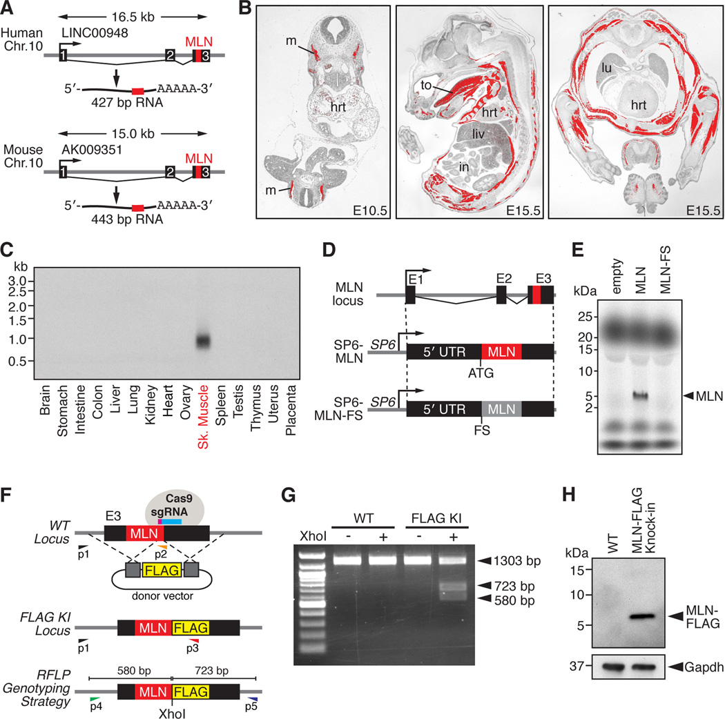
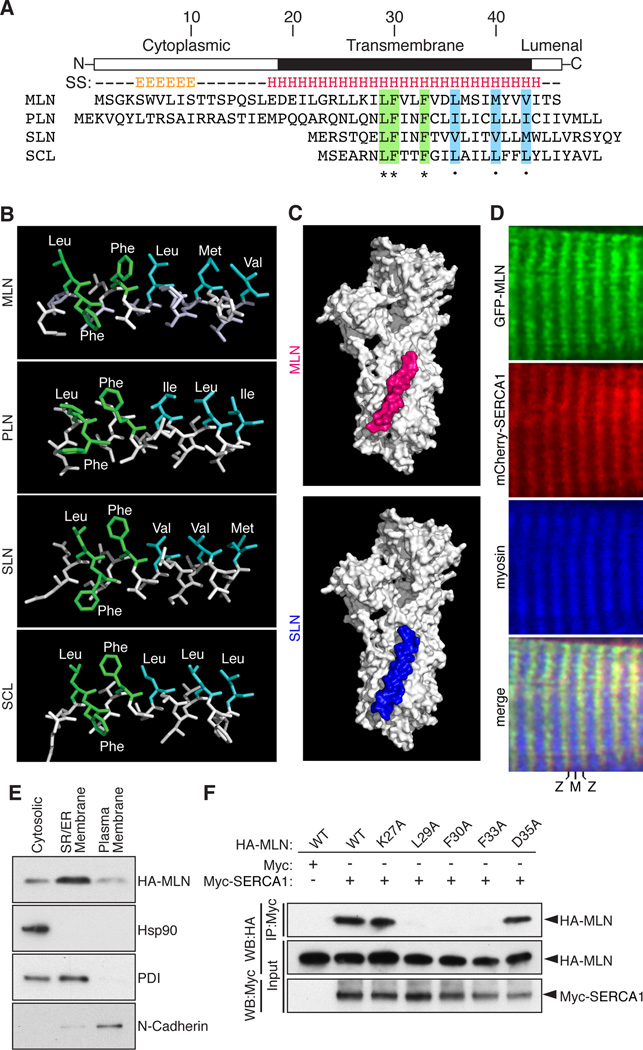
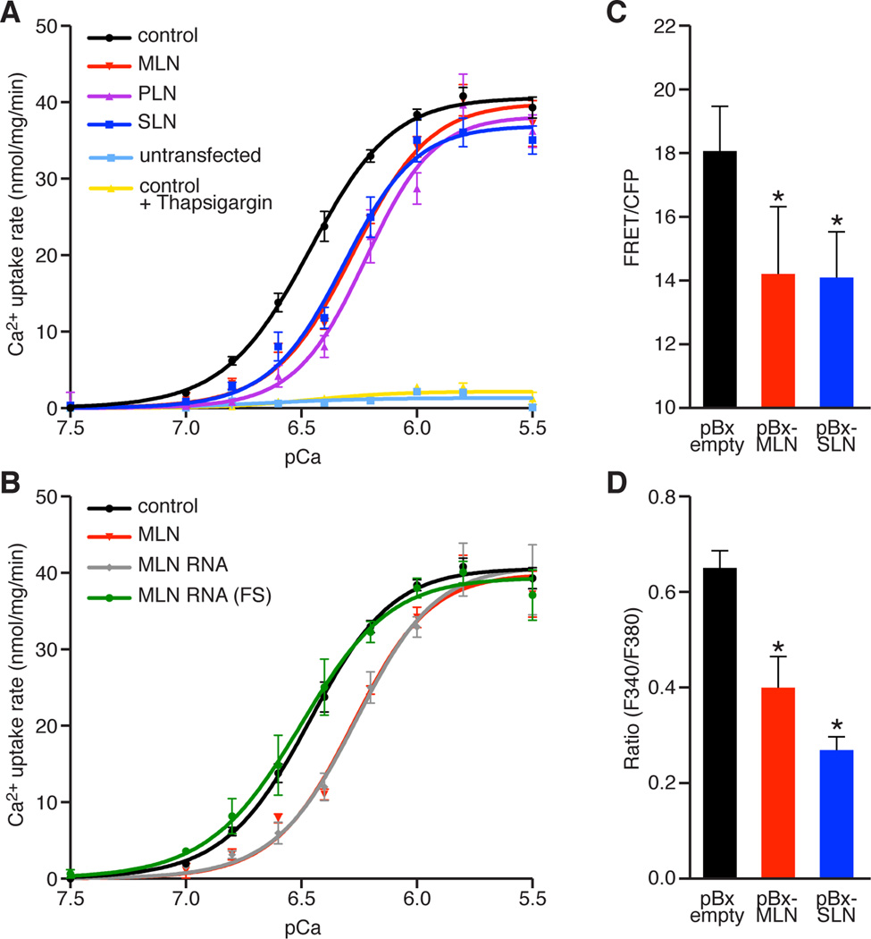
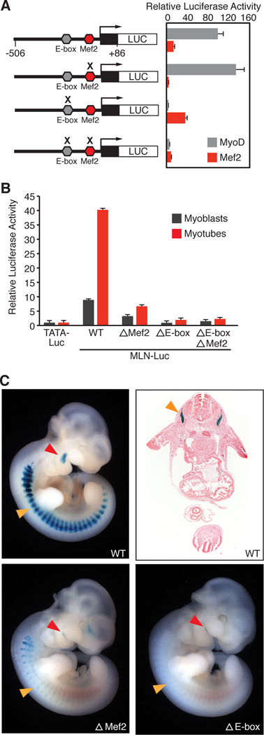
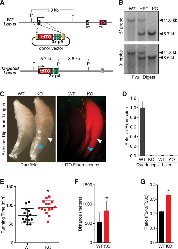
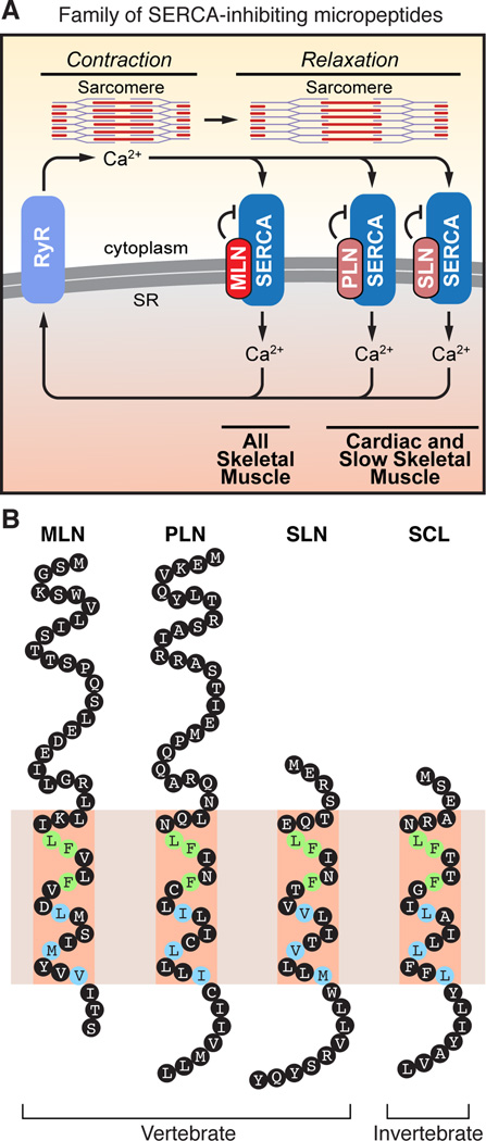
Functional micropeptides can be concealed within RNAs that appear to be noncoding. We discovered a conserved micropeptide, which we named myoregulin (MLN), encoded by a skeletal muscle-specific RNA annotated as a putative long noncoding RNA. MLN shares structural and functional similarity with phospholamban (PLN) and sarcolipin (SLN), which inhibit SERCA, the membrane pump that controls muscle relaxation by regulating Ca(2+) uptake into the sarcoplasmic reticulum (SR). MLN interacts directly with SERCA and impedes Ca(2+) uptake into the SR. In contrast to PLN and SLN, which are expressed in cardiac and slow skeletal muscle in mice, MLN is robustly expressed in all skeletal muscle. Genetic deletion of MLN in mice enhances Ca(2+) handling in skeletal muscle and improves exercise performance. These findings identify MLN as an important regulator of skeletal muscle physiology and highlight the possibility that additional micropeptides are encoded in the many RNAs currently annotated as noncoding.


Figures

Figure 1
Figure 1. Discovery of a Skeletal Muscle-specific Micropeptide
(A) A short ORF encoding a conserved micropeptide, that we named myoregulin (MLN), is contained within exon 3 of an annotated lncRNA in human and mouse genomes. The position of the MLN ORF is indicated in red. (B) In situ hybridization showing skeletal muscle-specific expression of MLN at the indicated embryonic time-points. (hrt, heart; in, intestine; liv, liver; lu, lung; m, myotome; to, tongue) (C) Northern blot of RNA isolated from adult mouse tissues using a probe specific to the full-length MLN transcript shows skeletal muscle-specific expression. (D) Diagram of the constructs used for in vitro translation of the MLN micropeptide. The full-length MLN RNA transcript was subcloned into the CS2 vector containing the SP6 phage RNA polymerase promoter (SP6-MLN). A frameshift mutation was introduced immediately after the endogenous ATG to disrupt the MLN ORF (SP6-MLN-FS). (E) Coupled in vitro transcription and translation reactions of the SP6-MLN vector using radiolabeled 35S-methionine produced a ∼5 kDa micropeptide, visualized by Tricine SDS-PAGE. The frame-shift mutation in the MLN ORF (SP6-MLN-FS) abolished any detectable expression. (F) Targeting strategy using CRISPR/Cas9-mediated homologous recombination to knock-in a FLAG epitope tag into the MLN locus in C2C12 cells. PCR-based genotyping using primers (P1-P3) or RFLP analysis of PCR products generated using primers (P4 and P5) were used to verify correct targeting. (G) RFLP analysis of WT C2C12 and heterozygous C2C12 myoblasts for the MLN-FLAG knock-in allele. (H) Western blot analysis showing endogenous expression of the MLN-FLAG fusion peptide in differentiated C2C12 myotubes, detected with an anti-FLAG antibody. See also Figure S1.
Figure 2
Figure 2. MLN Forms a Transmembrane Alpha Helix that Interacts with SERCA
(A) The secondary structure (SS) of MLN is predicted to contain an N-terminal beta sheet and C-terminal transmembrane alpha helix (E, beta sheet; H, alpha helix). Alignment of mouse MLN, PLN, SLN and the invertebrate ortholog SCL identified identical (green) and similar (blue) residues conserved in all four micropeptides. (B) The structure of the transmembrane helices of MLN, SLN, PLN and SCL was modeled using I-TASSER and showed a common spatial orientation of conserved residues. (C) Automated protein docking using ClusPro predicted that MLN occupies the same groove in SERCA1 that is recognized by SLN. (D) Expression of GFP-MLN and mCherry-SERCA1 fusion proteins in mature skeletal muscle fibers, showing co-localization in the SR, imaged using two-photon laser scanning confocal microscopy. (M, M-line; Z, Z-line) (E) Retroviral expression of an N-terminal HA-tagged MLN fusion peptide (HA-MLN) in C2C12 myoblasts was enriched in the subcellular fraction containing SR/ER membrane proteins. Enrichment for cytosolic, SR/ER membrane and plasma membrane proteins was verified by Western blot analysis for Hsp90, PDI and N-Cadherin, respectively. (F) Co-immunoprecipitation (CoIP) experiments with HA-tagged MLN alanine mutants and a Myc-tagged SERCA1 construct transfected into COS7 cells identified residues important for interaction of MLN with SERCA1. Interaction of MLN with SERCA1 was abolished by mutation of residues shared with PLN, SLN and SCL (L29A, F30A and F33A) but not by the charged residues K27A or D35A. Western blot performed with anti-HA (WB-HA) or anti-Myc (WB-Myc). See also Figure S2.
Figure 3
Figure 3. MLN Regulates SR Ca2+ Levels by Inhibiting SERCA Pump Activity
(A and B) The Ca2+-dependence of the relative rate of Ca2+ uptake is shown for homogenates from HEK 293 cells co-transfected with SERCA1 and the indicated constructs. Co-transfection with MLN, PLN or SLN resulted in a similar decrease in Ca2+ uptake, corresponding to a decreased affinity of SERCA for Ca2+, relative to empty vector (Control). For comparison, untransfected cells and SERCA1 expressing cells treated with the SERCA inhibitor thapsigargin (100nM) are shown. The activity of the full-length RNA transcript encoding the MLN ORF (MLN RNA) is abolished by a frameshift mutation in the MLN ORF (MLN-RNA FS). (C) Retroviral co-transduction of C2C12 myoblasts with the FRET-based Ca2+ sensor T1ER with MLN or SLN was used to directly measure the relative levels of SR Ca2+. Both MLN and SLN significantly decreased SR Ca2+ levels relative to an empty retroviral vector. (D) Retroviral over-expression of MLN or SLN in C2C12 myoblasts treated with 4-CMC and imaged with fura-2-AM showed decreased levels of SR Ca2+, measured by peak Ca2+ release from the SR. Data are presented as mean ± SEM. (* denotes p < 0.05 compared to pBx-empty). See also Table S1.
Figure 4
Figure 4. Developmental and Adult Expression of MLN, PLN and SLN in the Mouse
(A and B) Real-time PCR showing the relative expression of MLN, PLN, SLN and SERCA isoforms across multiple skeletal muscles, cardiac and non-muscle tissues isolated and pooled from three adult 8-week old C57Bl/6 male mice. See also Figure S3.
Figure 5
Figure 5. Regulation of MLN Transcription by MyoD and MEF2
(A) A fragment of the MLN promoter (−506 to +86, relative to the transcriptional start site) containing a highly conserved MyoD E-box (CACCTG) and MEF2 site (CTAATAACAG) was cloned in front of the luciferase reporter gene (MLN-Luc). The MLN-Luc reporter was robustly transactivated by the skeletal muscle transcription factors MyoD and MEF2 in COS7 cells (gray and red bars respectively). Mutation of the E-box (acCCgt) or Mef2 site (CTgggAACAG) (indicated by an X) abrogated transactivation by MyoD:E12 heterodimer or Mef2c, respectively. All luciferase values were normalized to the transactivation of a basal luciferase reporter (TATA-Luc) with MyoD or Mef2, respectively. (B) Transfection of the MLN-Luc reporter or mutant luciferase vectors into C2C12 myoblasts or myotubes showed they are essential for the transactivation of the MLN promoter. (C) X-gal and H&E staining of E10.5 mouse embryos harboring either the MLN promoter-lacZ transgene (WT) or mutations in the MLN promoter (ΔMef2 or ΔE-box). The MLN-promoter showed expression in the myotomal compartment of the somites (orange arrow) and premyogenic cells in the mandibular arch (red arrow). Mutation of the MEF2 or E-box sequences in the MLN promoter-lacZ transgene abrogated or abolished muscle specific expression. See also Figure S4.
Figure 6
Figure 6. Generation and Characterization of MLN Knockout Mice
(A) TALEN-mediated homologous recombination was used to insert a tdTomato fluorescent reporter and triple polyadenylation cassette into exon 1 of the MLN locus to generate a null allele. A schematic of the donor vector and targeting strategy is shown. (P, PvuII) (B) Southern blot analysis confirming correct targeting of the tdTO-triple polyadenylation cassette into exon 1 of the MLN locus using probes 5’ and 3’ to the TALEN cut site. (C) tdTomato fluorescence was specific to skeletal muscle (white arrowhead) of MLN KO mice and not detected in other tissues, such as tendon (blue arrowhead). (D) Real-time PCR using primers specific to exon 2 and 3 demonstrating absence of MLN transcripts in MLN KO muscle, as well as in liver. (E) Muscle performance was measured using forced treadmill running to exhaustion. MLN KO (N=15) mice ran ∼31% longer than WT littermates (N=14). (F) Comparison of distance run by MLN KO and WT mice in Figure 6E. (G) Myoblasts isolated and cultured from MLN KO hindlimb muscles were imaged using Fura-2-AM and treated with the RyR agonist 4-CMC in the absence of extracellular Ca2+, to indirectly measure SR Ca2+ levels. MLN KO myoblasts showed signigicantly increased SR Ca2+ levels, measured as peak Ca2+ release from the SR. Data are presented as mean ± SEM. (* denotes p < 0.05 compared to WT as in (E and F) and pBx-empty in (G)). See also Figure S5.
Figure 7
Figure 7. A Family of SERCA-inhibitory Micropeptides
(A) The Ca2+ pumps, RyR and SERCA, play a critical role in muscle contractility by controlling Ca2+ cycling between the cytosol and SR. MLN, PLN and SLN inhibit SERCA pump activity in different striated muscle types of vertebrates. (B) Illustration of the family of SERCA-inhibitory micropeptides. The discovery of MLN reveals that vertebrates encode three SERCA-inhibitory peptides that share conserved residues within their transmembrane alpha helices. Green shading denotes identical residues and blue denotes similar residues. MLN, myoregulin; PLN, phospholamban; SLN, sarcolipin; SCL, sarcolamban.

Comment in

References

    1. Abell E, Ahrends R, Bandara S, Park BO, Teruel MN. Parallel adaptive feedback enhances reliability of the Ca2+ signaling system. Proc Natl Acad Sci U S A. 2011;108:14485–14490. - PMC - PubMed
    1. Allen DG, Gervasio OL, Yeung EW, Whitehead NP. Calcium and the damage pathways in muscular dystrophy. Can J Physiol Pharmacol. 2010;88:83–91. - PubMed
    1. Anderson DM, Beres BJ, Wilson-Rawls J, Rawls A. The homeobox gene Mohawk represses transcription by recruiting the sin3A/HDAC co-repressor complex. Dev Dyn. 2009;238:572–580. - PubMed
    1. Anderson DM, George R, Noyes MB, Rowton M, Liu W, Jiang R, Wolfe SA, Wilson-Rawls J, Rawls A. Characterization of the DNA-binding properties of the Mohawk homeobox transcription factor. J Biol Chem. 2012;287:35351–35359. - PMC - PubMed
    1. Andrews SJ, Rothnagel JA. Emerging evidence for functional peptides encoded by short open reading frames. Nat Rev Genet. 2014;15:193–204. - PubMed

Publication types

MeSH terms