. 2015:125:331-51.
doi: 10.1016/bs.mcb.2014.10.010. Epub 2015 Jan 8.

Practical aspects of the cellular force inference toolkit (CellFIT)

Affiliations

Practical aspects of the cellular force inference toolkit (CellFIT)

Jim H Veldhuis et al. Methods Cell Biol. 2015.

Abstract

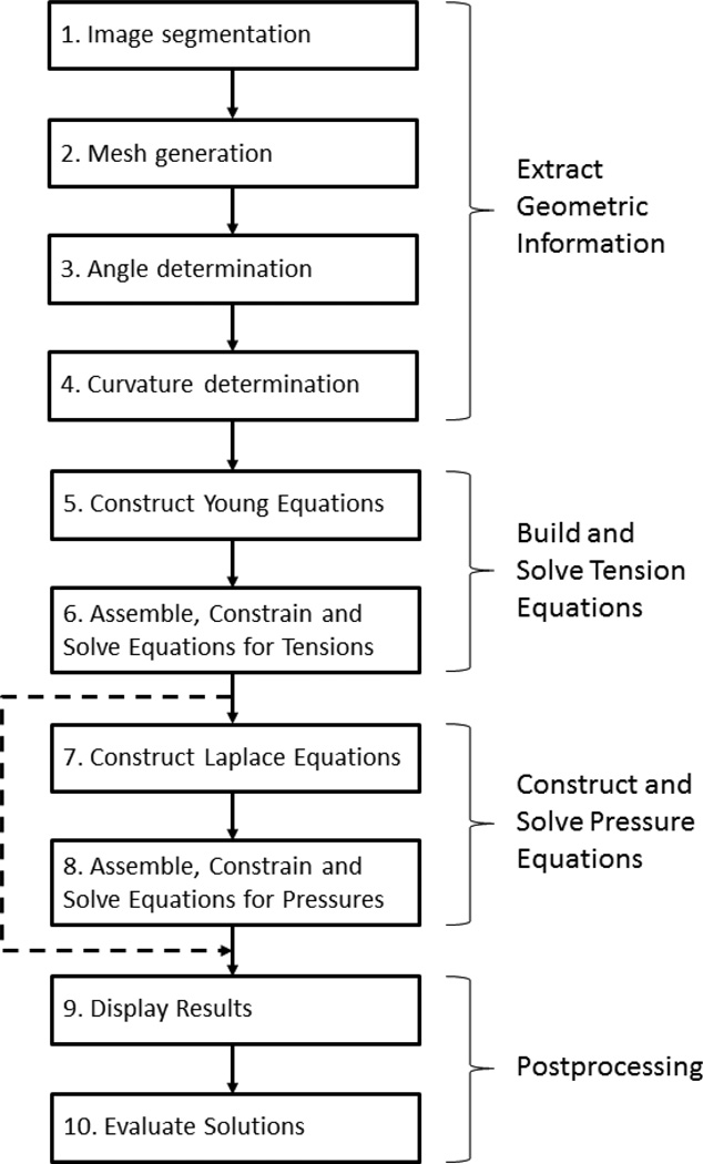
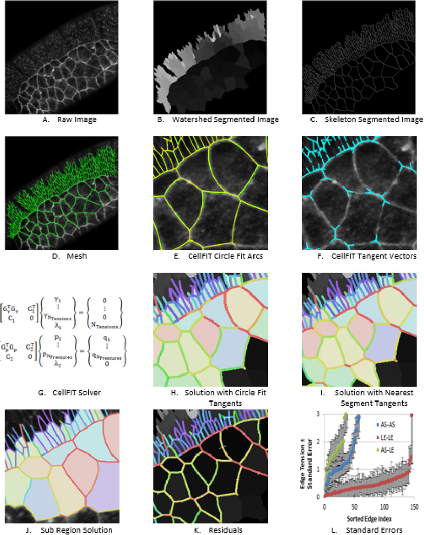
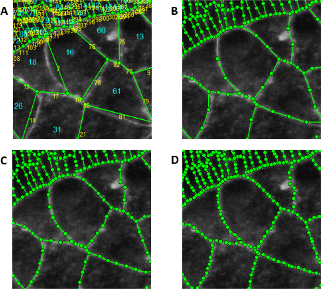
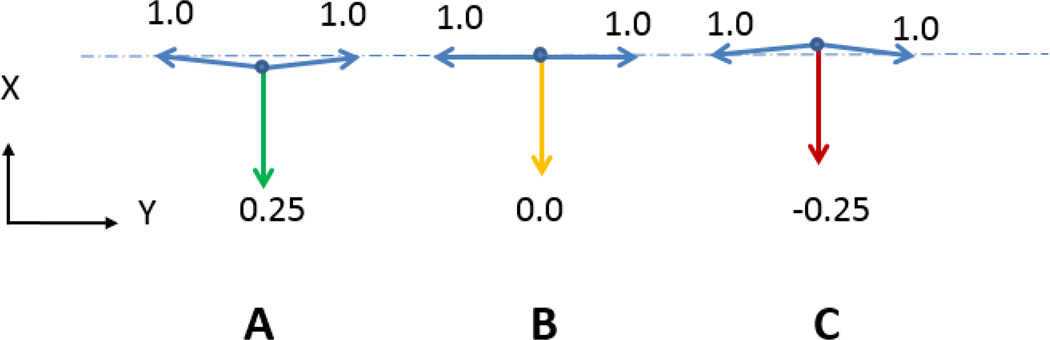
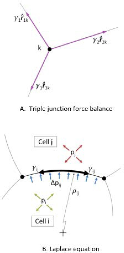
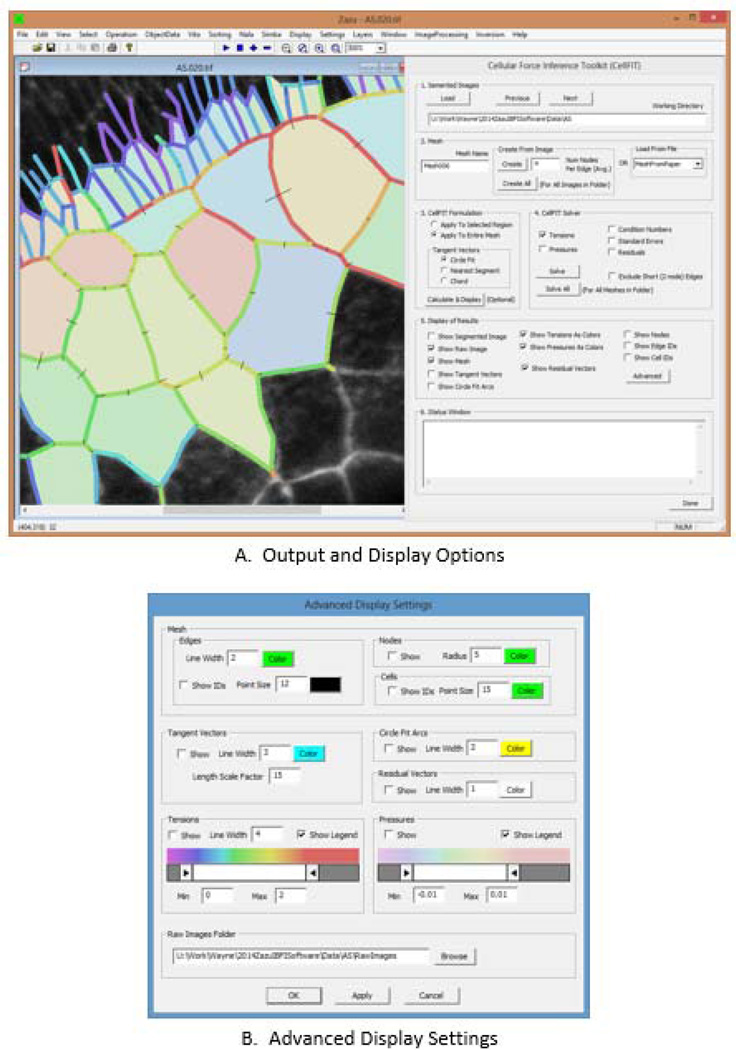
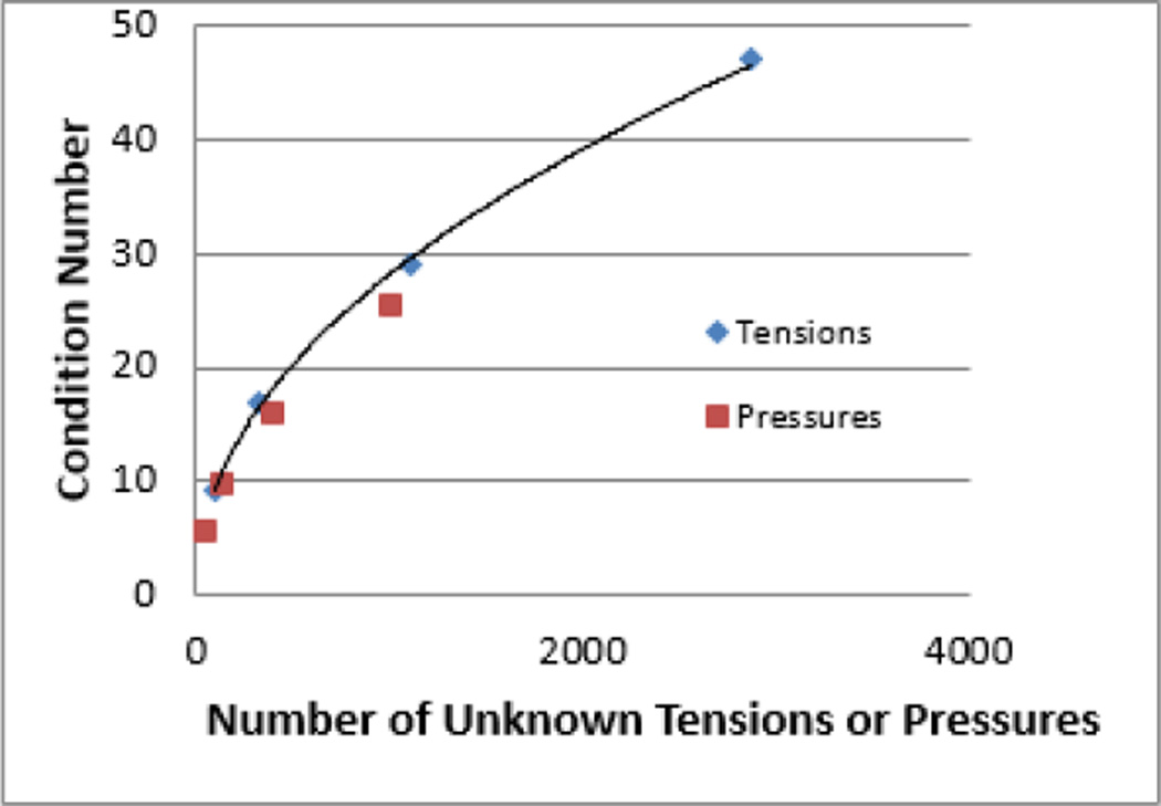
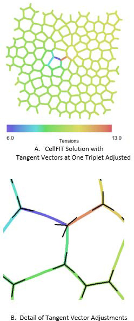
If we are to fully understand the reasons that cells and tissues move and acquire their distinctive geometries during processes such as embryogenesis and wound healing, we will need detailed maps of the forces involved. One of the best current prospects for obtaining this information is noninvasive force-from-images techniques such as CellFIT, the Cellular Force Inference Toolkit, whose various steps are discussed here. Like other current quasistatic approaches, this one assumes that cell shapes are produced by interactions between interfacial tensions and intracellular pressures. CellFIT, however, allows cells to have curvilinear boundaries, which can significantly improve inference accuracy and reduce noise sensitivity. The quality of a CellFIT analysis depends on how accurately the junction angles and edge curvatures are measured, and a software tool we describe facilitates determination and evaluation of this information. Special attention is required when edges are crenulated or significantly different in shape from a circular arc. Because the tension and pressure equations are overdetermined, a select number of edges can be removed from the analysis, and these might include edges that are poorly defined in the source image, too short to provide accurate angles or curvatures, or noncircular. The approach works well for aggregates with as many as 1000 cells, and introduced errors have significant effects on only a few adjacent cells. An understanding of these considerations will help CellFIT users to get the most out of this promising new technique.

Keywords: Cell mechanics; Cell shape; CellFIT; Force inference techniques; Force-from-shape methods; Interfacial tensions; Intracellular pressures; Morphogenetic forces.


Figures

Fig. 1
Fig. 1
CellFIT Flowchart. The flowchart shows the basic steps in the CellFIT procedure, and its numbers correspond to those used in the text.
Fig. 2
Fig. 2
Process Steps. A shows cells from the amnioserosa and adjacent lateral ectoderm (upper left corner) of a Bownes stage 13 Drosophila embryo (From (Brodland, Veldhuis et al. 2014). Watershedding was used to segment the image into cells as shown in B. C shows the cell boundaries in skeletonized form, while D shows the cell outlines as a mesh (see also Fig. 3). The circular arcs used to calculate the edge curvatures and edge angles at the triple junctions are shown in E and the resulting tangent angles in F. The resulting equation sets are shown in G and solutions based on circle- and nearest segment-edge fits are shown in H and I, respectively. J shows that analyzing a sub-region give results consistent with those produced when the whole region is analyzed, subject to offsets associate with the small sample size when the region of interest contains only a few cells. K shows the tension and pressure residuals resulting from with the analysis shown in H, and L shows the associated standard errors in the tensions, as calculated from the covariance matrix (Brodland, Veldhuis et al. 2014). As in the spectra shown in Fig. 7B, tensions and pressures shown in colours from the blue end of the spectrum have lower values and those from the red end higher ones.
Fig. 3
Fig. 3
Meshing details. A shows an enlargement of a portion of the tissue shown in Fig. 2. Cell numbers are shown in red and node numbers in blue. B–D show various average intermediate node spacings. The mesh in A has no intermediate nodes while those in B to D have 2, 3 and 4 intermediate nodes per edge on average, respectively. The meshes have been superimposed on the raw image from which they are derived so that their quality can be evaluated. We have found that meshes like that shown in C match the geometries of typical cells sufficiently without introducing significant extraneous nodes.
Fig. 4
Fig. 4
Cells with Crenulated Edges. One of the relatively rare situations where circle fits do not give good membrane approach angles is when an edge has a complex geometry, such as a crenulated shape. These cells are from the amnioserosa of a Bowne's Stage 13 Drosophila embryo. In cases like this, angle estimates based on the last points along the edge tend to be considerably more accurate. However, crenulated edges have been deemed to carry little if any tension, and so the exact direction used in the angle calculations might not be that important. Furthermore, such edges, especially if relatively sparse within a given tissue, might be removed from the analysis (see text).
Fig. 5
Fig. 5
Problematic Tangent Vectors. As noted in the text, cell edges that are nearly collinear as in A and B, can introduce numerical issues, especially if positional or angular errors arise and cause all of the edges to lie within an included angle smaller than 180 degrees, as in C. See text.
Fig. 6
Fig. 6
The Physical Basis of the Tension and Pressure Equations. The tension equations (Equation 1) arise from a force balance at each triple or higher-order junction in accordance with the vectors shown in A. As suggested by B, the pressure differences between cells are related to the membrane curvature and tension.
Fig. 7
Fig. 7
The CellFIT User Interface. The left side of part A shows a typical display window while the right side shows the primary user interface. This interface is designed to allow the user to control the various calculation steps in CellFIT and to make run-time choices. In contrast, the advanced interface shown in B is designed to facilitate data display and analysis. See text for details.
Fig. 8
Fig. 8
The Effect of Mesh Size on Condition Number. The condition numbers associated with the tension and pressure equations depend on the number of unknowns in those equations. Meshes consisting of 40, 125, 400 and 1000 cells were run and the resulting condition numbers for tensions (blue diagonal symbols) and pressures (red squares) are shown. The curve through the tension condition numbers is given by the exponential equation: [Condition Number] = 1.05 [Number of Unknown Tensions]0.48.
Fig. 9
Fig. 9
Error Influence Range. In order to assess the consequences of localized errors, the angles associated with one particular triple junction near the center of the cell aggregate shown in A were purposely given the erroneous orientations shown in B. Doing so caused the tensions field, which should be uniform, to be disrupted significantly for a distance of only one cell diameter from the introduced error, but it also caused an offset in the larger tension field, as described in the text.

References

    1. Anon E, Serra-Picamal X, Hersen P, Gauthier NC, Sheetz MP, Trepat X, Ladoux B. Cell crawling mediates collective cell migration to close undamaged epithelial gaps. Proceedings of the National Academy of Sciences of the United States of America. 2012;109(27):10891–10896. - PMC - PubMed
    1. Beer FP, Johnston ER, Dewolf JT. Mechanics of Materials. New York: McGraw-Hill Science; 2005.
    1. Borghi N, Sorokina M, Shcherbakova OG, Weis WI, Pruitt BL, Nelson WJ, Dunn AR. E-cadherin is under constitutive actomyosin-generated tension that is increased at cell-cell contacts upon externally applied stretch. Proc Natl Acad Sci. 2012;109:12568–12573. - PMC - PubMed
    1. Brodland GW, Veldhuis JH, Kim S, Perrone M, Mashburn D, Hutson MS. CellFIT: A Cellular Force-Inference Toolkit Using Curvilinear Cell Boundaries. PLoS One. 2014 in Press. - PMC - PubMed
    1. Brodland GW. The Differential Interfacial Tension Hypothesis (DITH): a comprehensive theory for the self-rearrangement of embryonic cells and tissues. Journal of Biomechanical Engineering. 2002;124(2):188–197. - PubMed

Publication types

LinkOut - more resources