. 2015 May;12(5):1027-35.
doi: 10.1016/j.hrthm.2015.01.045. Epub 2015 Jan 30.

Remodeling of stellate ganglion neurons after spatially targeted myocardial infarction: Neuropeptide and morphologic changes

Affiliations

Remodeling of stellate ganglion neurons after spatially targeted myocardial infarction: Neuropeptide and morphologic changes

Olujimi A Ajijola et al. Heart Rhythm. 2015 May.

Abstract

Background: Myocardial infarction (MI) induces remodeling in stellate ganglion neurons (SGNs).

Objective: We investigated whether infarct site has any impact on the laterality of morphologic changes or neuropeptide expression in stellate ganglia.

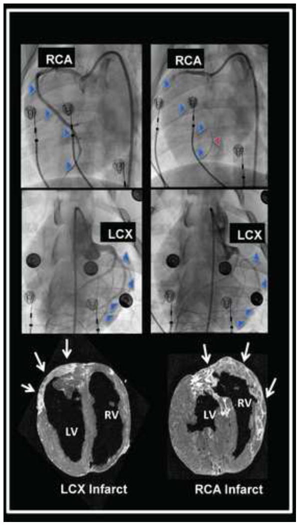
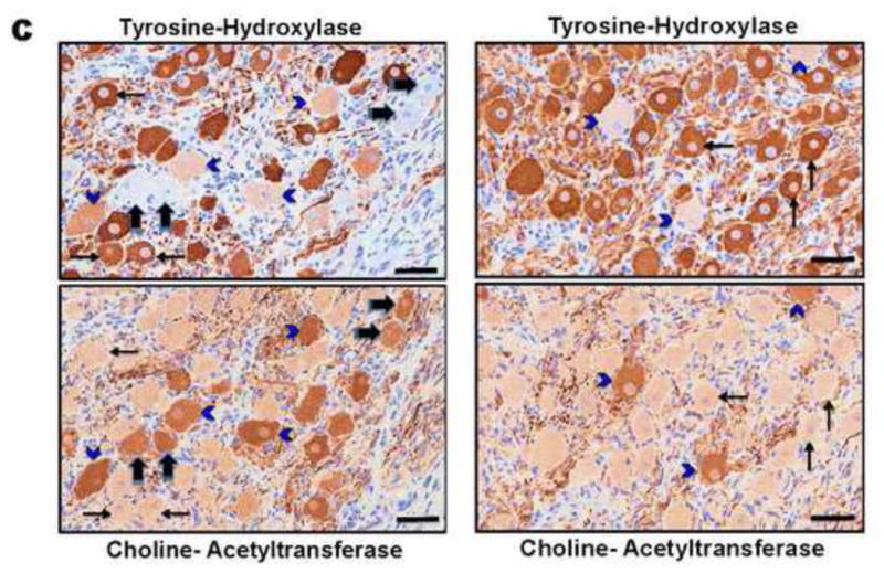
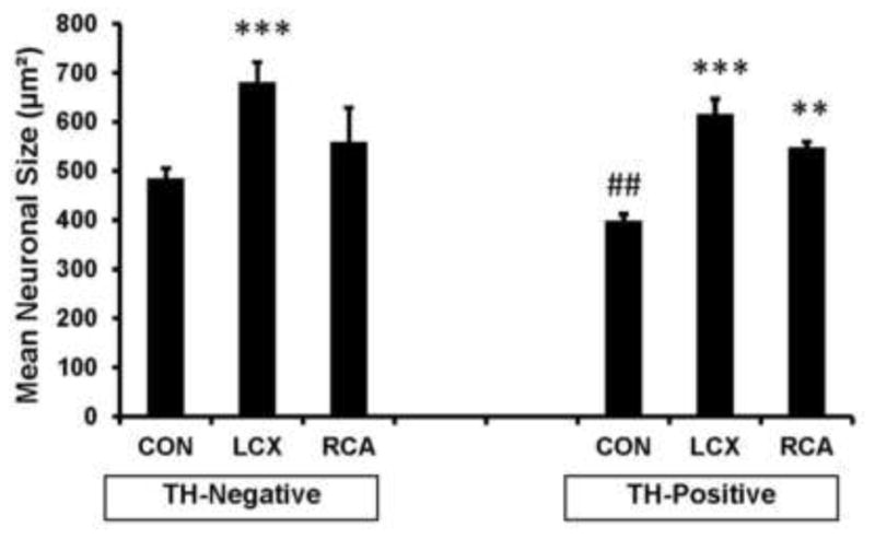
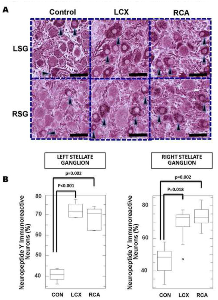
Methods: Yorkshire pigs underwent left circumflex coronary artery (LCX; n = 6) or right coronary artery (RCA; n = 6) occlusion to create left- and right-sided MI, respectively (control: n = 10). At 5 ± 1 weeks after MI, left and right stellate ganglia (LSG and RSG, respectively) were collected to determine neuronal size, as well as tyrosine hydroxylase (TH) and neuropeptide Y immunoreactivity.

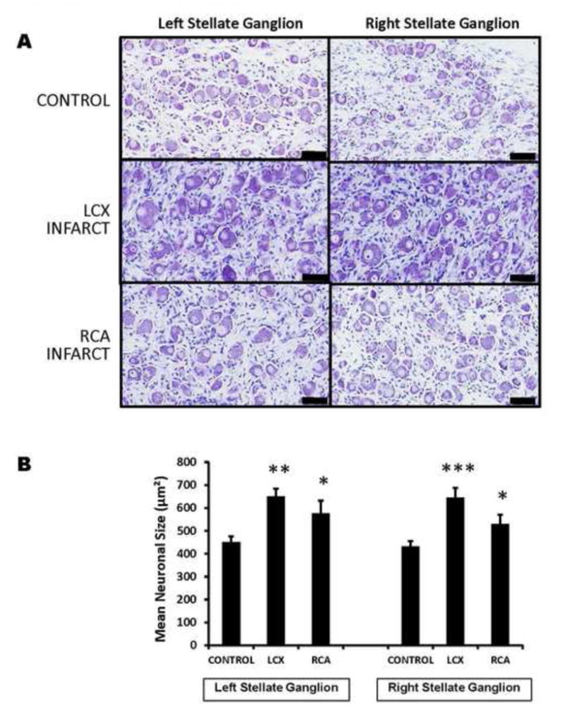
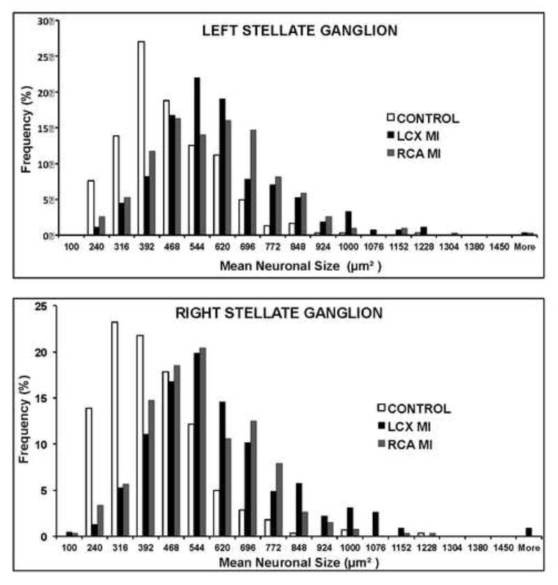
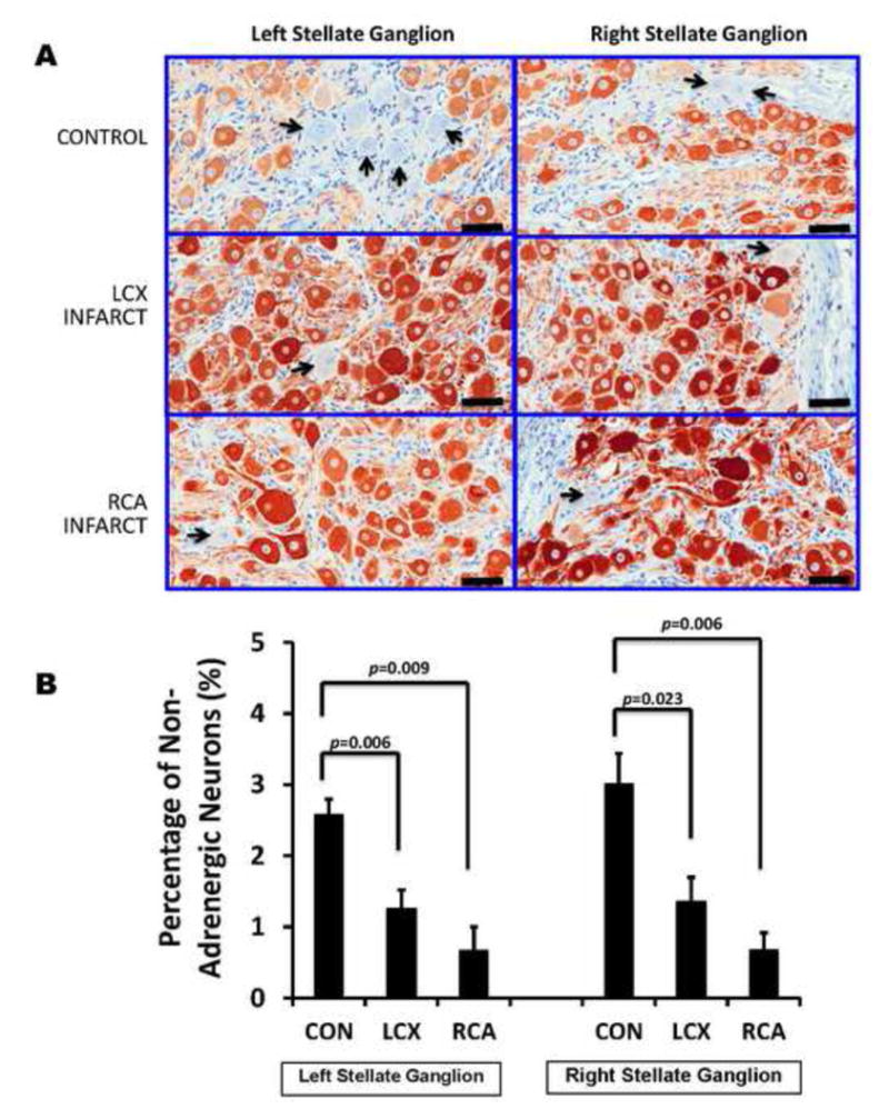
Results: Compared with control, LCX and RCA MIs increased mean neuronal size in the LSG (451 ± 25 vs 650 ± 34 vs 577 ± 55 μm(2), respectively; P = .0012) and RSG (433 ± 22 vs 646 ± 42 vs 530 ± 41 μm(2), respectively; P = .002). TH immunoreactivity was present in the majority of SGNs. Both LCX and RCA MIs were associated with significant decreases in the percentage of TH-negative SGNs, from 2.58% ± 0.2% in controls to 1.26% ± 0.3% and 0.7% ± 0.3% in animals with LCX and RCA MI, respectively, for LSG (P = .001) and from 3.02% ± 0.4% in controls to 1.36% ± 0.3% and 0.68% ± 0.2% in LCX and RCA MI, respectively, for RSG (P = .002). Both TH-negative and TH-positive neurons increased in size after LCX and RCA MI. Neuropeptide Y immunoreactivity was also increased significantly by LCX and RCA MI in both ganglia.

Conclusion: Left- and right-sided MIs equally induced morphologic and neurochemical changes in LSG and RSG neurons, independent of infarct site. These data indicate that afferent signals transduced after MI result in bilateral changes and provide a rationale for bilateral interventions targeting the sympathetic chain for arrhythmia modulation.

Keywords: Autonomic nervous system; Myocardial infarction; Neuronal remodeling; Neuropeptide remodeling; Sympathetic ganglia.


Conflict of interest statement

Conflict of Interest : The authors have no conflicts of interest to disclose.

Figures

FIGURE 1
FIGURE 1. Myocardial Infarct Models
Angiographic evidence of occlusion of the right coronary artery (RCA) and left circumflex artery (LCX) to create right and left-sided myocardial infarctions are shown in the top and middle panels, respectively. The blue arrowheads trace the course of the vessel at baseline, and indicate the missing vessel following microsphere occlusion. The red arrowhead identifies an intracoronary guidewire tracing out the course of the occluded RCA. The bottom panel shows delayed gadolinium magnetic resonance images of the RCA and LCX infarcts (bright tissue), also identified by white arrows. Short axis images of the ventricles are presented, viewed from the apex of the heart.
FIGURE 2
FIGURE 2. Myocardial Infarction Induces Stellate Neuronal Enlargement Without Relationship To Laterality
A. Shown are representative images of thionin-stained sections of right and left stellate ganglia from control animals, compared to left circumflex (LCX) artery and right coronary artery (RCA) occlusions to create left and right-sided myocardial infarctions. The larger neurons in LCX and RCA infarcts can be visually appreciated. (scale bar 50μm). B. Quantification of mean neuronal size in left and right stellate ganglia of control subjects compared to LCX and RCA infarcts are presented. *p<0.05, ** p<0.01, ***P<0.001 when compared to controls.
FIGURE 3
FIGURE 3. Distribution Of Stellate Ganglion Neuronal Size By Ganglion And Infarct Site
Histograms of neuronal size distribution from left and right stellate ganglia (LSG and RSG, respectively) from control animals subjects, and left circumflex (LCX) and right coronary artery (RCA) infarcts, creating left and right-sided infarcts respectively.
FIGURE 4
FIGURE 4. Myocardial Infarction Induces Neurochemical Remodeling Of Stellate Ganglion Neurons
A. Representative tyrosine hydroxylase (TH)-stained sections of right and left stellate ganglia from control animals, compared to left circumflex (LCX) artery and right coronary artery (RCA) occlusions to create left and right-sided myocardial infarctions. More TH negative neurons (indicated by black arrows)can be appreciated in control, compared to LCX and RCA infarcts. Larger neurons in infarcted ganglia can also be appreciated. Scale bar = 50μm. B. Quantification of the percentage of TH-negative neurons in left and right stellate ganglia of control subjects compared to LCX and RCA infarcts are presented. C. Examples of adjacent 4μm sections from a control animal (left panel) and LCX MI animal (right panel) stained with TH, and choline-acetyl transferase (ChAT) demonstrating neurons that stain only for TH (thin black arrows), only for ChAT (thick black arrows) or both (blue arrowheads), although these neurons stain less intensely for TH. Scale bar = 50μm.
FIGURE 5
FIGURE 5. Adrenergic and Non-Adrenergic Neurons Enlarge Following Myocardial Infarction
Graphical representation of mean neuronal size of tyrosine hydroxylase (TH) negative and positive neurons in ganglia from control subjects compared to LCX and RCA infarcts are presented. In control animals, TH positive neurons are smaller than TH-negative neurons. Not only are both populations of neurons larger in infarcted animals, the size difference seen in controls is lost. **p<0.01, ***P<0.001 compared to conrol. ##p<0.01 for Control TH-negative vs Control TH-positive.
FIGURE 6
FIGURE 6. Neuropeptide Y immunoreactivity increases following MI
A. Representative neuropeptide Y (NPY)-stained sections of left and right stellate ganglia (LSG and RSG, respectively) from control animals, compared to left circumflex (LCX) artery and right coronary artery (RCA) occlusions to create left and right-sided myocardial infarctions. Increased NPY immunoreactivity in LCX and RCA neurons can be identified. Darkly stained neurons are indicated by black arrow heads. Larger neurons in infarcted ganglia can also be appreciated. Scale bar = 50μm. B. Quantification of NPY immunoreactivity in left and right stellate ganglia of control subjects compared to LCX and RCA infarcts.

References

    1. Schwartz PJ. Cardiac sympathetic denervation to prevent life-threatening arrhythmias. Nature reviews Cardiology. 2014;11:346–353. - PubMed
    1. Shen MJ, Zipes DP. Role of the autonomic nervous system in modulating cardiac arrhythmias. Circulation Research. 2014;114:1004–1021. - PubMed
    1. Vaseghi M, Shivkumar K. The role of the autonomic nervous system in sudden cardiac death. Prog Cardiovasc Dis. 2008;50:404–419. - PMC - PubMed
    1. Bourke T, Vaseghi M, Michowitz Y, Sankhla V, Shah M, Swapna N, Boyle NG, Mahajan A, Narasimhan C, Lokhandwala Y, Shivkumar K. Neuraxial modulation for refractory ventricular arrhythmias: value of thoracic epidural anesthesia and surgical left cardiac sympathetic denervation. Circulation. 2010;121:2255–2262. - PMC - PubMed
    1. Ajijola OA, Lellouche N, Bourke T, Tung R, Ahn S, Mahajan A, Shivkumar K. Bilateral Cardiac Sympathetic Denervation for the Management of Electrical Storm. J Am Coll Cardiol. 2012;59:91–92. - PMC - PubMed

Publication types