Review
. 2015 Apr;22(3):183-96.
doi: 10.1111/micc.12190.

Vascular inward rectifier K+ channels as external K+ sensors in the control of cerebral blood flow

Affiliations
Review

Vascular inward rectifier K+ channels as external K+ sensors in the control of cerebral blood flow

Thomas A Longden et al. Microcirculation. 2015 Apr.

Abstract

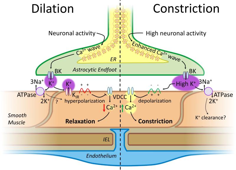
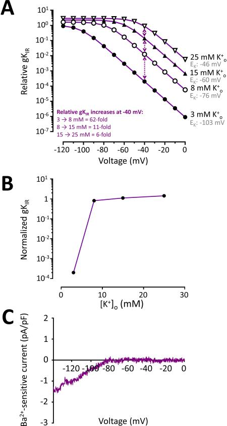
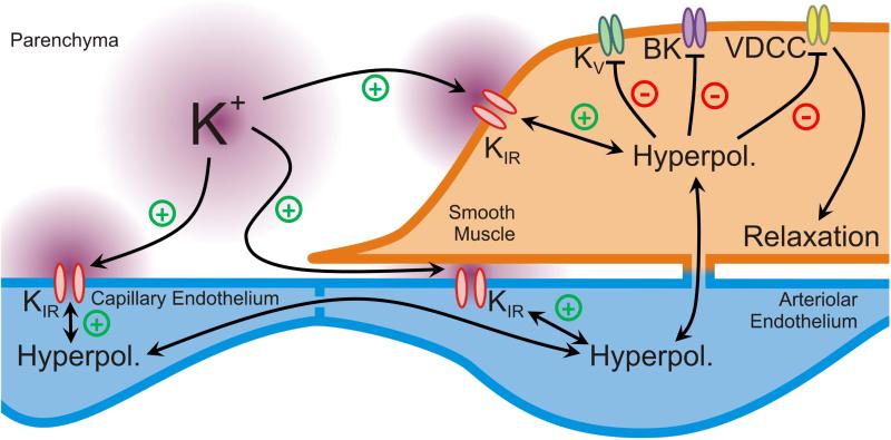
For decades it has been known that external K(+) ions are rapid and potent vasodilators that increase CBF. Recent studies have implicated the local release of K(+) from astrocytic endfeet-which encase the entirety of the parenchymal vasculature-in the dynamic regulation of local CBF during NVC. It has been proposed that the activation of KIR channels in the vascular wall by external K(+) is a central component of these hyperemic responses; however, a number of significant gaps in our knowledge remain. Here, we explore the concept that vascular KIR channels are the major extracellular K(+) sensors in the control of CBF. We propose that K(+) is an ideal mediator of NVC, and discuss KIR channels as effectors that produce rapid hyperpolarization and robust vasodilation of cerebral arterioles. We provide evidence that KIR channels, of the KIR 2 subtype in particular, are present in both the endothelial and SM cells of parenchymal arterioles and propose that this dual positioning of KIR 2 channels increases the robustness of the vasodilation to external K(+), enables the endothelium to be actively engaged in NVC, and permits electrical signaling through the endothelial syncytium to promote upstream vasodilation to modulate CBF.

Keywords: astrocytic endfoot; capillary; cerebral blood flow; endothelium; functional hyperemia; inward rectifier potassium channel; neurovascular coupling; parenchymal arteriole; smooth muscle.


Figures

Figure 1
Figure 1
K+ signaling mechanisms in NVC. K+-mediated dilation (left) begins with neuronal activity which is detected by astrocytic processes adjacent to synapses leading to phospholipase C (PLC)-mediated liberation of inositol 1,4,5-trisphosphate (IP3) and diacylglycerol (DAG) from membrane phosphatidylinositol 4,5-bisphosphate (PIP2) pools, ultimately resulting in an IP3-mediated Ca2+ wave which propagates into astrocytic endfeet enwrapping the cerebral microcirculation [115]. This engages large-conductance calcium (Ca2+)-activated K+ (BK) channels on the endfoot plasma membrane [26,27,97], and their activation leads to an increase in the concentration of K+ in the extracellular nanospace between the endfoot and SM. The rise in external K+ activates strong inward-rectifier K+ (KIR) channels on the smooth muscle, leading to membrane hyperpolarization, closure of voltage-dependent Ca2+ channels, vasorelaxation and increased blood flow [26]. During more intense neuronal activity (right), larger Ca2+ waves promote the release of higher concentrations of K+ from the endfoot, leading to membrane depolarization, VDCC activation and constriction [27]. Under both conditions, the Na+/K+ ATPase likely contributes to K+ clearance and may provide a brief accompanying hyperpolarizing current. Adapted from [67] and [27].
Figure 2
Figure 2
Inward-rectifier K+ channels are activated by increases in external K+. (A) In situ, raising K+ to <20 mM causes rapid and substantial vasodilation of pressurized (40 mm Hg) PAs due to KIR channel activation; further increases in K+ drive membrane depolarization and constriction. Trace from [67]. (B) The SM of PAs behaves as a K+ electrode with increasing concentrations of extracellular K+. Experimentally observed membrane potential (Vm) data (from [26,37,89]) are shown versus the potassium equilibrium potential (EK) predicted by the Nernst equation. At 3 mM K+, Vm is depolarized relative to EK due to myogenic inward cation currents. Raising K+ activates KIR channels, and the resultant K+ efflux effectively locks Vm at EK. (C) Hypothetical KIR current-voltage relationship (left) and illustration (right) showing that at basal extracellular K+ (3 mM) the pore of the KIR channel is blocked by Mg2+ or large cationic polyamines. Under these conditions (assuming 140 mM [K+]i), EK is -103, which is highly negative compared to the resting Vm of SM (approximately -35 to -40 mV) at the physiological intravascular pressure of 40 mmHg. The strong driving force for cation efflux under these conditions leads to blockade of the channel pore by the larger cations, resulting in very little channel activity. (D) When [K+]o is elevated to 8 mM (e.g., when released from the astrocytic endfoot during NVC), intracellular blockade of the channel is relieved. Channel unblock allows K+ to exit the cell, driving Vm to the new EK—where it will remain until the extracellular K+ is cleared—and leading to substantial vasodilation.
Figure 3
Figure 3
(A) KIR channel conductance over a range of Vm values with increasing concentrations of K+. With 3 mM [K+] o at -40 mV (the approximate resting Vm; dotted arrows), KIR channel conductance is very low. Raising K+ greatly increases KIR channel activity at a given Vm; channel activity is also increased by membrane hyperpolarization. Data were plotted according to the eq. 1 in the text. (B) The relationship between external K+ concentration and KIR channel conductance based on the K+- and voltage-dependence of KIR2 channels, calculated using eq. 1 (see text), and measured effects of external K+ on PA SM Vm [26,37]. Elevation of [K+]o from 3 mM to 8 mM causes an enormous, near-maximal increase in KIR channel conductance, with further elevations to 15 and 25 mM causing only small subsequent increases in conductance. (C) Subtracted 100-μM Ba2+-sensitive currents from a freshly dissociated EC from a rat PA in response to a voltage ramp from -140 to 0 mV indicating the presence of functional KIR2 channels in these cells. External K+ was 6 mM in this experiment.
Figure 4
Figure 4
Proposed scheme for KIR channels as K+ sensors to control cerebral blood flow. K+ ions released from astrocytic endfeet and neurons activates KIR2 channels present on SM cells and ECs in PAs and (possibly) on capillary ECs. Hyperpolarization caused by KIR engagement may then spread bi-directionally throughout the vascular syncytium, causing further KIR channel activation while at the same time deactivating KV and BK channels and VDCCs in the SM. This locks the membrane potential at EK until K+ is cleared and causes near maximal vasodilation and a substantial increase in cerebral blood flow.

References

    1. Attwell D, Buchan AM, Charpak S, Lauritzen M, MacVicar BA, Newman EA. Glial and neuronal control of brain blood flow. Nature. 2010;468:232–243. - PMC - PubMed
    1. Back T, Kohno K, Hossmann KA. Cortical negative DC deflections following middle cerebral artery occlusion and KCl-induced spreading depression: effect on blood flow, tissue oxygenation, and electroencephalogram. J Cereb Blood Flow Metab. 1994;14:12–19. - PubMed
    1. Bhandari S, Hunter M. Biophysical effects of pore mutations of ROMK1. Clin Sci. 2001;101:121–30. - PubMed
    1. Blanco G, Mercer RW. Isozymes of the Na-K-ATPase: heterogeneity in structure, diversity in function. Am J Physiol. 1998;275:F633–50. - PubMed
    1. Bradley KK, Jaggar JH, Bonev AD, Heppner TJ, Flynn ER, Nelson MT, Horowitz B. Kir2.1 encodes the inward rectifier potassium channel in rat arterial smooth muscle cells. J Physiol. 1999;515:639–651. - PMC - PubMed

Publication types

MeSH terms

Substances

LinkOut - more resources