Classification of histologically scored human knee osteochondral plugs by quantitative analysis of magnetic resonance images at 3T
- PMID: 25641500
- PMCID: PMC5875433
- DOI: 10.1002/jor.22810
Classification of histologically scored human knee osteochondral plugs by quantitative analysis of magnetic resonance images at 3T
Abstract
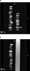
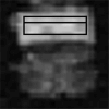
This work evaluates the ability of quantitative MRI to discriminate between normal and pathological human osteochondral plugs characterized by the Osteoarthritis Research Society International (OARSI) histological system. Normal and osteoarthritic human osteochondral plugs were scored using the OARSI histological system and imaged at 3 T using MRI sequences producing T1 and T2 contrast and measuring T1, T2, and T2* relaxation times, magnetization transfer, and diffusion. The classification accuracies of quantitative MRI parameters and corresponding weighted image intensities were evaluated. Classification models based on the Mahalanobis distance metric for each MRI measurement were trained and validated using leave-one-out cross-validation with plugs grouped according to OARSI histological grade and score. MRI measurements used for classification were performed using a region-of-interest analysis which included superficial, deep, and full-thickness cartilage. The best classifiers based on OARSI grade and score were T1- and T2-weighted image intensities, which yielded accuracies of 0.68 and 0.75, respectively. Classification accuracies using OARSI score-based group membership were generally higher when compared with grade-based group membership. MRI-based classification--either using quantitative MRI parameters or weighted image intensities--is able to detect early osteoarthritic tissue changes as classified by the OARSI histological system. These findings suggest the benefit of incorporating quantitative MRI acquisitions in a comprehensive clinical evaluation of OA.
Keywords: cartilage matrix; classification; imaging; osteoarthritis; quantitative MRI.
© 2015 Orthopaedic Research Society. Published by Wiley Periodicals, Inc.
Conflict of interest statement
Conflicts of interest: Michael Schär is an employee of Philips Healthcare, the manufacturer of equipment used in this study.
Figures


Similar articles
-
Machine learning classification of OARSI-scored human articular cartilage using magnetic resonance imaging.Osteoarthritis Cartilage. 2015 Oct;23(10):1704-12. doi: 10.1016/j.joca.2015.05.028. Epub 2015 Jun 9. Osteoarthritis Cartilage. 2015. PMID: 26067517 Free PMC article.
-
Histological Grade and Magnetic Resonance Imaging Quantitative T1rho/T2 Mapping in Osteoarthritis of the Knee: A Study in 20 Patients.Med Sci Monit. 2019 Dec 27;25:10057-10066. doi: 10.12659/MSM.918274. Med Sci Monit. 2019. PMID: 31881548 Free PMC article.
-
Associations of three-dimensional T1 rho MR mapping and three-dimensional T2 mapping with macroscopic and histologic grading as a biomarker for early articular degeneration of knee cartilage.Clin Rheumatol. 2017 Sep;36(9):2109-2119. doi: 10.1007/s10067-017-3645-2. Epub 2017 Apr 29. Clin Rheumatol. 2017. PMID: 28456927
-
Magnetic resonance imaging (MRI) of articular cartilage in knee osteoarthritis (OA): morphological assessment.Osteoarthritis Cartilage. 2006;14 Suppl A:A46-75. doi: 10.1016/j.joca.2006.02.026. Epub 2006 May 19. Osteoarthritis Cartilage. 2006. PMID: 16713720 Review.
-
The osteoarthritis initiative: report on the design rationale for the magnetic resonance imaging protocol for the knee.Osteoarthritis Cartilage. 2008 Dec;16(12):1433-41. doi: 10.1016/j.joca.2008.06.016. Epub 2008 Sep 10. Osteoarthritis Cartilage. 2008. PMID: 18786841 Free PMC article. Review.
Cited by
-
Functional MRI for evaluation of hyaline cartilage extracelullar matrix, a physiopathological-based approach.Br J Radiol. 2019 Nov;92(1103):20190443. doi: 10.1259/bjr.20190443. Epub 2019 Aug 23. Br J Radiol. 2019. PMID: 31433668 Free PMC article. Review.
-
Articular Cartilage-From Basic Science Structural Imaging to Non-Invasive Clinical Quantitative Molecular Functional Information for AI Classification and Prediction.Int J Mol Sci. 2023 Oct 7;24(19):14974. doi: 10.3390/ijms241914974. Int J Mol Sci. 2023. PMID: 37834422 Free PMC article. Review.
-
Ex vivo quantitative multiparametric MRI mapping of human meniscus degeneration.Skeletal Radiol. 2016 Dec;45(12):1649-1660. doi: 10.1007/s00256-016-2480-x. Epub 2016 Sep 17. Skeletal Radiol. 2016. PMID: 27639388
-
Quantitative OCT and MRI biomarkers for the differentiation of cartilage degeneration.Skeletal Radiol. 2016 Apr;45(4):505-16. doi: 10.1007/s00256-016-2334-6. Epub 2016 Jan 19. Skeletal Radiol. 2016. PMID: 26783011
-
Multiparametric Classification of Skin from Osteogenesis Imperfecta Patients and Controls by Quantitative Magnetic Resonance Microimaging.PLoS One. 2016 Jul 14;11(7):e0157891. doi: 10.1371/journal.pone.0157891. eCollection 2016. PLoS One. 2016. PMID: 27416032 Free PMC article.
References
-
- Burstein D, Gray ML, Hartman AL, et al. Diffusion of small solutes in cartilage as measured by nuclear-magnetic-resonance (NMR) spectroscopy and imaging. J Orthop Res. 1993;11:465–478. - PubMed
-
- Mlynarik V, Trattnig S, Huber M, et al. The role of relaxation times in monitoring proteoglycan depletion in articular cartilage. J Magn Reson Imaging. 1999;10:497–502. - PubMed
-
- Seo GS, Aoki J, Moriya H, et al. Hyaline cartilage: in vivo and in vitro assessment with magnetization transfer imaging. Radiology. 1996;201:525–530. - PubMed
-
- Wong CS, Yan CH, Gong NJ, et al. Imaging biomarker with T1rho and T2 mappings in osteoarthritis—in vivo human articular cartilage study. Eur J Radiol. 2013;82:647–650. - PubMed
Publication types
MeSH terms
Grants and funding
LinkOut - more resources
Full Text Sources
Other Literature Sources
Medical

