. 2015 May;33(5):1523-31.
doi: 10.1002/stem.1948.

HCELL Expression on Murine MSC Licenses Pancreatotropism and Confers Durable Reversal of Autoimmune Diabetes in NOD Mice

Affiliations

HCELL Expression on Murine MSC Licenses Pancreatotropism and Confers Durable Reversal of Autoimmune Diabetes in NOD Mice

Reza Abdi et al. Stem Cells. 2015 May.

Abstract

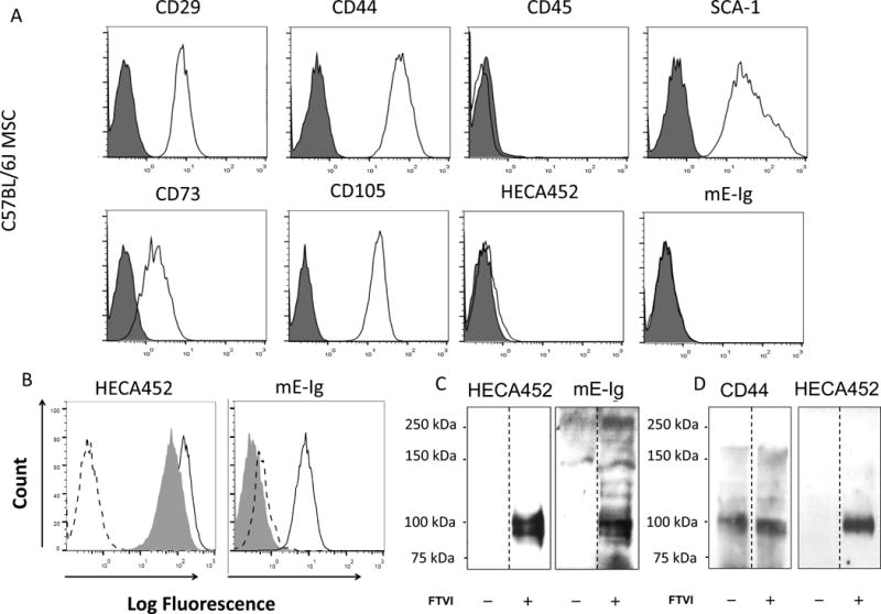
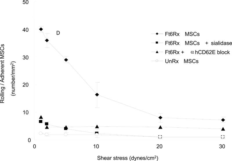
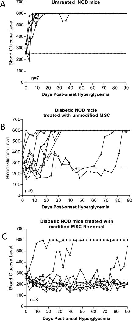
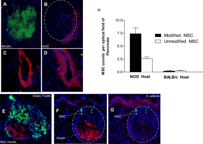
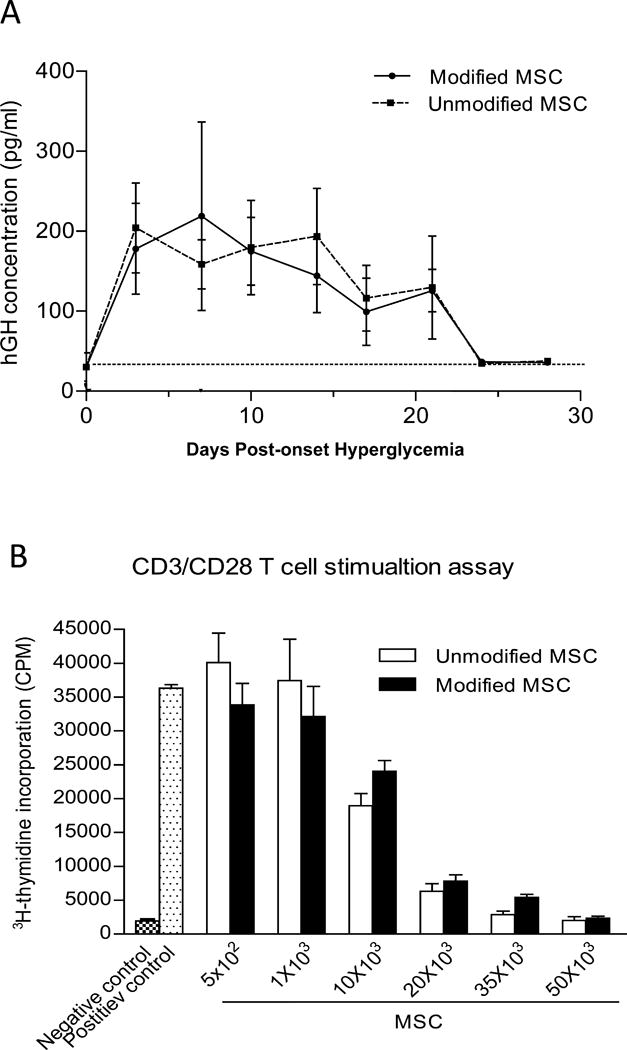
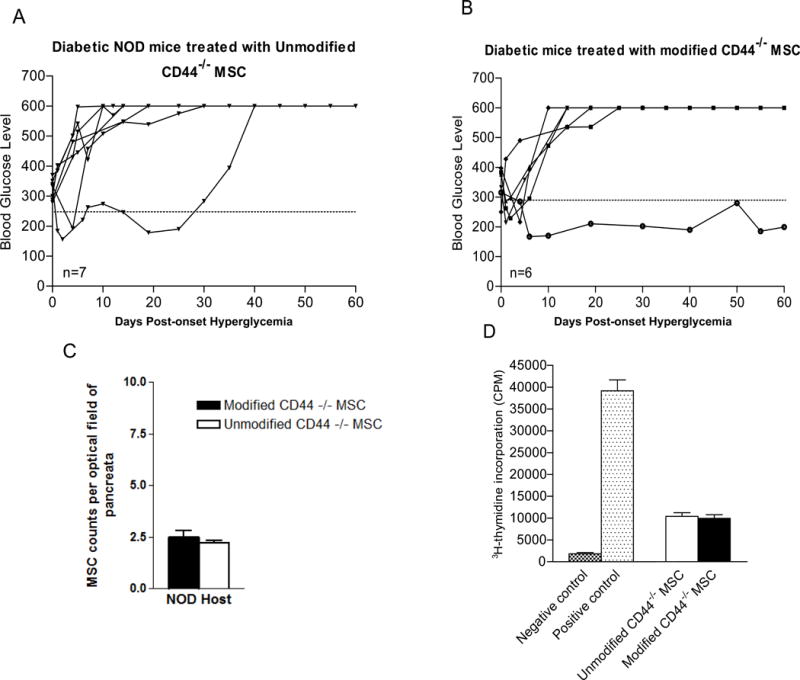
Type 1 diabetes (T1D) is an immune-mediated disease resulting in destruction of insulin-producing pancreatic beta cells. Mesenchymal stem cells (MSCs) possess potent immunomodulatory properties, garnering increasing attention as cellular therapy for T1D and other immunologic diseases. However, MSCs generally lack homing molecules, hindering their colonization at inflammatory sites following intravenous (IV) administration. Here, we analyzed whether enforced E-selectin ligand expression on murine MSCs could impact their effect in reversing hyperglycemia in nonobese diabetic (NOD) mice. Although murine MSCs natively do not express the E-selectin-binding determinant sialyl Lewis(x) (sLe(x) ), we found that fucosyltransferase-mediated α(1,3)-exofucosylation of murine MSCs resulted in sLe(x) display uniquely on cell surface CD44 thereby creating hematopoietic cell E-/L-selectin ligand (HCELL), the E-selectin-binding glycoform of CD44. Following IV infusion into diabetic NOD mice, allogeneic HCELL(+) MSCs showed threefold greater peri-islet infiltrates compared to buffer-treated (i.e., HCELL(-) ) MSCs, with distribution in proximity to E-selectin-expressing microvessels. Exofucosylation had no effect on MSC immunosuppressive capacity in in vitro assays; however, although engraftment was temporary for both HCELL(+) and HCELL(-) MSCs, administration of HCELL(+) MSCs resulted in durable reversal of hyperglycemia, whereas only transient reversal was observed following administration of HCELL(-) MSCs. Notably, exofucosylation of MSCs generated from CD44(-/-) mice induced prominent membrane expression of sLe(x) , but IV administration of these MSCs into hyperglycemic NOD mice showed no enhanced pancreatotropism or reversal of hyperglycemia. These findings provide evidence that glycan engineering to enforce HCELL expression boosts trafficking of infused MSCs to pancreatic islets of NOD mice and substantially improves their efficacy in reversing autoimmune diabetes. Stem Cells 2013;33:1523-1531.

Keywords: Diabetes; Glycan engineering; Glycosyltransferase programmed stereosubstitution; HCELL; Mesenchymal stem cell.


Figures

Figure 1
Figure 1. Effects of Exofucosylation (FTVI treatment) on E-selectin ligand expression by mouse MSC
(A) MSC derived from C57BL/6 marrow lacked expression of CD45 and expressed characteristic mouse MSC markers Sca-1, CD29, CD44, CD73 and CD105. Cells lacked reactivity with mAb HECA452 and with E-selectin-Ig chimera (mE-Ig) (istoype= red color and antibody = blue color). (B) FTVI-modified MSC (solid line) stained positive for mAbs HECA452 and were reactive with mE-Ig. Digestion of FTVI-modified MSC with bromelain and proteinase K (shaded histogram) significantly reduced mE-Ig reactivity, but not HECA452 staining, indicating that protease-sensitive glycoproteins serve as the principal E-selectin ligand(s). Dashed line represents staining controls (isotype control for HECA452 staining and calcium chelation with EDTA for mE-Ig staining). (C) Western blot analysis of HECA452 (left) and mE-Ig (right) reactivity of cell lysates of unmodified MSC (−) and FTVI-modified MSC (+). FTVI modification induced HECA452- and mE-Ig-reactive moieties predominantly on a doublet glycoprotein band of ~100 kDa. (D) CD44 was immunoprecipitated from equivalent amounts of cell lysate from FTVI-modified (+) or unmodified (−) MSCs. Immunoprecipitates were then electrophoresed and blotted with anti-CD44 mAbs (KM114 and IM7; left) and with mAb HECA452 (right).
Figure 2
Figure 2. Parallel plate flow chamber assay of FTVI-modified and unmodified wild type MSC adherence to TNF-α treated HUVEC
FTVI modification markedly improved MSC adhesion to HUVEC at 0.5 dynes/cm2. Treatment of HUVEC with anti-E-selectin (anti-CD62E) function-blocking mAb reduced rolling adhesive interactions of FTVI-modified MSC to levels similar to that of unmodified MSC. Similarly, removal of sLex determinants by sialidase treatment of FTVI-modified MSC reduced adhesion to levels equivalent to that of unmodified MSC.
Figure 3
Figure 3. Effects of intravenous administration of unmodified and FTVI-modified MSC on hyperglycemia in new onset diabetic NOD mice
(A) Hyperglycemic NOD injected with PBS (untreated control) showed no reversal of hyperglycemia (i.e., glucose levels consistently above 600 mg glucose/dL). As compared to infusion of unmodified MSCs (B), infusion of FTVI-modified MSCs (C) resulted in a marked increase in number of mice with reversion to normoglycemia and in the durability of diabetes reversal.
Figure 4
Figure 4. Immunofluorescence staining of islets to assess expression of E-selectin and localization of MSC in the pancreas
Figures (A) and (B): Pancreatic islets of (A) diabetic-resistant BALB/c mice and (B) NOD mice were stained for expression of insulin (green) and E-selectin (red). Islets are demarcated by dashed line. Compared to BALB/c (A), NOD mice (B) show diminished insulin production due to insulitis. In Figures (C)–(G), cryostat sections of pancreas from MSC-treated NOD mice are stained with DAPI to identify nuclei (blue). Figures (C) and (D): Staining of sequential sections of NOD pancreas demonstrates co-localization of endothelial marker CD31 (C) and E-selectin (D), confirming the presence of E-selectin on peri-islet endothelial cells. Figure (E): Co-staining of NOD islet with T-cell marker CD3 (green) and insulin (red) reveals characteristic T-cell infiltration at the margins of the islet. Figures (F) and (G): Immunofluorescence images of a cryostat section stained for infiltrating MSC (visualized with FITC-conjugated anti-sLex mAb HECA452; green), islet (F) (visualized by APC-conjugated anti-insulin mAb; red) and E-selectin-expressing microvessel (G) (visualized with PE-conjugated anti-E-selectin mAb; red). Staining identifies HECA452+ MSC in zones of insulitis, in proximity to E-selectin-expressing microvessels in the peri-islet area. Figure (H): Pancreatic infiltration of intravenously administered MSC into NOD and BALB/c hosts. Accumulation of FTVI-modified MSC into pancreata of NOD mice is 3-fold higher compared to that of unmodified MSC (p<0.01), whereas no difference in pancreatic infiltrates is observed in BALB/c host (n=3 mice per group; minimum 30 fields counted per group at 60X magnification).
Figure 5
Figure 5. FTVI-modification of MSC does not affect cell survival or immunosuppressive capacity
(A) Similar levels of hGH were detected in the serum of NOD mice at different time points following injection with pHRST-hGH-transduced FTVI-modified or unmodified MSC. (B) FTVI-modified and unmodified MSC equally suppressed proliferation of NOD CD4+ T cells stimulated with CD3/CD28.
Figure 6
Figure 6. Lack of CD44 expression abrogates the anti-diabetic effect of systemically administered FTVI-modified MSC
(A) As compared to unmodified wild type MSC (Figure 3B), administration of CD44-deficient MSC shows modest anti-diabetic effect. Only 1 NOD mouse (out of 7) receiving unmodified CD44KO MSC showed reversal of hyperglycemia which was transient (diabetes recurrence at ~day 30), and 6 out of 7 diabetic NOD mice remained hyperglycemic. (B) As compared to results in mice receiving FTVI-modified wild-type MSC (Figure 3C), administration of FTVI-modified CD44KO MSC conferred minimal anti-diabetic effects, with only 1 out of 6 NOD mice showing reversal of hyperglycemia. (C) Accumulation of MSC in NOD pancreata was no different in mice receiving FTVI-modified CD44KO MSC (white bar) compared to mice receiving unmodified CD44KO MSC (black bar) (p<0.01), and in each case was similar to that receiving unmodified MSC (Figure 4H). MSC infiltrates were quantified at X60 magnification. (D) Both FTVI-modified and unmodified CD44KO MSC possess immunosuppressive capacity, similarly dampening T cell proliferation in the CD3/CD28 T cell stimulation assay.

Similar articles

Cited by

References

    1. Bresson D, von Herrath M. Immunotherapy for the prevention and treatment of type 1 diabetes: optimizing the path from bench to bedside. Diabetes Care. 2009;32:1753–1768. - PMC - PubMed
    1. Ludvigsson J, Krisky D, Casas R, et al. GAD65 antigen therapy in recently diagnosed type 1 diabetes mellitus. N Engl J Med. 2012;366:433–442. - PubMed
    1. Bonifacio E. Immunotherapy in type 1 diabetes: a shorter but more winding road? Diabetes. 2012;61:2214–2215. - PMC - PubMed
    1. Prockop DJ, Brenner M, Fibbe WE, et al. Defining the risks of mesenchymal stromal cell therapy. Cytotherapy. 2010;12:576–578. - PubMed
    1. Dazzi F, van Laar JM, Cope A, et al. Cell therapy for autoimmune diseases. Arthritis Res Ther. 2007;9:206. - PMC - PubMed

Publication types

MeSH terms