

Diffusion-perfusion mismatch: an opportunity for improvement in cortical function
- PMID: 25642208
- PMCID: PMC4294157
- DOI: 10.3389/fneur.2014.00280
Diffusion-perfusion mismatch: an opportunity for improvement in cortical function
Abstract
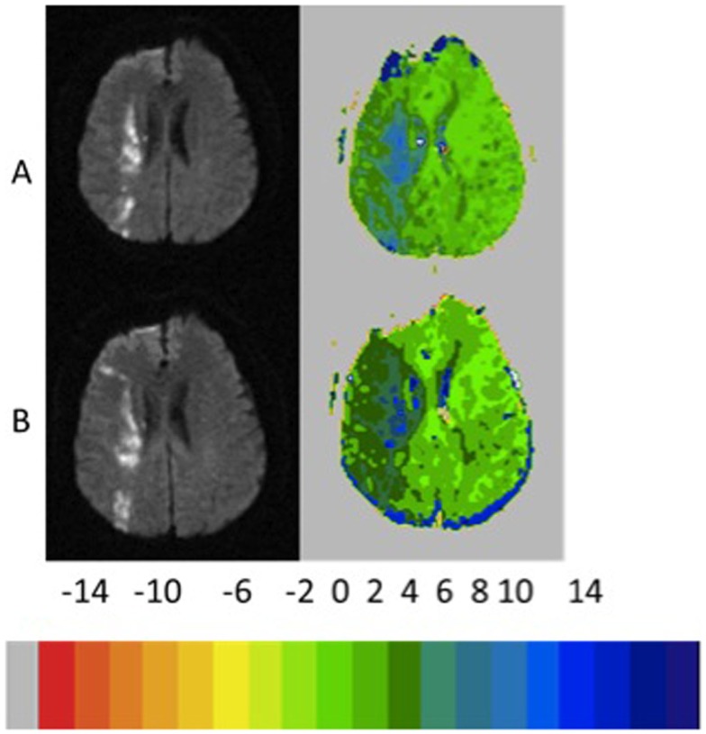
Objective: There has been controversy over whether diffusion-perfusion mismatch provides a biomarker for the ischemic penumbra. In the context of clinical stroke trials, regions of the diffusion-perfusion mismatch that do not progress to infarct in the absence of reperfusion are considered to represent "benign oligemia." However, at least in some cases (particularly large vessel stenosis), some of this hypoperfused tissue may remain dysfunctional for a prolonged period without progressing to infarct and may recover function if eventually reperfused. We hypothesized that patients with persistent diffusion-perfusion mismatch using a hypoperfusion threshold of 4-5.9 s delay on time-to-peak (TTP) maps at least sometimes have persistent cognitive deficits relative to those who show some reperfusion of this hypoperfused tissue.
Methods: We tested this hypothesis in 38 patients with acute ischemic stroke who had simple cognitive tests (naming or line cancelation) and MRI with diffusion and perfusion imaging within 24 h of onset and again within 10 days, most of whom had large vessel stenosis or occlusion.
Results: A persistent perfusion deficit of 4-5.9 s delay in TTP on follow up MRI was associated with a persistent cognitive deficit at that time point (p < 0.001). When we evaluated only patients who did not have infarct growth (n = 14), persistent hypoperfusion (persistent mismatch) was associated with a lack of cognitive improvement compared with those who had reperfused. The initial volume of hypoperfusion did not correlate with the later infarct volume (progression to infarct), but change in volume of hypoperfusion correlated with change in cognitive performance (p = 0.0001). Moreover, multivariable regression showed that the change in volume of hypoperfused tissue of 4-5.9 s delay (p = 0.002), and change in volume of ischemic tissue on diffusion weighted imaging (p = 0.02) were independently associated with change in cognitive function.
Conclusion: Our results provide additional evidence that non-infarcted tissue with a TTP delay of 4-5.9 s may be associated with persistent deficits, even if it does not always result in imminent progression to infarct. This tissue may represent the occasional opportunity to intervene to improve function even days after onset of symptoms.
Keywords: NIHSS; acute ischemic stroke; diffusion–perfusion mismatch; functional outcome; penumbra.
Figures


Similar articles
-
SB 234551 selective ET(A) receptor antagonism: perfusion/diffusion MRI used to define treatable stroke model, time to treatment and mechanism of protection.Exp Neurol. 2008 Jul;212(1):53-62. doi: 10.1016/j.expneurol.2008.03.011. Epub 2008 Mar 25. Exp Neurol. 2008. PMID: 18462720
-
Combined SPECT and diffusion-weighted MRI as a predictor of infarct growth in acute ischemic stroke.J Nucl Med. 2000 May;41(5):788-94. J Nucl Med. 2000. PMID: 10809193
-
Diffusion- and perfusion-weighted MRI. The DWI/PWI mismatch region in acute stroke.Stroke. 1999 Aug;30(8):1591-7. doi: 10.1161/01.str.30.8.1591. Stroke. 1999. PMID: 10436106
-
Extension of therapeutic window in ischemic stroke by selective mismatch imaging.Int J Stroke. 2019 Jun;14(4):351-358. doi: 10.1177/1747493019840936. Epub 2019 Apr 1. Int J Stroke. 2019. PMID: 30935350 Review.
-
Validation of MRI Determination of the Penumbra by PET Measurements in Ischemic Stroke.J Nucl Med. 2017 Feb;58(2):187-193. doi: 10.2967/jnumed.116.185975. Epub 2016 Nov 22. J Nucl Med. 2017. PMID: 27879370 Review.
Cited by
-
Evaluation of Enhanced Learning Techniques for Segmenting Ischaemic Stroke Lesions in Brain Magnetic Resonance Perfusion Images Using a Convolutional Neural Network Scheme.Front Neuroinform. 2019 May 29;13:33. doi: 10.3389/fninf.2019.00033. eCollection 2019. Front Neuroinform. 2019. PMID: 31191282 Free PMC article.
-
Understanding recovery of language after stroke: insights from neurovascular MRI studies.Front Lang Sci. 2023;2:1163547. doi: 10.3389/flang.2023.1163547. Epub 2023 Jun 5. Front Lang Sci. 2023. PMID: 38162928 Free PMC article.
-
Magnetic Resonance Perfusion Imaging Provides a Significant Tool for the Identification of Cardioembolic Stroke.Curr Neurovasc Res. 2016;13(4):271-276. doi: 10.2174/1567202613666160901143040. Curr Neurovasc Res. 2016. PMID: 27586679 Free PMC article.
-
Variability in Motor and Language Recovery during the Acute Stroke Period.Cerebrovasc Dis Extra. 2016 Mar 22;6(1):12-21. doi: 10.1159/000444149. eCollection 2016 Jan-Apr. Cerebrovasc Dis Extra. 2016. PMID: 27099611 Free PMC article.
-
Remapping and Reconnecting the Language Network after Stroke.Brain Sci. 2024 Apr 24;14(5):419. doi: 10.3390/brainsci14050419. Brain Sci. 2024. PMID: 38790398 Free PMC article. Review.
References
Grants and funding
LinkOut - more resources
Full Text Sources
Other Literature Sources