

Review
doi: 10.1088/0957-4484/26/7/074004.
Epub 2015 Feb 2.
Challenges in DNA motion control and sequence readout using nanopore devices
Affiliations
- PMID: 25642629
- PMCID: PMC4710574
- DOI: 10.1088/0957-4484/26/7/074004
Item in Clipboard
Review
Challenges in DNA motion control and sequence readout using nanopore devices
Nanotechnology.
.
Abstract
Nanopores are being hailed as a potential next-generation DNA sequencer that could provide cheap, high-throughput DNA analysis. In this review we present a detailed summary of the various sensing techniques being investigated for use in DNA sequencing and mapping applications. A crucial impasse to the success of nanopores as a reliable DNA analysis tool is the fast and stochastic nature of DNA translocation. We discuss the incorporation of biological motors to step DNA through a pore base-by-base, as well as the many experimental modifications attempted for the purpose of slowing and controlling DNA transport.
Figures
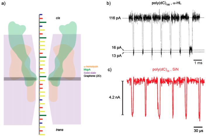
Ionic current detection of ssDNA in protein and solid-state nanopores. (a) An illustration comparing the relative sizes of α-hemolysin (αHL) (orange), MspA (green), solid-state (purple), and graphene nanopores (gray). The reduced thickness of MspA and graphene pores allows for the greatest spatial resolution, which is the motivation behind their usage towards DNA sequencing applications. (b) A concatenated current trace of 100 mer cytosine homopolymer translocations through an αHL channel in 1.0 M KCl solution when applying a transmembrane voltage of 120 mV. The restriction of the pore diameter and low voltage application allows for transport times on the order of 100 μs [2]. (c) A concatenated current trace of poly(dC) 30 mer translocations through an ultrathin silicon nitride nanopore (diameter of 1.4 nm) in 1.0 M KCl when applying a voltage bias of 1 V. While these small, thin pores can provide an improvement in structural details and signal to noise ratio, the reduced thickness and higher applied voltage causes the average dwell time to be on the order of 10 μs [109].
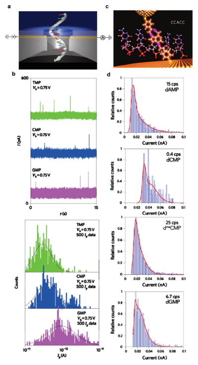
Nucleic acid base detection by transverse tunneling current read-out. (a) A representation of ssDNA translocating through a gold tunneling junction sandwiched inside a solid-state membrane. (b) Top: sample tunneling current traces recorded for the detection of the RNA monomers TMP (green), CMP (blue), and GMP (purple) at a trans-electrode bias of 0.75 V. Bottom: histograms of the measured tunneling current Ip of 500 data points for each nucleotide. When fitting the distributions to Gaussian functions, the peaks indicate signature mean currents for each of the three bases [104]. (c) An illustration depicting the binding of a short oligomer CCACC between a gold probe and surface, both functionalized with the reagent 4-mercaptobenzamide. (d) Current histograms that show differentiation between the DNA nucleotides dAMP, dCMP, dmeCMP, and dGMP due to distinction in both tunneling current amplitude and the current spikes per second. The red solid lines are double Gaussian fits in the logarithm of the current, implying two binding conformations for each nucleotide [35].
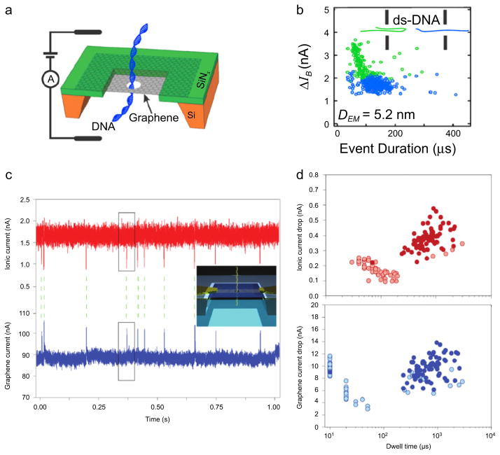
Graphene nanopores for ionic current and electric field detection of DNA. (a) A schematic of a dsDNA molecule translocating through a graphene nanopore. (b) A sample scatter plot of current blockade ΔIB versus the event duration for 10 kbp DNA transport through a 5.2 nm pore (160 mV applied bias, 3 M KCl, pH 10). A pore of this size permits two types of translocation events: unfolded, linear events (blue dots) and folded events (green dots). This heterogeneity of events can be eliminated by raising the solution pH or fabricating pores of smaller diameter [26]. (c) Graphene nanoribbon (GNR) for field-effect sensing of translocating DNA molecules through a nearby nanopore. By monitoring both the transmembrane ion current (red trace) and GNR transistor current (blue trace), individual translocations of circular pNEB DNA (2713 bp) are detected by and correlated between the two signals. Inset: an illustration of DNA translocation a nanopore embedded inside a GNR. (d) Scatter plots of ionic current drop and graphene current drop versus dwell time. Solid-colored circles are translocation events (n = 125) that were correlated between the ion and graphene current signals (i.e., 56% were successfully correlated). The majority of uncorrelated events occur at faster time scales, perhaps due to DNA collisions [103].
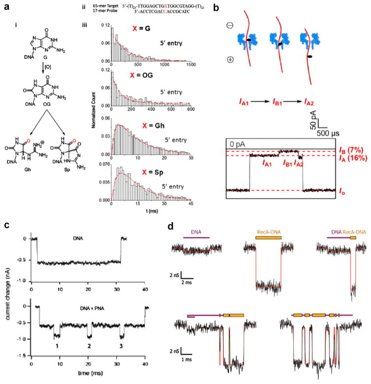
Mapping of DNA damage and DNA–protein complexes using nanopores. (a) Using αHL pores to unzip DNA duplexes containing a single oxidized guanine. (i) The molecular structure of guanine (G) and it’s oxidatively-damaged products 8-oxo-8,8-dihydroguanine (OG), guanidinohydantoid (Gh), and spiroiminodihydantoid (Sp). (ii) The hybridized DNA sequence used experimentally to detect a single guanine base lesion. (iii) The unzipping duration histograms for all the guanine base modifications. Two different distribution shapes emerge, one shape for G and OG and another for Gh and Sp, which indicates two distinct unzipping processes. The G and OG distributions fit to a single exponential decay, which points to a single-step unzipping process, whereas the Gh and Sp distributions fit to a more complex double-exponential function, which indicates a two-step unzipping process [40, 85]. (b) Detection of an abasic site in DNA by the selective attachment of 2-aminomethyl-18-crown-6, which causes a noticeable increase in current blockade when translocating an αHL pore. This approach can be expanded to detect multiple abasic sites in a single DNA strand [4]. (c) The binding of sequence-specific peptide nucleic acids (PNA) to a DNA molecule makes it possible to map these regions in a DNA strand by monitoring the current signal while a single DNA electrophoreses through the pore [90]. (d) The non-specific binding of the protein RecA to DNA results in a larger current blockade in regions where it coats the bare DNA. The five sample current traces show events with two distinct blockade levels caused by alternating regions of DNA and RecA–DNA complex translocating a 30 nm nanopore. RecA is a DNA-stiffening protein that has shown promise in DNA-tagging for sequence mapping (Nabsys) [45].
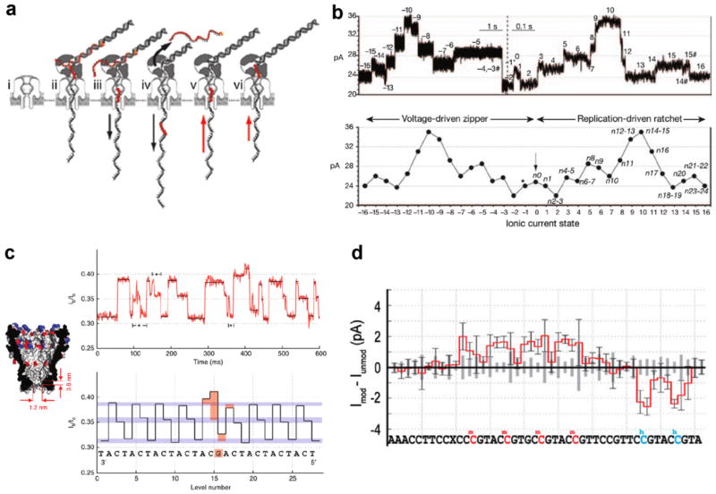
Enzyme-mediated transport of DNA through protein nanopores. (a) A step-by-step schematic describing sequencing by phi29 synthesis of a DNA sequence containing an a basic region. (i) An αHL pore embedded in a lipid bilayer before DNA capture. (ii) The capture of a DNA–polymerase complex by the protruding electric field. (iii) The electrophoretic force starts the unzipping of the blocking oligomer, which prevented DNA digestion and synthesis in the bulk solution. (iv) Once completely unzipped, the blocking oligo diffuses away from the pore. (v) DNA synthesis by phi29 reverses the direction of DNA motion against the applied bias. (vi) Once the abasic region of the DNA template reaches the polymerase, synthesis concludes and the DNA–polymerase complex disassociates from the pore mouth. (b) Top: a sample current trace of a single DNA molecule being unzipped by the electrophoretic force, then synthesized by the phi29 DNAP. Bottom: when averaging multiple current traces, multiple current levels are discerned that represent an averaged combination of the bases lying within the pore constriction [18]. (c) Using phi29 DNAP to ratchet a repeating CAT DNA sequence with a single substitution of G through a modified MspA nanopore. Left: the narrower and shorter constriction of MspA yield greater spatial resolution for base recognition. Right: despite the inherent stochastic motion of the DNA sequence, the raw and analyzed current traces detect an increase in fractional current blockade at the location of the guanine substitution [63]. (d) By driving DNA through a modified MspA nanopore by polymerase catalysis, the epigenetic modifications methylcytosine (mC) and hydroxymethylcytosine (hmC) can be distinguished from cytosine (C), despite not having single base resolution [50].
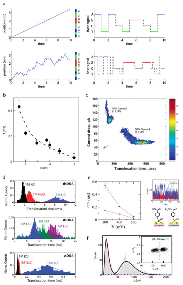
Slowing down and controlling voltage-driven DNA translocation through solid-state nanopores. (a) Simulating the effect of random velocity fluctuations in DNA sequencing in an ideal single-base resolution nanopore device. Top: when a 10 nt ssDNA translocates the pore with a constant velocity, the current signal shows perfect identification of each base. Bottom: conversely, when realistic, random fluctuations are introduced to the system, the DNA sequence is detected incorrectly with multiple, erroneous base insertions and deletions [60]. (b) Reducing pore diameter has the effect of slowing down DNA transport due to greater DNA–pore interactions [115]. (c) The addition of glycerol to the electrolyte solution has the effect of unfolding DNA molecules and increasing DNA dwell time in the pore. For a 3 kbp DNA, an increase from 10% glycerol to 50% glycerol results in a four-fold increase in dwell time [24]. (d) The salt type and concentration used in electrolyte buffer has an effect on translocation time. The Dekker group observed that NaCl and LiCl resulted in longer dwell times for λ-DNA and further found that increasing the concentration of LiCl to 4 M gave even slower translocation times. An even more drastic increase in dwell time was seen for an M13mp18 ssDNA when switching the salt type to LiCl [46]. (e) Hydrophobic (blue) and hydrophilic (red) monolayer coating of pores effect voltage-driven DNA translocation. Under a moderate applied voltage, the hydrophobic monolayer causes DNA to translocate faster than in the case of a hydrophilic monolayer. By conducting MD simulations, it was seen that slower transport when using hydrophilic monolayer was due to the DNA molecule fluctuating radially inside the pore because of its great affinity to the hydrophilic pore surface [111]. (f) Using a 2.5 nm pore allows for the distinction between 100 bp and 500 bp in a mixture with an accuracy >98% [14].
References
Publication types
MeSH terms
Substances
Grants and funding
LinkOut - more resources
Full Text Sources
Other Literature Sources