Nasal associated lymphoid tissue of the Syrian golden hamster expresses high levels of PrPC
- PMID: 25642714
- PMCID: PMC4314084
- DOI: 10.1371/journal.pone.0117935
Nasal associated lymphoid tissue of the Syrian golden hamster expresses high levels of PrPC
Abstract
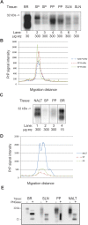
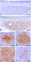
The key event in the pathogenesis of the transmissible spongiform encephalopathies is a template-dependent misfolding event where an infectious isoform of the prion protein (PrPSc) comes into contact with native prion protein (PrPC) and changes its conformation to PrPSc. In many extraneurally inoculated models of prion disease this PrPC misfolding event occurs in lymphoid tissues prior to neuroinvasion. The primary objective of this study was to compare levels of total PrPC in hamster lymphoid tissues involved in the early pathogenesis of prion disease. Lymphoid tissues were collected from golden Syrian hamsters and Western blot analysis was performed to quantify PrPC levels. PrPC immunohistochemistry (IHC) of paraffin embedded tissue sections was performed to identify PrPC distribution in tissues of the lymphoreticular system. Nasal associated lymphoid tissue contained the highest amount of total PrPC followed by Peyer's patches, mesenteric and submandibular lymph nodes, and spleen. The relative levels of PrPC expression in IHC processed tissue correlated strongly with the Western blot data, with high levels of PrPC corresponding with a higher percentage of PrPC positive B cell follicles. High levels of PrPC in lymphoid tissues closely associated with the nasal cavity could contribute to the relative increased efficiency of the nasal route of entry of prions, compared to other routes of infection.
Conflict of interest statement
Figures


References
-
- Stahl N, Borchelt DR, Hsiao K, Prusiner SB (1987) Scrapie prion protein contains a phosphatidylinositol glycolipid. Cell 51: 229–240. - PubMed
-
- Bendheim PE, Brown HR, Rudelli RD, Scala LJ, Goller NL, et al. (1992) Nearly ubiquitous tissue distribution of the scrapie agent precursor protein. Neurology 42: 149–156. - PubMed
-
- Ford MJ, Burton LJ, Morris RJ, Hall SM (2002) Selective expression of prion protein in peripheral tissues of the adult mouse. Neuroscience 113: 177–192. - PubMed
-
- Fournier JG, Escaig-Haye F, Billette de Villemeur T, Robain O, Lasmézas CI, et al. (1998) Distribution and submicroscopic immunogold localization of cellular prion protein (PrPC) in extracerebral tissues. Cell Tissue Res 292: 77–84. - PubMed
-
- Horiuchi M, Yamazaki N, Ikeda T, Ishiguro N, Shinagawa M (1995) A cellular form of prion protein (PrPC) exists in many non-neuronal tissues of sheep. J Gen Virol 76: 2583–2587. - PubMed
Publication types
MeSH terms
Substances
Grants and funding
LinkOut - more resources
Full Text Sources
Other Literature Sources

