Inactivation of DAP12 in PMN inhibits TREM1-mediated activation in rheumatoid arthritis
- PMID: 25642940
- PMCID: PMC4313943
- DOI: 10.1371/journal.pone.0115116
Inactivation of DAP12 in PMN inhibits TREM1-mediated activation in rheumatoid arthritis
Abstract
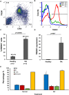
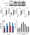
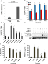
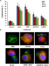
Rheumatoid arthritis (RA) is an autoimmune disease characterized by dysregulated and chronic systemic inflammatory responses that affect the synovium, bone, and cartilage causing damage to extra-articular tissue. Innate immunity is the first line of defense against invading pathogens and assists in the initiation of adaptive immune responses. Polymorphonuclear cells (PMNs), which include neutrophils, are the largest population of white blood cells in peripheral blood and functionally produce their inflammatory effect through phagocytosis, cytokine production and natural killer-like cytotoxic activity. TREM1 (triggering receptor expressed by myeloid cells) is an inflammatory receptor in PMNs that signals through the use of the intracellular activating adaptor DAP12 to induce downstream signaling. After TREM crosslinking, DAP12's tyrosines in its ITAM motif get phosphorylated inducing the recruitment of Syk tyrosine kinases and eventual activation of PI3 kinases and ERK signaling pathways. While both TREM1 and DAP12 have been shown to be important activators of RA pathogenesis, their activity in PMNs or the importance of DAP12 as a possible therapeutic target have not been shown. Here we corroborate, using primary RA specimens, that isolated PMNs have an increased proportion of both TREM1 and DAP12 compared to normal healthy control isolated PMNs both at the protein and gene expression levels. This increased expression is highly functional with increased activation of ERK and MAPKs, secretion of IL-8 and RANTES and cytotoxicity of target cells. Importantly, based on our hypothesis of an imbalance of activating and inhibitory signaling in the pathogenesis of RA we demonstrate that inhibition of the DAP12 signaling pathway inactivates these important inflammatory cells.
Conflict of interest statement
Figures


Similar articles
-
Role of TREM1-DAP12 in renal inflammation during obstructive nephropathy.PLoS One. 2013 Dec 16;8(12):e82498. doi: 10.1371/journal.pone.0082498. eCollection 2013. PLoS One. 2013. PMID: 24358193 Free PMC article.
-
The TREM-1/DAP12 pathway.Immunol Lett. 2008 Mar 15;116(2):111-6. doi: 10.1016/j.imlet.2007.11.021. Epub 2007 Dec 26. Immunol Lett. 2008. PMID: 18192027 Review.
-
The immunoreceptor tyrosine-based activation motif (ITAM) -related factors are increased in synovial tissue and vasculature of rheumatoid arthritic joints.Arthritis Res Ther. 2012 Nov 12;14(6):R245. doi: 10.1186/ar4088. Arthritis Res Ther. 2012. PMID: 23146195 Free PMC article.
-
Non-T cell activation linker (NTAL) negatively regulates TREM-1/DAP12-induced inflammatory cytokine production in myeloid cells.J Immunol. 2007 Feb 15;178(4):1991-9. doi: 10.4049/jimmunol.178.4.1991. J Immunol. 2007. PMID: 17277102
-
Structure, expression pattern and biological activity of molecular complex TREM-2/DAP12.Hum Immunol. 2013 Jun;74(6):730-7. doi: 10.1016/j.humimm.2013.02.003. Epub 2013 Feb 28. Hum Immunol. 2013. PMID: 23459077 Review.
Cited by
-
Inhibition of Lung Tumor Development in ApoE Knockout Mice via Enhancement of TREM-1 Dependent NK Cell Cytotoxicity.Front Immunol. 2019 Jun 18;10:1379. doi: 10.3389/fimmu.2019.01379. eCollection 2019. Front Immunol. 2019. PMID: 31275318 Free PMC article.
-
Genome-wide CRISPR/Cas9 knockout screening uncovers a novel inflammatory pathway critical for resistance to arginine-deprivation therapy.Theranostics. 2021 Jan 25;11(8):3624-3641. doi: 10.7150/thno.51795. eCollection 2021. Theranostics. 2021. PMID: 33664852 Free PMC article.
-
GPR84 and TREM-1 Signaling Contribute to the Pathogenesis of Reflux Esophagitis.Mol Med. 2016 Aug;21(1):1011-1024. doi: 10.2119/molmed.2015.00098. Epub 2016 May 9. Mol Med. 2016. PMID: 26650186 Free PMC article.
-
Identification and Exploration of Pyroptosis-Related Genes in Macrophage Cells Reveal Necrotizing Enterocolitis Heterogeneity Through Single-Cell and Bulk-Sequencing.Int J Mol Sci. 2025 Apr 24;26(9):4036. doi: 10.3390/ijms26094036. Int J Mol Sci. 2025. PMID: 40362275 Free PMC article.
-
Transcription factor CEBPB inhibits the proliferation of osteosarcoma by regulating downstream target gene CLEC5A.J Clin Lab Anal. 2019 Nov;33(9):e22985. doi: 10.1002/jcla.22985. Epub 2019 Jul 31. J Clin Lab Anal. 2019. PMID: 31364785 Free PMC article.
References
MeSH terms
Substances
Grants and funding
LinkOut - more resources
Full Text Sources
Other Literature Sources
Medical
Miscellaneous

