. 2015 Feb 2;10(2):e0116270.
doi: 10.1371/journal.pone.0116270. eCollection 2015.

Alpha-enolase is upregulated on the cell surface and responds to plasminogen activation in mice expressing a ∆133p53α mimic

Affiliations

Alpha-enolase is upregulated on the cell surface and responds to plasminogen activation in mice expressing a ∆133p53α mimic

Sonal Sawhney et al. PLoS One. .

Abstract

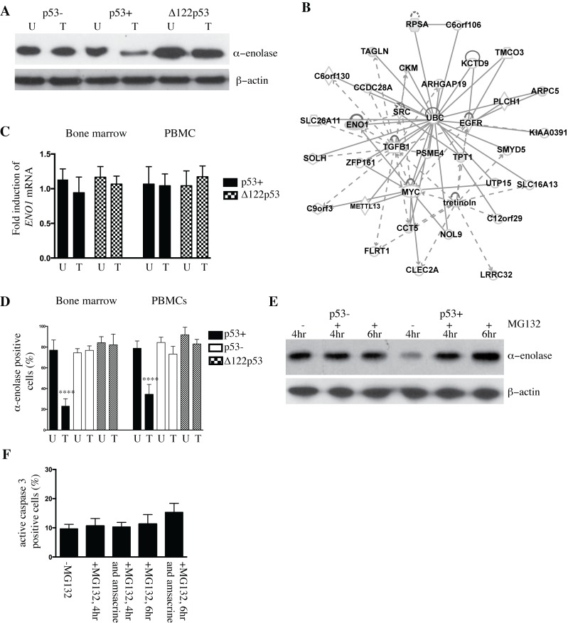
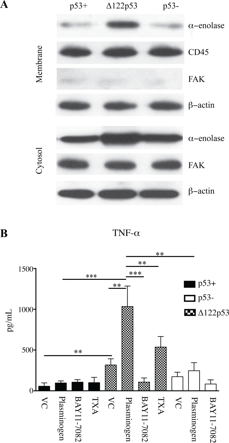
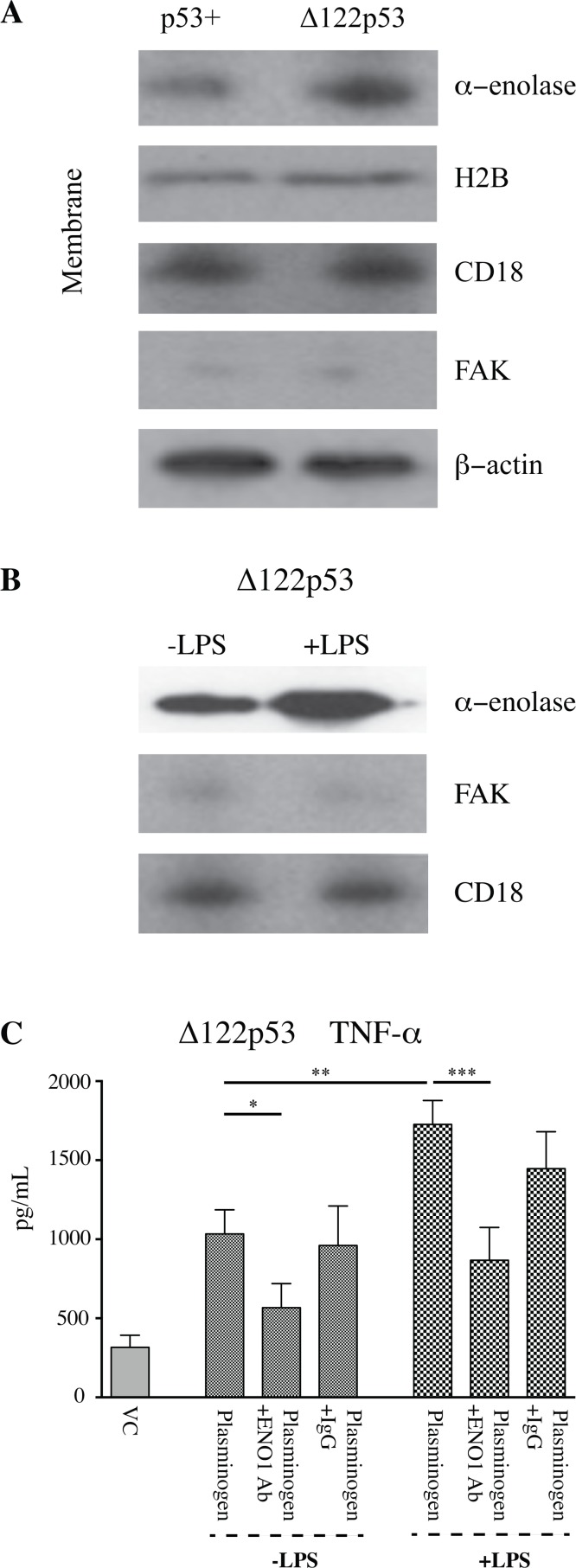
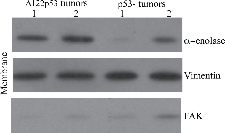
The p53 protein is a master regulator of the stress response. It acts as a tumor suppressor by inducing transcriptional activation of p53 target genes, with roles in apoptosis, cell cycle arrest and metabolism. The discovery of at least 12 isoforms of p53, some of which have tumor-promoting properties, has opened new avenues of research. Our previous work studied tumor phenotypes in four mouse models with different p53 backgrounds: wild-type p53, p53 null, mutant p53 lacking the proline domain (mΔpro), and a mimic for the human Δ133p53α p53 isoform (Δ122p53). To identify the major proteins affected by p53 function early in the response to DNA damage, the current study investigated the entire proteome of bone marrow, thymus, and lung in the four p53 models. Protein extracts from untreated controls and those treated with amsacrine were analyzed using two-dimensional fluorescence difference gel electrophoresis. In the bone marrow, reactive proteins were universally decreased by wild-type p53, including α-enolase. Further analysis of α-enolase in the p53 models revealed that it was instead increased in Δ122p53 hematopoietic and tumor cell cytosol and on the cell surface. Alpha-enolase on the surface of Δ122p53 cells acted as a plasminogen receptor, with tumor necrosis factor alpha induced upon plasminogen stimulation. Taken together, these data identified new proteins associated with p53 function. One of these proteins, α-enolase, is regulated differently by wild-type p53 and Δ122p53 cells, with reduced abundance as part of a wild-type p53 response and increased abundance with Δ122p53 function. Increased cell surface α-enolase on Δ122p53 cells provides a possible explanation for the model's pro-inflammatory features and suggests that p53 isoforms may direct an inflammatory response by increasing the amount of α-enolase on the cell surface.


Conflict of interest statement

Competing Interests: JR is a member of the PLOS ONE Editorial Board. This does not alter the authors’ adherence to PLOS ONE policies on sharing data and materials. On behalf of all authors, there are no other potential competing financial or non-financial, professional, or personal interests.

Figures

Figure 1
Figure 1. Altered abundance of alpha-enolase in p53+ and Δ122p53 bone marrow.
A. Alpha-enolase is decreased in wild-type p53 (p53+) bone marrow upon DNA damage with amsacrine treatment and increased in Δ122p53 bone marrow cells irrespective of amsacrine treatment compared with p53 null (p53-) cells by western blotting. Amsacrine treated (T) or untreated (U). B. Network analysis of the differentially expressed proteins in p53+ bone marrow untreated and p53+ bone marrow treated with amsacrine from two-dimensional fluorescence difference gel electrophoresis using Ingenuity Pathways Analysis software. Gray shading indicates proteins identified in the current study. Solid and dashed lines indicate direct and indirect interactions, respectively. C. The decrease in α-enolase in p53+ treated bone marrow was not due to reduced expression of ENO1. No significant differences were found in the amounts of ENO1 transcript expressed in p53+ and Δ122p53 bone marrow and peripheral blood mononuclear cells (PBMCs) in untreated cells (U) and cells treated with amsacrine (T) using real-time PCR. The results are expressed as the mean ± SD, from 3 mice per genotype and represent the fold increase in ENO1 expression, normalized for beta2-M expression. D. The percentage of alpha-enolase positive cells was reduced in p53+ bone marrow and peripheral blood mononuclear cells (PBMCs) treated with amsacrine. Bone marrow and PBMCs from five mice per genotype were extracted and treated with amsacrine (T) or left untreated (U). Following short-term culture, cells were fixed and cell clots sectioned. Alpha-enolase was detected using immunohistochemistry. Positive cells were identified by light microscopy and the percentage of positive cells per total cell count (500 cells) was compared between treated and untreated cells; the results are expressed as the mean ± SD (n = 6 mice per genotype), ****, P < 0.0001. E. The ubiquitin-associated proteasome inhibitor MG132 was added to p53+ and p53- bone marrow treated with amsacrine for 4 or 6 hours. Post-treatment, the amount of alpha-enolase in cell lysates was compared between cells treated with amsacrine or left untreated, by western blotting. F. MG132 treatment did not lead to a statistically significant increase in the percentage of apoptotic cells in p53+ bone marrow at 4 and 6 hours post-treatment with MG132 alone or with MG132 and amsacrine. Apoptotic cells were determined from counting the percentage of active caspase-3-positive cells in bone marrow from immunohistochemistry-stained sections and light microscopy. The percentage of positive cells per total cell count (500 cells) was compared between treated and untreated cells; the results are expressed as the mean ± SD (n = 3).
Figure 2
Figure 2. Increased alpha-enolase on the Δ122p53 PBMC cell membrane.
A. Increased alpha-enolase is present on the cell membrane of Δ122p53 PBMCs compared to that on p53- and p53+ PBMCs. The cell membrane and cytosolic fractions of untreated PBMCs from p53+, p53-, and Δ122p53 mice were separated and subjected to western blotting with an antibody to alpha-enolase. β-actin, FAK, and CD45 were used as loading controls for total protein, cytosolic, and cell membrane fractions, respectively. B. Increased TNF-alpha was released from Δ122p53 PBMCs following plasminogen stimulation compared with that from p53+ and p53- PBMCs. PBMCs were pre-incubated with plasminogen (lys-plasminogen) with or without the NFκB inhibitor BAY 11–7082 (2.5 μM for 90 minutes), or the inhibitor to plasmin activation (TXA, 10 mM), or vehicle-treated only (VC). Following pre-incubation and prior to TNF-alpha measurement, tissue plasminogen activator (3 nM) was added, and the amount of TNF-alpha secreted in culture media was measured by ELISA. The results represent the mean ± SD (n = 3 for each measurement). ***, P < 0.001, **, P < 0.01, P < 0.05.
Figure 3
Figure 3. Increased α-enolase function on the Δ122p53 cell membrane.
A. Alpha-enolase was not the only plasminogen receptor increased on the Δ122p53 cell membrane. The cell membrane and cytosolic fractions of untreated PBMCs from p53+ and Δ122p53 mice were separated and subjected to western blotting with an antibody to histone H2B and alpha-enolase. β-actin, FAK, and CD18 were used as loading controls for total protein, cytosolic, and cell membrane fractions, respectively. B. LPS was added to enhance the amount of alpha-enolase on the Δ122p53 PBMCs cell membrane. The cell membrane and cytosolic fractions of untreated Δ122p53 PBMCs or those incubated with LPS (5 μg/mL for 6 hours) were separated and subjected to western blotting with an antibody to alpha-enolase. FAK and CD18 were used as loading controls for cytosolic and cell membrane fractions, respectively. C. TNF-alpha released from Δ122p53 following pre-incubation with lys-plasminogen and LPS (5 μg/mL for 6 hours) with or without two anti-alpha-enolase antibodies (each at 15 μg/mL) to block plasminogen binding or rabbit IgG as a control (30 μg/mL). Prior to TNF-alpha measurement, tissue plasminogen activator (3 nM) was added and the amount of TNF-alpha secreted in culture media was measured by ELISA. The results represent the mean ± SD (n = 3 for each measurement) ***, P < 0.001, **, P < 0.01.
Figure 4
Figure 4. Increased α-enolase on the Δ122p53 tumor cell membrane.
Tumors from Δ122p53 mice had increased alpha-enolase at the cell surface compared to tumors from p53- mice. Sarcomas were dissected from Δ122p53 and p53- mice at necropsy, the cytosolic and cell membrane fractions were separated, and these fractions were subjected to western blotting using an antibody to alpha-enolase. The cell membrane results from two tumors per genotype (1 and 2) are shown. Western blotting for vimentin was used as a loading control for the mesenchymal cell membrane fraction and FAK was included to control for cytosolic contamination in cell membrane preparations.

Similar articles

Cited by

References

    1. Oren M (2003) Decision making by p53: life, death and cancer. Cell Death Differ 10: 431–442. - PubMed
    1. Levine AJ, Tomasini R, McKeon FD, Mak TW, Melino G (2011) The p53 family: guardians of maternal reproduction. Nat Rev Mol Cell Biol 12: 259–265. 10.1038/nrm3086 - DOI - PubMed
    1. Pant V, Quintas-Cardama A, Lozano G (2012) The p53 pathway in hematopoiesis: lessons from mouse models, implications for humans. Blood 120: 5118–5127. 10.1182/blood-2012-05-356014 - DOI - PMC - PubMed
    1. Quadrato G, Di Giovanni S (2012) Gatekeeper between quiescence and differentiation: p53 in axonal outgrowth and neurogenesis. Int Rev Neurobiol 105: 71–89. 10.1016/B978-0-12-398309-1.00005-6 - DOI - PubMed
    1. Maddocks OD, Vousden KH (2011) Metabolic regulation by p53. J Mol Med (Berl) 89: 237–245. 10.1007/s00109-011-0735-5 - DOI - PMC - PubMed

Publication types