The Dilator Naris Muscle as a Reporter of Facial Nerve Regeneration in a Rat Model
- PMID: 25643189
- PMCID: PMC4520777
- DOI: 10.1097/SAP.0000000000000273
The Dilator Naris Muscle as a Reporter of Facial Nerve Regeneration in a Rat Model
Abstract
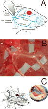
Objective: Many investigators study facial nerve regeneration using the rat whisker pad model, although widely standardized outcomes measures of facial nerve regeneration in the rodent have not yet been developed. The intrinsic whisker pad "sling" muscles producing whisker protraction, situated at the base of each individual whisker, are extremely small and difficult to study en bloc. Here, we compare the functional innervation of 2 potential reporter muscles for whisker pad innervation: the dilator naris (DN) and the levator labii superioris (LLS), to characterize facial nerve regeneration.
Methods: Motor supply of the DN and LLS was elucidated by measuring contraction force and compound muscle action potentials during stimulation of individual facial nerve branches, and by measuring whisking amplitude before and after DN distal tendon release.
Results: The pattern of DN innervation matched that of the intrinsic whisker pad musculature (ie, via the buccal and marginal mandibular branches of the facial nerve), whereas the LLS seemed to be innervated almost entirely by the zygomatic branch, whose primary target is the orbicularis oculi muscle.
Conclusions: Although the LLS has been commonly used as a reporter muscle of whisker pad innervation, the present data show that its innervation pattern does not overlap substantially with the muscles producing whisker protraction. The DN muscle may serve as a more appropriate reporter for whisker pad innervation because it is innervated by the same facial nerve branches as the intrinsic whisker pad musculature, making structure/function correlations more accurate, and more relevant to investigators studying facial nerve regeneration.
Figures


Similar articles
-
The convergence of facial nerve branches providing whisker pad motor supply in rats: implications for facial reanimation study.Muscle Nerve. 2012 May;45(5):692-7. doi: 10.1002/mus.23232. Muscle Nerve. 2012. PMID: 22499096 Free PMC article.
-
Recovery of original nerve supply after hypoglossal-facial anastomosis causes permanent motor hyperinnervation of the whisker-pad muscles in the rat.J Comp Neurol. 1993 Dec 8;338(2):214-24. doi: 10.1002/cne.903380206. J Comp Neurol. 1993. PMID: 8308168
-
Whisking recovery after automated mechanical stimulation during facial nerve regeneration.JAMA Facial Plast Surg. 2014 Mar-Apr;16(2):133-9. doi: 10.1001/jamafacial.2013.2217. JAMA Facial Plast Surg. 2014. PMID: 24407357 Free PMC article.
-
Reanimation of the paralyzed face.Otolaryngol Clin North Am. 1992 Jun;25(3):649-67. Otolaryngol Clin North Am. 1992. PMID: 1625868 Review.
-
Trigeminal Sensory Supply Is Essential for Motor Recovery after Facial Nerve Injury.Int J Mol Sci. 2022 Dec 1;23(23):15101. doi: 10.3390/ijms232315101. Int J Mol Sci. 2022. PMID: 36499425 Free PMC article. Review.
Cited by
-
Dual Convergence of Facial Nerve Branches Innervating Whisker Pad in Rats.Curr Med Sci. 2018 Dec;38(6):982-988. doi: 10.1007/s11596-018-1973-3. Epub 2018 Dec 7. Curr Med Sci. 2018. PMID: 30536059
-
Selective Denervation of the Facial Dermato-Muscular Complex in the Rat: Experimental Model and Anatomical Basis.Front Neuroanat. 2021 Mar 22;15:650761. doi: 10.3389/fnana.2021.650761. eCollection 2021. Front Neuroanat. 2021. PMID: 33828465 Free PMC article.
References
-
- Coulson SE, O’Dwyer NJ, Adams RD, Croxson GR. Expression of emotion and quality of life after facial nerve paralysis. Otol Neurotol. 2004;25(6):1014–1019. - PubMed
-
- Sinno H, Thibaudeau S, Izadpanah A, et al. Utility outcome scores for unilateral facial paralysis. Ann Plast Surg. 2012;69(4):435–438. - PubMed
-
- Willand MP, Holmes M, Bain JR, Fahnestock M, De Bruin H. Electrical muscle stimulation after immediate nerve repair reduces muscle atrophy without affecting reinnervation. Muscle Nerve. 2013;48(2):219–225. - PubMed
Publication types
MeSH terms
Grants and funding
LinkOut - more resources
Full Text Sources
Other Literature Sources
Research Materials

