. 2015 Jan 29;4(1):71-83.
doi: 10.3390/cells4010071.

ELISPOT Assays in 384-Well Format: Up to 30 Data Points with One Million Cells

Affiliations

ELISPOT Assays in 384-Well Format: Up to 30 Data Points with One Million Cells

Jodi Hanson et al. Cells. .

Abstract

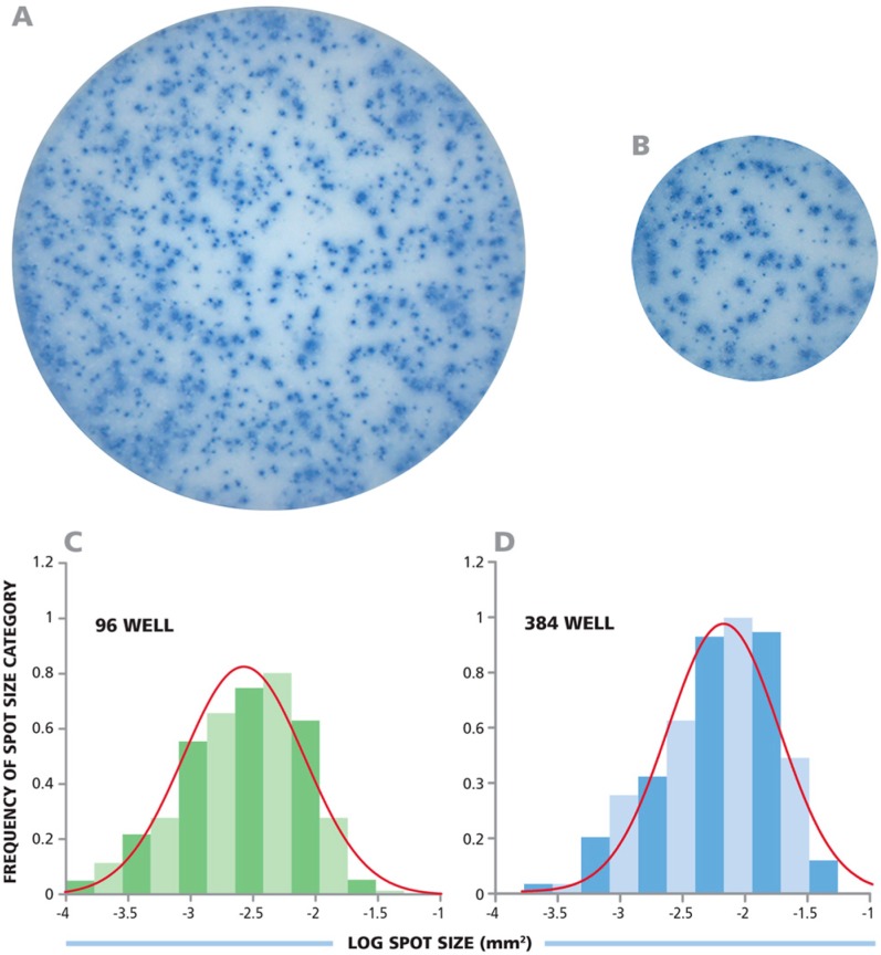
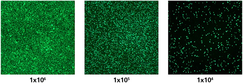
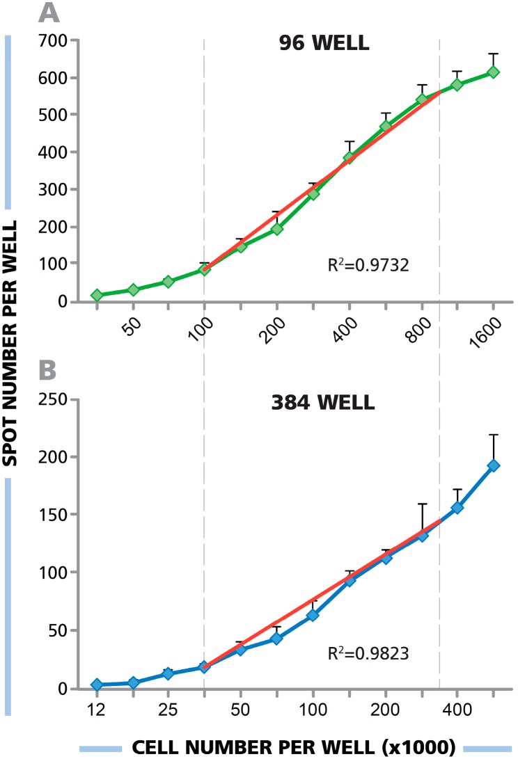
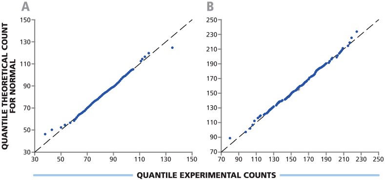
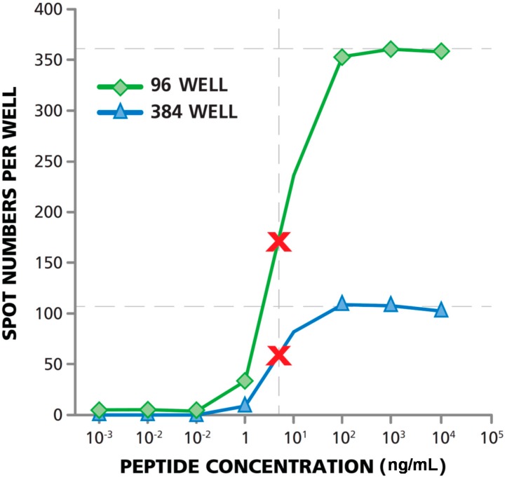
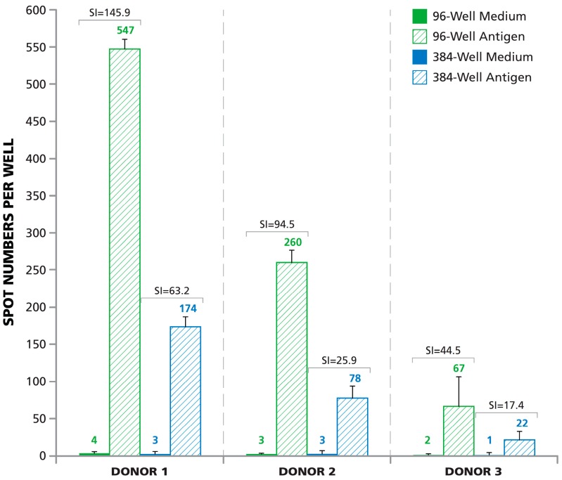
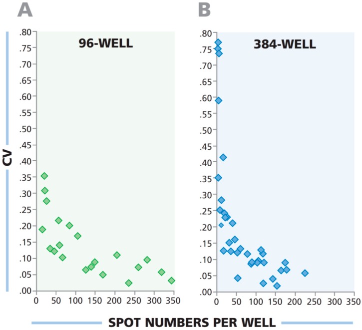
Comprehensive immune monitoring requires that frequencies of T cells, producing different cytokines, are measured to establish the magnitude of Th1, Th2, and Th17 components of cell-mediated immunity. Antigen titration provides additional information about the affinity of T cell response. In tumor immunity, it is also advisable to account for determinant spreading by testing multiple epitopes. Efforts for comprehensive immune monitoring would require substantial numbers of PBMC to run the above tests systematically, which in most test cases is limiting. Immune monitoring with ELISPOT assays have been performed, thus far, in a 96-well format. In this study we show that one can increase cell utilization by performing the assay in 384-well plates whose membrane surface area is one third that of 96-well plates. Systematic testing of PBMC for antigen-specific T cell response in the two formats demonstrated that the 384-well assay corresponds to a one-in-three miniaturization of the 96-well assay. The lowest number of cells that can be used in the 384-well format, while allowing for sufficient contact with APC, is 33,000 PBMC/well. Therefore, with one million PBMC typically obtained from 1 mL of blood, a 30 well T cell ELISPOT assay can be performed in a 384-well format.


Figures

Figure 1
Figure 1
Identical IFN-γ spot sizes and size distributions in the 96- and 384-well format plates. (A,B) Respective images from a 96-well and a 384-well plate were captured with identical optical and digital resolution; (C,D) The spot sizes from both wells are plotted as a histogram with a fitted Log Normal curve overlaid in red.
Figure 2
Figure 2
PBMC were stained with fluorescent Calcein AM dye and the specified numbers of cells were plated into respective wells of a 96-well plate. The cells were visualized on an ImmunoSpot® S6 Ultimate Reader by using 480 nm as the excitation wavelength, and detecting fluorescence with a 520 ± 20 nm emission wavelength filter.
Figure 3
Figure 3
(A,B) PBMC were plated in an ELISPOT assay in serial dilution at the numbers specified, and HCMV pp65 was added to elicit IFN-γ production by the specific CD8+ cells. The number of PBMC added to a (B) 384-well plate was one-third the number in the (A) 96-well plate. The ideal linear function is shown by the superimposed red line. For the 96-well plate, the experimental data approximated linearity (R2 = 0.9782) between 1.0 × 10⁵ cells per well and 1 × 10⁶ cells per well. For the 384-well plate, linearity was approximated (R2 = 0.9823) between 3.3 × 10⁴ PBMC per well and 3.5 × 10⁵ cells per well.
Figure 4
Figure 4
Spot count variation among replicate wells of 384-well plate follows Normal distribution. PBMC were plated at (A) 0.5 × 10⁵ cells per well in 384 replicate wells; and (B) 1.0 × 10⁵ cells per well in 192 replicate wells. The response to HCMV pp65 antigen in an IFN-γ ELISPOT assay was measured. Normality was determined using Shapiro Wilk test with α value of 0.05 and p-values of 0.708 and 0.388 respectively. Q-Q plots for experimental vs. theoretical counts are shown.
Figure 5
Figure 5
T cell functional affinity measurements in 384- and 96-well formats provide identical results. HCMV peptide pp65-induced IFN-γ production by CD8+ cells was measured in PBMC in both 96-well and 384-well plate types. The highest peptide concentration tested was 10 μg/mL with subsequent 1:10 serial dilutions. The numbers of PBMC plated in the 96-well format was 3 × 10⁵/well (green line), and 1 × 10⁵/well for the 384-well plate (blue line). Fifty percent maximal activation is marked with the red X.
Figure 6
Figure 6
Signal-to-noise performance of 96-well vs. 384-well plate. HCMV pp65 antigen-induced responses (hatched bars) and medium controls (solid bars—Mostly too small to be seen) are shown for three donors with high, medium, and low response levels. PBMC numbers were 3 × 10⁵ for the 96-well plate and 1.0 × 10⁵ for the 384-well plate. The mean spot count/well calculated from three replicate wells for each condition is specified above the bars. The stimulation index (SI) is defined as the number of ELISPOTs induced by an antigen divided by the number of spots in the medium background. SI for the corresponding medium control/antigen pairs is specified.
Figure 7
Figure 7
The variability in spot counts among replicate wells is higher in 384-well plates than in 96-well plates when low frequency T cells are being detected. PBMC from three donors were serially diluted and tested, in quadruplicate wells for each donor and cell dilution, for antigen-elicited IFN-γ response. The PBMC were plated in serial dilution between 1.0 × 10⁶ and 1.0 × 10⁵ cells per well in the 96-well plate, and at 3 × 10⁵ and 3 × 10⁴ cells per well in the 384-well plate. The CVs of the quadruplicate wells were plotted against mean spot numbers for the corresponding test results.

References

    1. Lehmann P.V., Sundararaman S. When results of T-cell immune monitoring match/do not match clinical outcomes of tumor vaccine trials: What more could and should we measure? In: Shurin M.R., Umansky V., Malyguine A., editors. The Tumor Immunoenvironment. Springer Dordrecht; New York, NY, USA: 2013. pp. 725–740. Chapter 32.
    1. Karulin A.Y., Hesse M.D., Tary-Lehmann M., Lehmann P.V. Single-cytokine-producing cd4 memory cells predominate in type 1 and type 2 immunity. J. Immunol. 2000;164:1862–1872. doi: 10.4049/jimmunol.164.4.1862. - DOI - PubMed
    1. Kuerten S., Nowacki T.M., Kleen T.O., Asaad R.J., Lehmann P.V., Tary-Lehmann M. Dissociated production of perforin, granzyme b, and ifn-gamma by hiv-specific cd8(+) cells in hiv infection. AIDS Res. Hum. Retrov. 2008;24:62–71. doi: 10.1089/aid.2007.0125. - DOI - PubMed
    1. Nowacki T.M., Kuerten S., Zhang W., Shive C.L., Kreher C.R., Boehm B.O., Lehmann P.V., Tary-Lehmann M. Granzyme b production distinguishes recently activated cd8(+) memory cells from resting memory cells. Cell. Immunol. 2007;247:36–48. doi: 10.1016/j.cellimm.2007.07.004. - DOI - PMC - PubMed
    1. Targoni O.S., Lehmann P.V. Endogenous myelin basic protein inactivates the high avidity t cell repertoire. J. Exp. Med. 1998;187:2055–2063. doi: 10.1084/jem.187.12.2055. - DOI - PMC - PubMed

LinkOut - more resources