. 2015 Mar;22(3):242-7.
doi: 10.1038/nsmb.2956. Epub 2015 Feb 2.

DNA interstrand cross-link repair requires replication-fork convergence

Affiliations

DNA interstrand cross-link repair requires replication-fork convergence

Jieqiong Zhang et al. Nat Struct Mol Biol. 2015 Mar.

Abstract

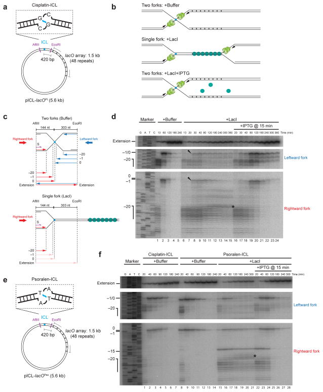
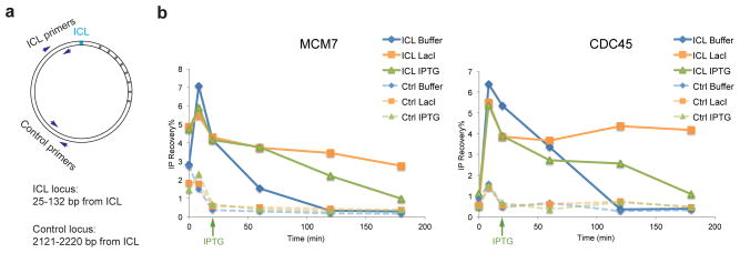
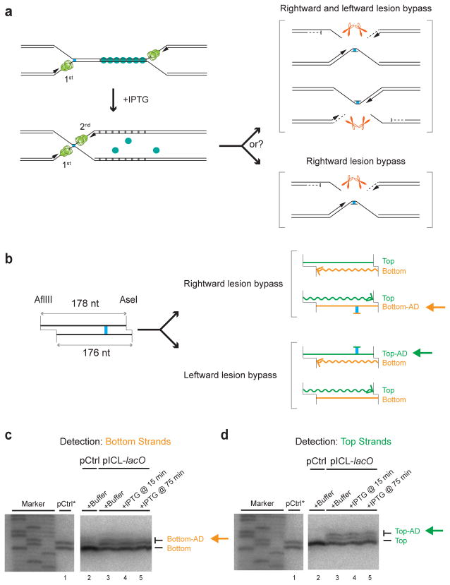
DNA interstrand cross-links (ICLs) prevent strand separation during DNA replication and transcription and therefore are extremely cytotoxic. In metazoans, a major pathway of ICL repair is coupled to DNA replication, and it requires the Fanconi anemia pathway. In most current models, collision of a single DNA replication fork with an ICL is sufficient to initiate repair. In contrast, we show here that in Xenopus egg extracts two DNA replication forks must converge on an ICL to trigger repair. When only one fork reaches the ICL, the replicative CMG helicase fails to unload from the stalled fork, and repair is blocked. Arrival of a second fork, even when substantially delayed, rescues repair. We conclude that ICL repair requires a replication-induced X-shaped DNA structure surrounding the lesion, and we speculate on how this requirement helps maintain genomic stability in S phase.


Figures

Figure 1
Figure 1. A single fork stalled at an ICL fails to undergo approach
(a) Cartoon of the cisplatin containing plasmid, pICL-lacOPt. (b) Expected outcomes of pICL-lacO replication in the presence of buffer, LacI, and LacI + IPTG. (c) Schematic illustration of nascent leading strands generated in the experiment shown in (d). (d) Nascent strand analysis of pICL-lacO during replication-coupled repair, with or without LacI and IPTG, as indicated. Nascent strands, together with the sequencing ladder, were separated on a polyacrylamide gel and visualized by autoradiography. Top panel: extension products. Middle panel: nascent strands of the leftward fork. Bottom panel: nascent strands of the rightward fork. The ladder was generated from extension of primer S (shown in c, purple) on a control plasmid lacking the ICL. Black arrowheads: a small fraction of leading strands approaching to −1. Asterisk (*), background bands described in Supplementary Fig. 3d. Uncropped image of Fig. 1d is presented in Supplementary Data Set 1a. (e) Cartoon of the psoralen-ICL containing plasmid, pICL-lacOPso. (f) Nascent strand analysis of pICL-lacOPt (containing the cisplatin-ICL used throughout the paper) and pICL-lacOPso, with or without LacI and IPTG, as indicated. Asterisk (*), background bands described in Supplementary Fig. 3d. The −15 arrest of the leading strands in repair might come from specific stalling of the CMG helicase when it encounters a psoralen-ICL. Uncropped image of Fig. 1f is presented in Supplementary Data Set 1b.
Figure 2
Figure 2. CMG is not evicted from a single fork stalled at an ICL
(a) Schematic of primers used in chromatin immunoprecipitation (ChIP). (b) MCM7 and CDC45 ChIP anaylsis at different time points during repair. pICL-lacO was replicated with or without LacI, and IPTG was added immediately before the 20 minute time point, as indicated (green arrow). A repetition of this experiment is shown in Supplementary Fig. 5a.
Figure 3
Figure 3. A single, stalled fork does not undergo incisions
(a) Schematic of repair intermediates expected in panel (b). (b) Replication intermediates of pICL-lacO separated on a native agarose gel. pICL-lacO and an internal control plasmid lacking the lacO array (pQuant) were pre-incubated with buffer or LacI and replicated in the presence of [α-32P]dATP. IPTG was added as indicated. Sc: supercoiled. Oc: open circular. Blue arrowhead: “Figure 8” DNA structure. Green arrowhead: “Theta” DNA structure. (c) Schematic of incision assay. Before dual incisions, single cutting with BlpI yields X-shaped products (blue strands, upper cartoon), whereas after dual incisions, BlpI digestion yields linear molecules (blue strands, lower cartoon). (d) Incision assay. pICL-lacO was replicated with or without LacI and IPTG, as indicated. The repair intermediates were digested with BlpI, separated on an alkaline (denaturing) gel, and visualized by Southern blotting to detect parental strands. Unreplicated pICL-lacO was used to generate size markers for the X-shaped structure and linear structure, which came from a small fraction of uncrosslinked background plasmids in the pICL-lacO preparations. Uncropped image of Fig. 3d is presented in Supplementary Data Set 1c. (e) XPF and SLX4 ChIP analysis. pICL-lacO and pQuant were replicated with or without LacI, and IPTG was added immediately before the 20 minute time point where indicated (green arrow). At different times, samples were withdrawn for XPF and SLX4 ChIP using primer pairs for the ICL locus (Fig. 2a) or pQuant (Ctrl). A repetition of this experiment is shown in Supplementary Fig. 5c.
Figure 4
Figure 4. The order of replisome arrival at an ICL does not determine which leading strand undergoes lesion bypass
(a) Scheme to determine whether the order in which the two forks arrive at an ICL dictates which parental strand is used as the lesion bypass template. (b) Schematic depiction of final repair products after AflIII and AseI digestion, depending on which leading strand undergoes lesion bypass. Note that AflIII and AseI generate different sized overhangs, allowing us to differentiate top (178 nt) and bottom (176 nt) strands. Top-AD or Bottom-AD: top or bottom strand containing an adduct. (c, d) Strand-specific Southern blotting to detect the adducts. pCtrl or pICL-lacO was replicated in the presence of buffer or LacI, and IPTG was added at the indicated times. After 6 hours, repair products were digested with AflIII and AseI, separated on a sequencing gel, and analyzed with strand-specific Southern blotting to visualize the bottom (c) or top (d) strands. To generate size markers for the top (178 nt) and bottom (176 nt) strands (lane 1), pCtrl was replicated in the presence of [α-32P]dATP (pCtrl*), and was analyzed on the same sequencing gel as the strand-specific Southern after AflIII and AseI digestion. The fact that no top (c, lane 2) or bottom strand (d, lane 2) was detected in Southern blotting of pCtrl established the strand-specificity of the blotting protocol. Uncropped images of Figs. 4c and 4d are presented in Supplementary Data Set 1d.

References

    1. Lawley PD, Phillips DH. DNA adducts from chemotherapeutic agents. Mutat Res. 1996;355:13–40. - PubMed
    1. Clauson C, Schärer OD, Niedernhofer L. Advances in understanding the complex mechanisms of DNA interstrand cross-link repair. Cold Spring Harb Perspect Med. 2013;3:a012732. - PMC - PubMed
    1. Garaycoechea JI, Patel KJ. Why does the bone marrow fail in Fanconi anemia? Blood. 2014;123:26–34. - PubMed
    1. Zhang J, Walter JC. Mechanism and regulation of incisions during DNA interstrand cross-link repair. DNA Repair (Amst) 2014;19:135–142. - PMC - PubMed
    1. Deans AJ, West SC. DNA interstrand crosslink repair and cancer. Nat Rev Cancer. 2011;11:467–480. - PMC - PubMed

Publication types

LinkOut - more resources