. 2015 Mar;22(3):230-7.
doi: 10.1038/nsmb.2961. Epub 2015 Feb 2.

Mechanism of microhomology-mediated end-joining promoted by human DNA polymerase θ

Affiliations

Mechanism of microhomology-mediated end-joining promoted by human DNA polymerase θ

Tatiana Kent et al. Nat Struct Mol Biol. 2015 Mar.

Abstract

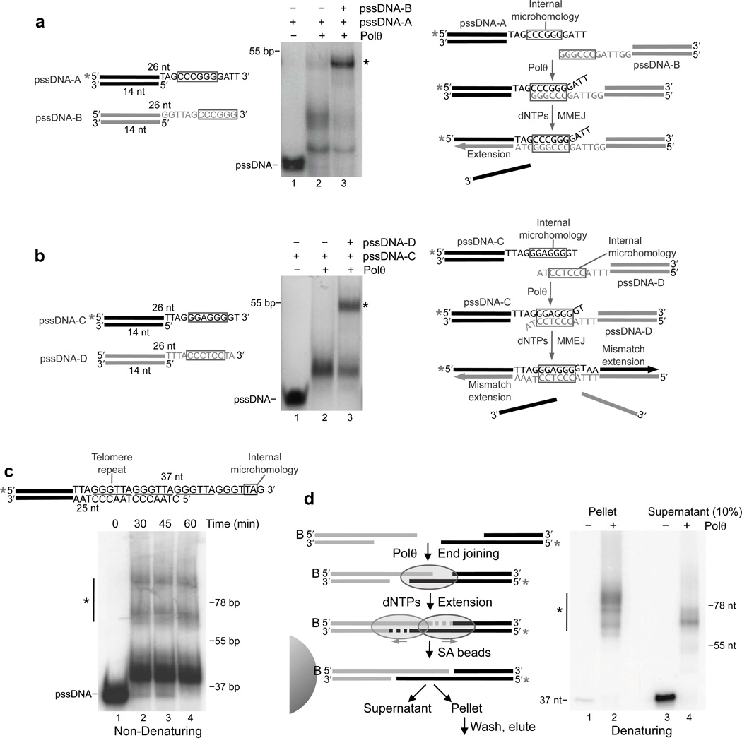
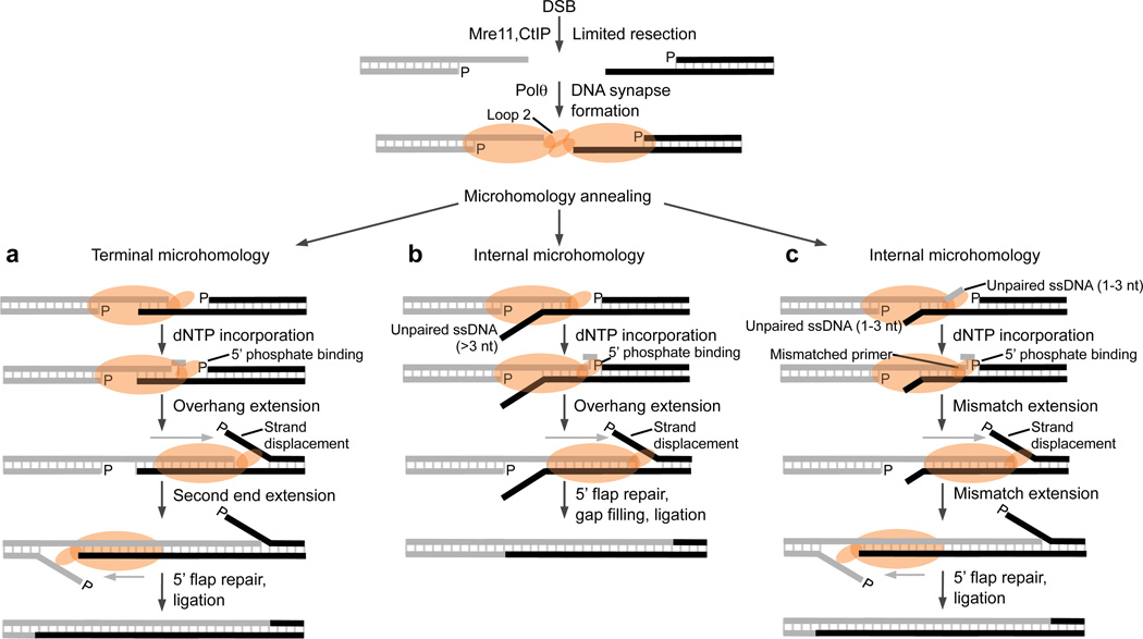
Microhomology-mediated end-joining (MMEJ) is an error-prone alternative double-strand break-repair pathway that uses sequence microhomology to recombine broken DNA. Although MMEJ has been implicated in cancer development, the mechanism of this pathway is unknown. We demonstrate that purified human DNA polymerase θ (Polθ) performs MMEJ of DNA containing 3' single-strand DNA overhangs with ≥2 bp of homology, including DNA modeled after telomeres, and show that MMEJ is dependent on Polθ in human cells. Our data support a mechanism whereby Polθ facilitates end-joining and microhomology annealing, then uses the opposing overhang as a template in trans to stabilize the DNA synapse. Polθ exhibits a preference for DNA containing a 5'-terminal phosphate, similarly to polymerases involved in nonhomologous end-joining. Finally, we identify a conserved loop domain that is essential for MMEJ and higher-order structures of Polθ that probably promote DNA synapse formation.


Figures

Figure 1
Figure 1. Polθ promotes microhomology-mediated end-joining in vitro and in vivo
a, Model of Polθ involvement in MMEJ. Polθ is proposed to extend paired 3’ overhangs at DNA synapses. b, pssDNA and ssDNA substrates. * = 32P. (c,d) Non-denaturing gels showing MMEJ reactions with pssDNA (c) and ssDNA (d). * = MMEJ products. e, Schematic of solid-phase MMEJ assay (left). (Left panel) Denaturing gel showing DNA purified from pellet and supernatant fractions following MMEJ in the presence (lanes 2 and 4) and absence (lanes 1 and 3) of Polθ. (Right panel) Denaturing gel showing MMEJ reactions with pssDNA-4 (lanes 1 and 2) and ssDNA-4 (lanes 3 and 4). * = MMEJ products. f, Non-denaturing gel showing MMEJ reactions with the indicated Pols. * = MMEJ products. g, MMEJ GFP reporter assay. Schematic of GFP reporter with microhomology, I-SceI site, stop codon and GFP gene indicated (top). Plot of % GFP cells following transient expression of I-SceI and transfection with Polθ siRNA and scrambled siRNA. Error bars, s.d. (n = 9 independent experiments)(left). Western blots of Polθ and β-actin following transfection with Polθ siRNA (lane 2) and scrambled siRNA (lane 1)(right).
Figure 2
Figure 2. Polθ uses the opposing overhang as a template in trans which stabilizes the DNA synapse
a, Model of Polθ overhang extension of pssDNA-4. Microhomology is outlined. b, Non-denaturing (left) and denaturing (center, right) gels showing MMEJ reactions in the presence of indicated dNTPs. Lane 3 in right panel represents a 48 nt marker based on model in panel a. * = MMEJ products. c, Model of Polθ strand displacement synthesis during MMEJ of pssDNA-4 conjugated with Cy3 and black-hole quencher (BHQ). Plot of fluorescence intensity following MMEJ in the presence (grey) and absence (black) of dNTPs. RU = relative units. Error bars, s.d. (n = 3 independent experiments).
Figure 3
Figure 3. Template preferences for Polθ MMEJ
a, Schematic of pssDNA-4 substrates with variable length overhangs (left). (Middle panels) Non-denaturing gels showing MMEJ reactions with the indicated pssDNA. (Right) Plot of % MMEJ products. b, Plot of % MMEJ products generated from pssDNA-4 with (black) and without (grey) a 5’-terminal phosphate for the indicated times. c, Non-denaturing gels showing a time course of MMEJ reactions with pssDNA-2 containing CG (left) and AT (right) 3’ terminal microhomology. * = MMEJ products.
Figure 4
Figure 4. Polθ promotes MMEJ of DNA containing internal microhomology
(a,b) Schematic of pssDNA substrates with microhomology outlined (left). Non-denaturing gel showing MMEJ reactions with the indicated pssDNA (middle). Model of MMEJ (right). * = 32P, * = MMEJ products. c, MMEJ of pssDNA modeled after telomeres. (Top) pssDNA with telomere repeats underlined and microhomology outlined. (Bottom) Non-denaturing gel showing a time course of MMEJ. d, Schematic of solid-phase MMEJ assay (left). Denaturing gel of DNA purified from supernatant and pellet fractions following MMEJ reactions in the presence (lane 2 and 4) and absence (lanes 1 and 3) of Polθ. 10% of supernatant was analyzed (right). * = 32P, * = MMEJ products.
Figure 5
Figure 5. Polθ promotes DNA synapse formation and strand annealing separately from its replication function
a, Schematic of DNA synapse assay (left). (Left panel) Plot of relative fluorescence intensity following Polθ synapse formation in the presence of Cy5 pssDNA with (grey) and without (black) Cy3 pssDNA. (Right panel) Plot of relative fluorescence intensity following Polθ synapse formation in the presence of Cy3 and Cy5 pssDNA with (grey) and without (black) 4 bp of microhomology. RU = relative units. Error bars, s.d. (n = 3 independent experiments). (b,c) Schematic of annealing assay (left). (Middle) Non-denaturing gel showing ssDNA (b) and pssDNA (c) annealing in the presence (lane 3) and absence (lane 2) of Polθ. ssDNA (b) and pssDNA (c) marker (lane 1). (Right) Plot of % annealing. % annealing = (intensity of upper band)/(sum of the intensities of upper and lower bands). Error bars, s.d.. (n = 3 independent experiments).
Figure 6
Figure 6. Insertion loop 2 promotes microhomology-mediated end-joining, DNA binding and polymerase complexes
a, Schematic of Polθ with polymerase domains and insertion loops indicated. b, Superposition of Bacillus Pol I structure (blue; PDB code 4DQQ) in complex with primer-template (orange) and Polθ model (grey; residues 1944–2590) assembled by Swiss Model server using Bacillus Pol I:primer-template structure (PDB code 4DQQ) as a template. c, Non-denaturing gel showing MMEJ reactions with Polθ WT (lane 2) and Polθ L2 (lane 3). d, Denaturing gel showing primer-template extension with Polθ WT (lane 2), Polθ L2 (lane 3), and Polδ (lane 4). (e,f) EMSA with Polθ WT (left) and Polθ L2 (right) on pssDNA-4 (e) and primer-template (f). g, Plot of % DNA bound calculated from EMSA in panels e and f. % bound = intensity of upper band/sum of the intensities of upper and lower bands.
Figure 7
Figure 7. Models of Polθ MMEJ
Following limited resection of a DSB by Mre11 and CtIP, Polθ dimers promoted by loop 2 facilitate DNA synapse formation (top). Polθ promotes annealing of terminal (a) or internal (b,c) microhomology following DNA synapse formation. Polθ extends the annealed overhang by using the opposing overhang as a template in trans which stabilizes the DNA synapse. Overhang extension is facilitated by Polθ binding to the 5’ terminal phosphate on the opposing DNA and results in strand displacement. a, Polθ extends both overhangs in the case of terminal microhomology. b, Polθ only extends the terminally paired overhang when internal microhomology is located relatively far from the 3’ terminus on the opposing strand. c, Polθ performs mismatch extension of overhangs that contain internal microhomology relatively close to their 3’ terminus. Last, 5’ flap repair is presumably required prior to ligation in each case.

Similar articles

Cited by

References

    1. McVey M, Lee SE. MMEJ repair of double-strand breaks (director's cut): deleted sequences and alternative endings. Trends Genet. 2008;24:529–538. - PMC - PubMed
    1. Bennardo N, Cheng A, Huang N, Stark JM. Alternative-NHEJ is a mechanistically distinct pathway of mammalian chromosome break repair. PLoS Genet. 2008;4 - PMC - PubMed
    1. Truong LN, et al. Microhomology-mediated End Joining and Homologous Recombination share the initial end resection step to repair DNA double-strand breaks in mammalian cells. Proceedings of the National Academy of Sciences of the United States of America. 2013;110:7720–7725. - PMC - PubMed
    1. Deriano L, Roth DB. Modernizing the nonhomologous end-joining repertoire: alternative and classical NHEJ share the stage. Annu Rev Genet. 2013;47:433–455. - PubMed
    1. Corneo B, et al. Rag mutations reveal robust alternative end joining. Nature. 2007;449:483–486. - PubMed

METHODS REFERENCES

    1. Gunn A, Stark JM. I-SceI-based assays to examine distinct repair outcomes of mammalian chromosomal double strand breaks. Methods Mol Biol. 2012;920:379–391. - PubMed
    1. Schwede T, Kopp J, Guex N, Peitsch MC. SWISS-MODEL: An automated protein homology-modeling server. Nucleic acids research. 2003;31:3381–3385. - PMC - PubMed
    1. Wang W, Wu EY, Hellinga HW, Beese LS. Structural factors that determine selectivity of a high fidelity DNA polymerase for deoxy-, dideoxy-, and ribonucleotides. The Journal of biological chemistry. 2012;287:28215–28226. - PMC - PubMed
    1. Guex N, Peitsch MC. SWISS-MODEL and the Swiss-PdbViewer: an environment for comparative protein modeling. Electrophoresis. 1997;18:2714–2723. - PubMed
    1. DeLano WL. Unraveling hot spots in binding interfaces: progress and challenges. Curr Opin Struct Biol. 2002;12:14–20. - PubMed

Publication types

Substances