. 2015 Feb 2;11(2):e1004648.
doi: 10.1371/journal.ppat.1004648. eCollection 2015 Feb.

CXCL9 contributes to antimicrobial protection of the gut during citrobacter rodentium infection independent of chemokine-receptor signaling

Affiliations

CXCL9 contributes to antimicrobial protection of the gut during citrobacter rodentium infection independent of chemokine-receptor signaling

Sarah A Reid-Yu et al. PLoS Pathog. .

Abstract

Chemokines have been shown to be effective bactericidal molecules against a variety of bacteria and fungi in vitro. These direct antimicrobial effects are independent of their chemotactic activities involving immunological receptors. However, the direct biological role that these proteins may play in host defense, particularly against intestinal pathogens, is poorly understood. Here, we show that CXCL9, an ELR- chemokine, exhibits direct antimicrobial activity against Citrobacter rodentium, an attaching/effacing pathogen that infects the gut mucosa. Inhibition of this antimicrobial activity in vivo using anti-CXCL9 antibodies increases host susceptibility to C. rodentium infection with pronounced bacterial penetration into crypts, increased bacterial load, and worsened tissue pathology. Using Rag1(-/-) mice and CXCR3(-/-) mice, we demonstrate that the role for CXCL9 in protecting the gut mucosa is independent of an adaptive response or its immunological receptor, CXCR3. Finally, we provide evidence that phagocytes function in tandem with NK cells for robust CXCL9 responses to C. rodentium. These findings identify a novel role for the immune cell-derived CXCL9 chemokine in directing a protective antimicrobial response in the intestinal mucosa.


Conflict of interest statement

The authors have declared that no competing interests exist.

Figures
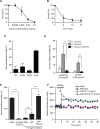
Fig 1
Fig 1. Citrobacter rodentium is sensitive to CXCL9-directed bacterial killing.
(A) Dose response survival to increasing concentrations of CXCL9. Wild type C. rodentium was exposed to the various concentrations of CXCL9 for 2 h and survival was expressed as a percentage compared to buffer-only controls. Data are the means with standard error from 3 experiments. (B) C. rodentium time kill curves were plotted in response to 0.39 μg/ml CXCL9. Data are plotted as a percent survival compared to buffer controls without chemokine addition. Data are the means with standard error from 3 experiments. (C) Bacterial killing assays were performed in tandem with C. rodentium (CR), enterohemorrhagic E. coli (EHEC), enteropathogenic E. coli (EPEC), and Salmonella enterica serovar Typhimurium (STM) in the presence of 0.39 μg/ml CXCL9. Data is plotted as a percent survival compared to buffer controls. (D) PhoP-PhoQ is required for partial resistance to CXCL9-directed killing. The indicated strains were incubated with CXCL9 or α-defensin and survival was determined by viable colony counting. Data are the means with standard error from 3 experiments. (E) Anti-CXCL9 antibody blocks the killing activity of CXCL9. C. rodentium was incubated with CXCL9 in the presence of either PBS, IgG control, or anti-CXCL9 antibody. CXCL9-specific antibody blocked 100% of the killing activity. Data are the means with standard error from 3 experiments. (F) Membrane permeability in response to CXCL9 or the membrane-disrupting antimicrobial peptide polymyxin B was measured by ANS release. Arrow indicates time of CXCL9 or antimicrobial peptide injection. Data is representative of 3 independent experiments. Statistical significance was assessed utilizing an ANOVA with Newman-Keuls post-test.
Fig 2
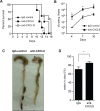
Fig 2. Loss of CXCL9 results in increased C. rodentium burden and worsened host outcome.
(A) Rag1-/- mice were infected with C. rodentium and administered control rabbit IgG, anti-CXCL9 antibody, or anti-CXCL10 antibody. Survival was monitored for 15 days. Data are from 2 independent experiments. * P <0.05 (Gehan-Breslow-Wilcox) compared to IgG control. (B) Rag1-/- mice were infected as above and viable C. rodentium counts were determined in fecal pellets. Data are the means with standard errors from 5 animals. * P <0.05 compared to IgG control. (C) Representative images of gross pathology in the cecum and colon of Rag1-/- mice infected with C. rodentium for 10 days. Note increased water content in mice depleted of CXCL9, emptied and shrunken cecum and constricted colon. Arrowhead indicates hematoma, which was common in infected CXCL9-depleted animals. (D) Water content in fecal output was determined at day 10 after infection. Data is pooled from three separate experiments (9 animals) * P<0.05. Statistical significance was assessed utilizing the t-test.
Fig 3
Fig 3. Mice depleted of CXCL9 have greater C. rodentium-induced pathology.
(A) Rag1-/- mice were infected with C. rodentium and administered IgG control antibody or anti-CXCL9 antibody. Representative histopathology images (200x) of the distal colon are shown from animals infected for 10 days. (B) Quantification of pathology in colon. Data is pooled from two experiments, n = 6 per group. (C) Localization of C. rodentium in Rag1-/- mouse colon by immunohistochemistry. Mice were infected with C. rodentium for 10 days. Images (200x) are representative of two separate experiments, n = 6 per group. (D) Quantification of crypt invasion by C. rodentium in Rag1-/- mice. Data are the means with standard errors. (E) Localization of C. rodentium in the distal colon in C57BL/6 mice and IFN-γ-/- mice by immunohistochemistry. Mice were assessed on day 10 after C. rodentium infection. Images (200x) are representative of two separate experiments, n = 5 per group. (F) Quantification of crypt invasion by C. rodentium in IFNγ-/- mice. Data are the means with standard errors. Statistical significance was assessed utilizing the t-test. **P<0.01; ***P<0.001.
Fig 4
Fig 4. CXCL9-mediated host protection against C. rodentium is independent of CXCR3.
(A) CXCR3-/- mice were infected with C. rodentium and tissue-associated bacterial burden was assessed on day 10 after anti-CXCL9 treatment, or control IgG treatment. Data are the means with standard error from 3 experiments. (B) Representative H&E-stained colon sections (200x) from C. rodentium-infected CXCR3-/- mice. Images are representative from 2 experiments, n = 4 per group. (C) Pathology scores in the distal colon of CXCR3-/- mice infected with C. rodentium. (D) Localization of C. rodentium in uncontrived CXCR3-/- mice (left panel) or mice depleted of CXCL9 (right panel). Immunohistochemistry images (200x) are representative of 2 experiments, n = 4 per group. (E) Quantification of crypt invasion by C. rodentium in CXCR3-/- mice. Data are the means with standard errors. Statistical significance was assessed utilizing the t-test. *P<0.05; **P<0.01.
Fig 5
Fig 5. Macrophages require NK cells and IFNγ for optimal CXCL9 production.
(A). Bone marrow-derived macrophages (BMDM), or dendritic cells (BMDC) were incubated for 24 h at 37°C with either 1 ng/mL IFNγ, and/or heat killed (hk) C. rodentium. CXCL9 release was determined from cell culture supernatants by ELISA. Data is the mean with standard error from three experiments. (B). Co-culture of BMDM or BMDC with NK cells leads to increased release of CXCL9. Data is the mean with standard error from three experiments. (C and D). 5x10⁴ BMDM, or BMDC from C57BL/6 (WT) or IFNγ-/- were incubated for 24 h at 37°C with C57BL/6 (WT) or IFNγ-/- NK cells, hk C. rodentium, or media alone, as indicated. Data is the mean with standard error from three experiments. (E). CXCL9 expression was determined from cecal or colonic explant supernatants from Citrobacter-infected C57BL/6 (WT), or IFNγ-/- mice, day 10 p.i. Explant data is from 5 separate animals per group. Significance was determined using an ANOVA with a Newman Keuls post test (A-C), or a Student’s t test (D-E). *P<0.05; **P<0.01; ***P<0.001.

Similar articles

Cited by

References

    1. Griffith JW, Sokol CL, Luster AD (2014) Chemokines and chemokine receptors: positioning cells for host defense and immunity. Annu Rev Immunol 32: 659–702. 10.1146/annurev-immunol-032713-120145 - DOI - PubMed
    1. Yang D (2003) Many chemokines including CCL20/MIP-3 display antimicrobial activity. J Leukocyte Biol 74: 448–455. - PubMed
    1. Cole AM, Ganz T, Liese AM, Burdick MD, Liu L, et al. (2001) Cutting edge: IFN-inducible ELR- CXC chemokines display defensin-like antimicrobial activity. J Immunol 167: 623–627. - PubMed
    1. Hancock RE, Sahl HG (2006) Antimicrobial and host-defense peptides as new anti-infective therapeutic strategies. Nat Biotechnol 24: 1551–1557. - PubMed
    1. Zasloff M (2002) Antimicrobial peptides of multicellular organisms. Nature 415: 389–395. - PubMed

Publication types

LinkOut - more resources