. 2015 Feb 2;10(2):e0116486.
doi: 10.1371/journal.pone.0116486. eCollection 2015.

Amoebal endosymbiont Parachlamydia acanthamoebae Bn9 can grow in immortal human epithelial HEp-2 cells at low temperature; an in vitro model system to study chlamydial evolution

Affiliations

Amoebal endosymbiont Parachlamydia acanthamoebae Bn9 can grow in immortal human epithelial HEp-2 cells at low temperature; an in vitro model system to study chlamydial evolution

Chikayo Yamane et al. PLoS One. .

Abstract

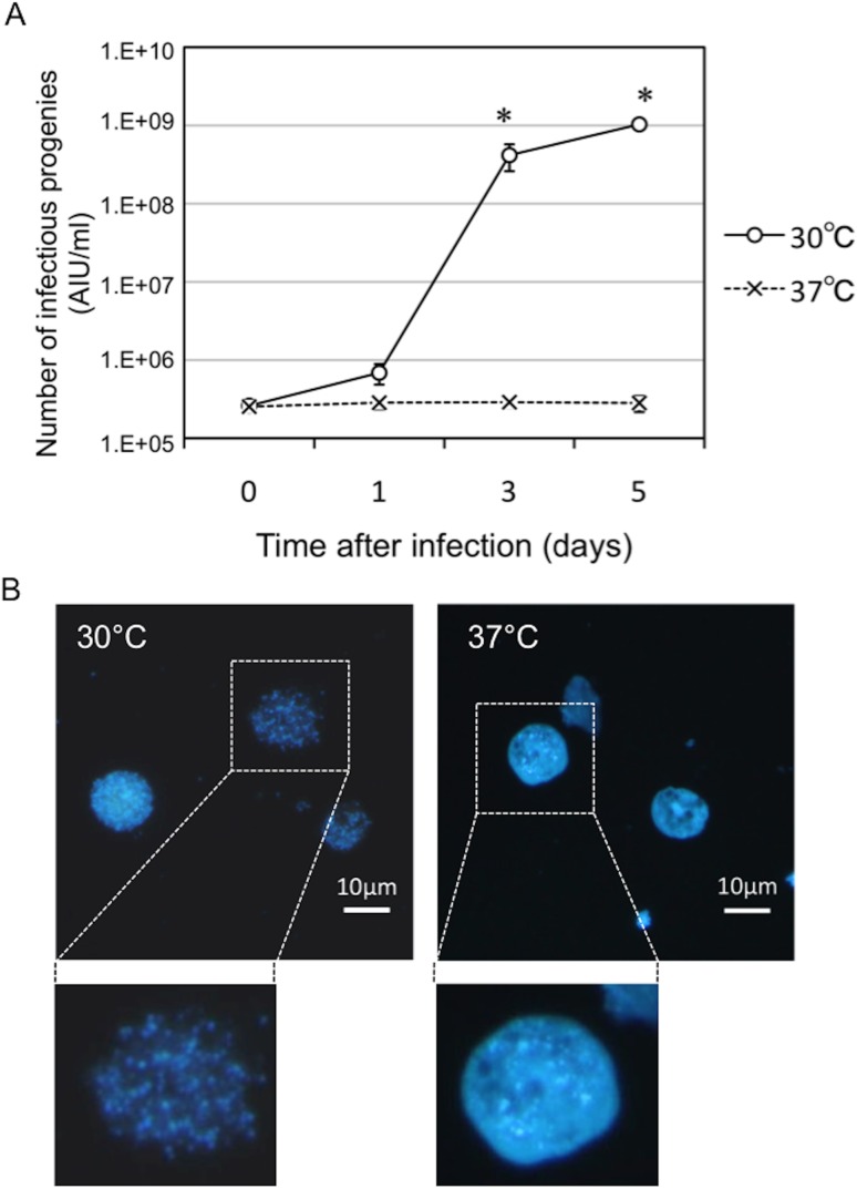
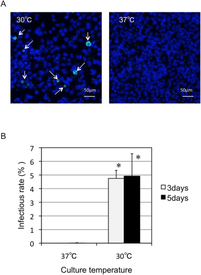
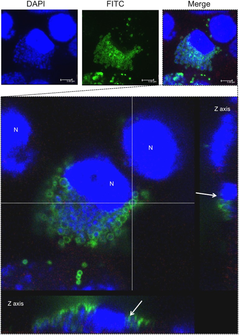
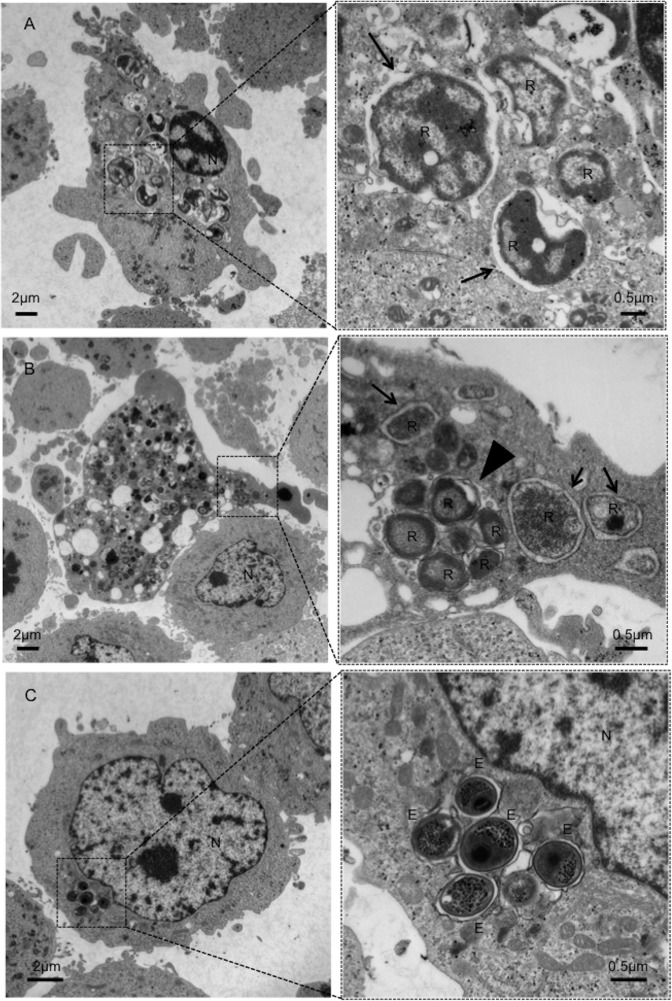
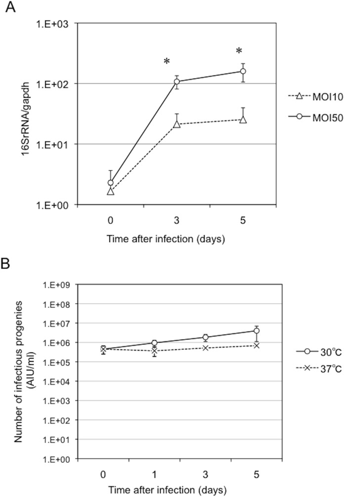
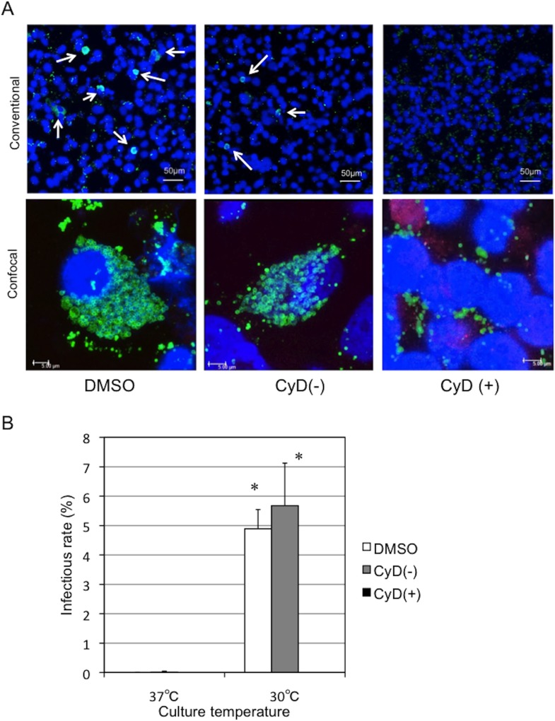
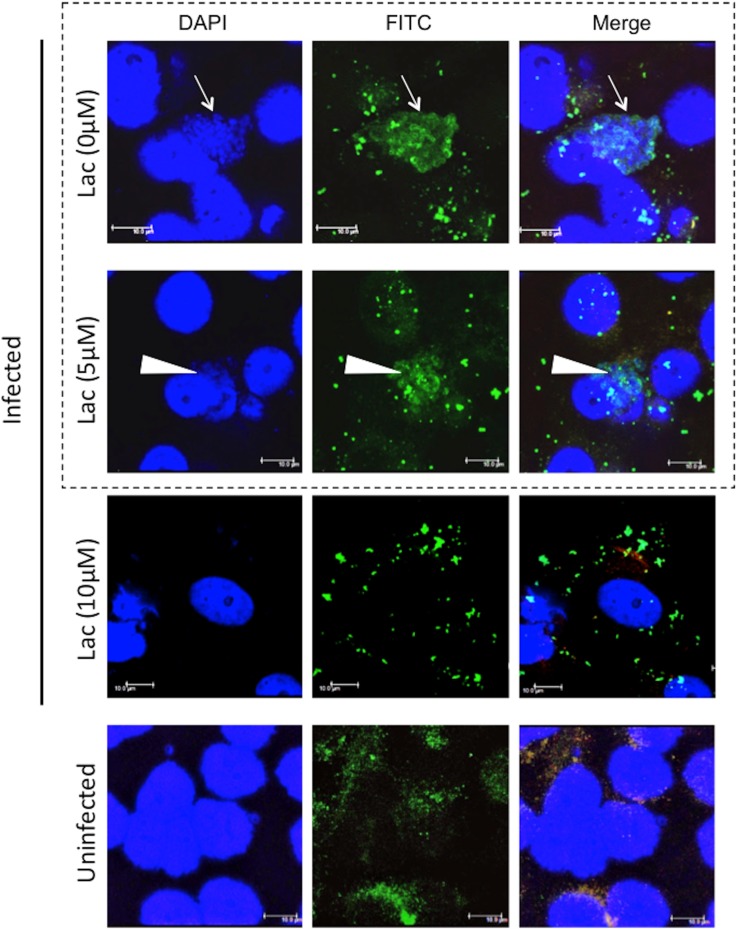
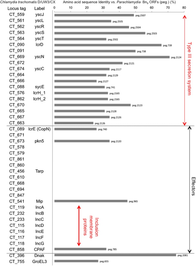
Ancient chlamydiae diverged into pathogenic and environmental chlamydiae 0.7-1.4 billion years ago. However, how pathogenic chlamydiae adapted to mammalian cells that provide a stable niche at approximately 37 °C, remains unknown, although environmental chlamydiae have evolved as endosymbionts of lower eukaryotes in harsh niches of relatively low temperatures. Hence, we assessed whether an environmental chlamydia, Parachlamydia Bn9, could grow in human HEp-2 cells at a low culture temperature of 30 °C. The assessment of inclusion formation by quantitative RT-PCR revealed that the numbers of bacterial inclusion bodies and the transcription level of 16SrRNA significantly increased after culture at 30 °C compared to at 37 °C. Confocal microscopy showed that the bacteria were located close to HEp-2 nuclei and were actively replicative. Transmission electron microscopy also revealed replicating bacteria consisting of reticular bodies, but with a few elementary bodies. Cytochalasin D and rifampicin inhibited inclusion formation. Lactacystin slightly inhibited bacterial inclusion formation. KEGG analysis using a draft genome sequence of the bacteria revealed that it possesses metabolic pathways almost identical to those of pathogenic chlamydia. Interestingly, comparative genomic analysis with pathogenic chlamydia revealed that the Parachlamydia similarly possess the genes encoding Type III secretion system, but lacking genes encoding inclusion membrane proteins (IncA to G) required for inclusion maturation. Taken together, we conclude that ancient chlamydiae had the potential to grow in human cells, but overcoming the thermal gap was a critical event for chlamydial adaptation to human cells.


Conflict of interest statement

Competing Interests: The authors declare that they have no competing interests.

Figures

Figure 1
Figure 1. Changes in the growth rate of Parachlamydia Bn9 in C3 amoebae at 30 or 37°C.
The amoebae were infected with the bacteria (MOI 10), and then incubated for 5 days at 30 or 37°C. The bacterial growth was assessed by conventional fluorescence microscopy. (A) Number of infection progenies in the C3 amoebae infected with Parachlamydia Bn9. The bacterial numbers were estimated by AIU assays [13]. Data are the means ± SD from at least three experiments. *P < 0.05 vs. each culture (30 or 37°C) at immediately after infection. (B) Representative DAPI images of the C3 amoebae infected with Parachlamydia Bn9 at 3 days after infection. Squares surrounded by dotted lines are enlarged below.
Figure 2
Figure 2. Changes in the infectious rate of Parachlamydia Bn9 in HEp-2 cells at 30 or 37°C.
The HEp-2 cells were infected with the bacteria (MOI 50), and then incubated for 5 days at 30 or 37°C. Inclusion formation was assessed by conventional fluorescence microscopy. (A) Representative inclusion images of HEp-2 cells infected with Parachlamydia Bn9 at 3 days after infection. Arrows show the inclusion bodies (Green). Blue, DAPI staining. (B) Changes in the inclusion formation rate of Parachlamydia Bn9 in HEp-2 cells. Data are the means + SD from at least three experiments. *P < 0.05 vs. each culture period (3 or 5 days) at 37°C.
Figure 3
Figure 3. Localization of inclusion bodies formed in the HEp-2 cells infected with Parachlamydia Bn9 at 30°C.
The HEp-2 cells were infected with the bacteria (MOI 10) and then incubated for 5 days at 30°C. The inclusion bodies were assessed 3 days after infection using confocal laser microscopy. The top three images show a representative inclusion body formed in the infected HEp-2 cells. The image with a square surrounded by dotted lines is enlarged below. Arrows in the Z axis panels show bacterial location close to the nucleus of the HEp-2 cells. Blue, DAPI. Green, bacterial cluster. N, HEp-2 nucleus.
Figure 4
Figure 4. Representative TEM image showing the ultrastructure of Parachlamydia Bn9 in HEp-2 cells (A-C).
The squares surrounded by dotted lines are enlarged right panels. Arrows show the bacterial RBs surrounded by plasma membrane. Arrowhead shows an inclusion body. E, EB. R, RB. N, nucleus.
Figure 5
Figure 5. Changes in bacterial growth in the HEp-2 cells infected with Parachlamydia Bn9 at 30 or 37°C.
The HEp-2 cells were infected with the bacteria (MOI 10 or 50), and then incubated for 5 days at 30 or 37°C. Bacterial growth was assessed using qRT-PCR and AIU assays [13]. The cells infected with the bacteria at MOI 50 alone were used for the AIU assay. (A) Change in the level of bacterial 16SrRNA transcripts in infected HEp-2 cells. Each value shows a ratio of 16SrRNA transcripts to housekeeping gapdh transcripts. Data are the means ± SD from at least three experiments. *P < 0.05 vs. culture with MOI 50. (B) Change in amount of infectious bacterial progeny in infected HEp-2 cells. Each value shows the amount of infection progeny as an AIU value, using a co-culture of the C3 amoebae, as described previously [13]. Data are the means ± SD from at least three experiments.
Figure 6
Figure 6. Effect of cytochalasin D on inclusion formation in HEp-2 cells infected with Parachlamydia Bn9.
The HEp-2 cells were infected with the bacteria (MOI 10), in the presence or absence of cytochalasin D (2.5 µg/ml) and then incubated for 5 days at 30°C. Inclusion formation was assessed using conventional and confocal fluorescence microscopy. (A) Representative images showing inclusion formation in infected HEp-2 cells in the presence or absence of cytochalasin D. The images were captured 3 days after infection. Conventional, Observation using conventional fluorescence microscopy. Confocal, Observation using confocal fluorescence microscopy. DMSO, a solvent control. CyD(+), cytochalasin D. CyD(-), medium alone. (B) Change in inclusion formation rate in infected HEp-2 cells in the presence or absence of cytochalasin D. See above. Data are the means + SD from at least three experiments. *P < 0.05 vs. each culture [DMSO, CyD(-), CyD(+)] at 37°C.
Figure 7
Figure 7. Effect of rifampicin on inclusion formation in HEp-2 cells infected with Parachlamydia Bn9.
The HEp-2 cells were infected with the bacteria (MOI 10), in the presence or absence of rifampicin (0.5 µM) and then incubated for 5 days at 30°C. Inclusion formation was assessed using conventional and confocal fluorescence microscopy. (A) Representative images showing inclusion formation in infected HEp-2 cells in the presence or absence of rifampicin. The images were captured 3 days after infection. Conventional, Observation using conventional fluorescence microscopy. Confocal, Observation using confocal fluorescence microscopy. DMSO, a solvent control. Rif(+), rifampicin. Rif(-), medium alone. (B) Change in inclusion formation rate in infected HEp-2 cells in the presence or absence of rifampicin. See above. Data are the means + SD from at least three experiments. *P < 0.05 vs. each culture [DMSO, Rif(-), Rif(+)] at 37°C.
Figure 8
Figure 8. Predicted three-dimensional structure of Parachalmydia Bn9 CPAF (query: RAST gene ID peg.785).
The structure was constructed by alignment with Chlamydia trachomatis chain A, crystal structure of mature CPAF (3DOR_A). Both serine and histidine (yellow), which are critical amino acids of the active site of pathogenic chlamydial CPAF (Bednar et al., 2011), are well conserved. Red line, identical sequences. Blue line, similar sequences. Gray, non-conserved sequences. See S2 Fig.
Figure 9
Figure 9. Effect of lactacystin on inclusion formation in HEp-2 cells infected with Parachlamydia Bn9.
The HEp-2 cells were infected with the bacteria (MOI 10), in the presence or absence of lactacystin (5 or 10 µM) and then incubated for 5 days at 30°C. Inclusion formation was assessed using confocal laser microscopy. The images were captured 3 days after infection. Arrows show healthy inclusion formation. Arrowheads show disrupted inclusion in the presence of lactacystin (5 µM). Addition of 10 µM lactacystin completely abolished the formation of typical inclusion bodies in HEp-2 cells. For showing more conclusive image, the images surrounded by dashed line were enlarged into S9 Fig.
Figure 10
Figure 10. Amino-acid sequence identity of type III secretion system-associated and effector proteins of the C. trachomatis D/UW3/CX into Parachlamydia Bn9.
Each of the values (top hit alone) shows percentage identity (%) of C. trachomatis D/UW3/CX (NC_000117.1) into Parachlamydia Bn9 (BAWW01000001-BAWW 01000072). “peg” numbers reveal locus tags for the Parachlamydia annotated by using RAST server. See Materials and Methods “Genome sequencing and annotation” and S1 Table. Cut off, E value < 1.0E-10.

Similar articles

Cited by

References

    1. Greub G, Raoult D (2003) History of the ADP/ATP-translocase-encoding gene, a parasitism gene transferred from a Chlamydiales ancestor to plants 1 billion years ago. Appl Environ Microbiol 69: 5530–5535. 10.1128/AEM.69.9.5530-5535.2003 - DOI - PMC - PubMed
    1. Horn M, Collingro A, Schmitz-Esser S, Beier CL, Purkhold U, et al. (2004) Illuminating the evolutionary history of chlamydiae. Science 304: 728–730. 10.1126/science.1096330 - DOI - PubMed
    1. Da Ros CT, Schmitt Cda S (2008) Global epidemiology of sexually transmitted diseases. Asian J Androl 10: 110–114. 10.1111/j.1745-7262.2008.00367.x - DOI - PubMed
    1. Burillo A, Bouza E (2010) Chlamydophila pneumoniae. Infect Dis Clin North Am 24: 61–71. 10.1016/j.idc.2009.10.002 - DOI - PubMed
    1. Matsuo J, Kobayashi M, Nakamura S, Mizutani Y, Yao T, et al. (2010a) Stability of Chlamydophila pneumoniae in a harsh environment without a requirement for acanthamoebae. Microbiol Immunol 54: 63–73. 10.1111/j.1348-0421.2009.00182.x - DOI - PubMed

Publication types

LinkOut - more resources