. 2015 Apr;48(2):249-58.
doi: 10.1111/cpr.12161. Epub 2015 Jan 21.

Silencing dishevelled-1 sensitizes paclitaxel-resistant human ovarian cancer cells via AKT/GSK-3β/β-catenin signalling

Affiliations

Silencing dishevelled-1 sensitizes paclitaxel-resistant human ovarian cancer cells via AKT/GSK-3β/β-catenin signalling

Kun Zhang et al. Cell Prolif. 2015 Apr.

Abstract

Objectives: Expression of dishevelled-1 (DVL1) has recently been linked to cancer progression, however, its role in resistance to cancer therapy is unclear. In this study, we aimed to explore the function of DVL1 in paclitaxel-resistant human ovarian cancer cells.

Materials and methods: The MTT assay was used to assess effects of DVL1 silencing on sensitivity of cells that were otherwise resistant to paclitaxel (Taxol). Western blotting and immunofluorescence staining were used to examine effects of DVL1 on AKT/GSK-3β/β-catenin signalling.

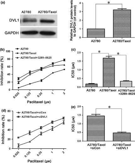
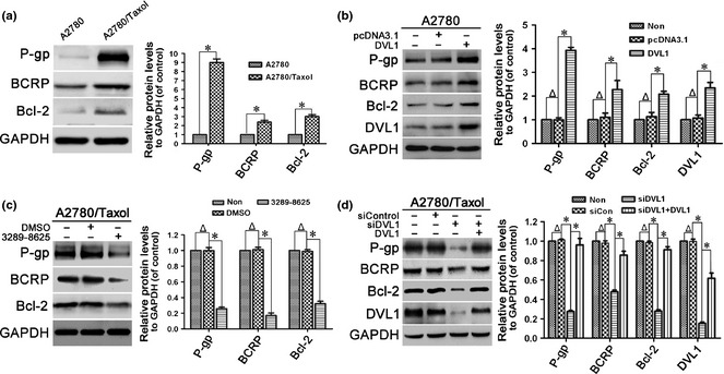
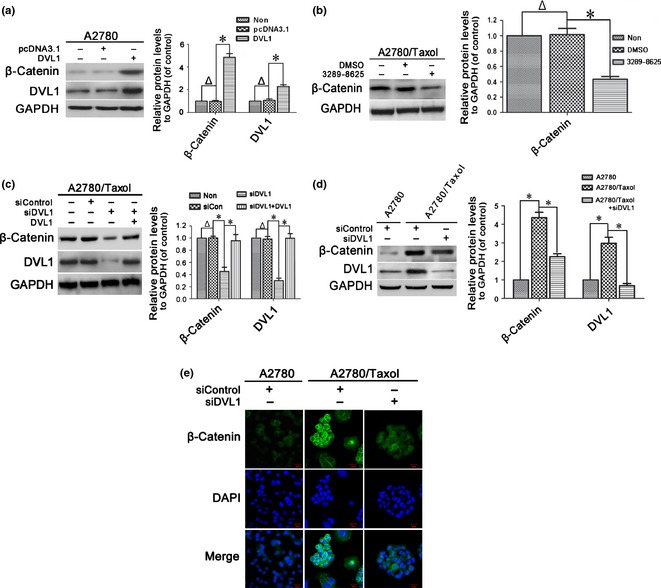
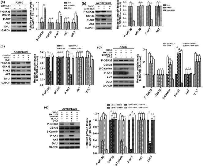
Results: Dishevelled-1 was found to be over-expressed in a paclitaxel-resistant cell line derived from human ovarian cancer cell line A2780 (A2780/Taxol line) as well as parental A2780 cells. Down-regulation of DVL1 (using the inhibitor 3289-8625 or siRNA (siDVL1) against DVL1) sensitized A2780/Taxol cells to paclitaxel. Over-expression of DVL1 in A2780 cells increased protein levels of P-gp, BCRP and Bcl-2, which are known targets of β-catenin. Silencing DVL1 in A2780/Taxol cells also reduced levels of these proteins, and led to accumulation of β-catenin. In addition, DVL1 aberrantly activated AKT/GSK-3β/β-catenin signalling. Inactivation of AKT signalling attenuated DVL1-mediated inhibition of GSK-3β and accumulation of β-catenin, in both A2780 and A2780/Taxol cells.

Conclusions: Taken together, these results suggest that silencing DVL1 sensitized A2780/Taxol cells to paclitaxel, by down-regulating AKT/GSK-3β/β-catenin signalling, providing a novel strategy for chemosensitization of ovarian cancer to paclitaxel-induced cytotoxicity.


Figures

Figure 1
Figure 1
Silencing DVL1 enhanced the sensitivity of A2780/Taxol ovarian cancer cells to paclitaxel. (a) Western blotting of DVL1 expression in A2780 and A2780/Taxol cells. GAPDH was used as the internal control. The left panel shows representative Western blots, and the right panel shows quantitation of the Western blots to show the relative DVL1 expression normalized to GAPDH control. (b) The drug sensitivity of A2780, A2780/Taxol and compound 3289‐8625‐treated A2780/Taxol cells was assessed using MTT assays to measure cell viability. (c) The IC50 of paclitaxel in A2780, A2780/Taxol and compound 3289‐8625‐treated A2780/Taxol cells. The IC50 of paclitaxel was calculated from the survival curves generated in (b) using the Bliss method. (d) The drug sensitivity of A2780/Taxol cells transfected with siDVL1 or siControl. (e) The IC50 of paclitaxel in transfected A2780/Taxol cells, which was calculated from the survival curves in (d) using the Bliss method. Each experiment was performed in triplicate. *P < 0.05.
Figure 2
Figure 2
Effect of DVL1 on drug resistance‐related proteins. Western blotting for the drug resistance‐related proteins P‐gp, BCRP and Bcl‐2 in (a) A2780 and A2780/Taxol cells, (b) A2780 cells transfected with pcDNA3.1‐DVL1 or pcDNA3.1 for 72 h, (c) A2780/Taxol cells treated with or without 100 μM compound 3289‐8625 for 72 h, and (d) A2780/Taxol cells transfected with siControl, siDVL1 or siDVL1 plus pcDNA3.1‐DVL1 for 72 h. GAPDH was used as the internal control. The left panels show representative Western blots, and the right panels show relative quantitation of the blots after normalization to GAPDH. In B and D, DVL1 blots were included to confirm either the overexpression of DVL1 from pcDNA3.1‐DVL1 or the silencing of DVL1 by siDVL1. Each experiment was performed in triplicate. Δ P > 0.05; *P < 0.05.
Figure 3
Figure 3
The effect of DVL1 on the accumulation and nuclear translocation of β‐catenin. (a–d) Western blotting for β‐catenin in (a) A2780 cells transfected with pcDNA3.1‐DVL1 or pcDNA3.1 for 72 h, (b) A2780/Taxol cells treated with or without 100 μm compound 3289‐8625 for 72 h, (c) A2780/Taxol cells transfected with siControl, siDVL1 or siDVL1 plus pcDNA3.1‐DVL1 for 72 h, and (d) A2780 cells transfected with siControl and A2780/Taxol cells transfected with siControl or siDVL1 for 72 h. GAPDH was used as the internal control. Left panels show representative Western blots, and right panels show the relative quantification after normalization to GAPDH. In (a, c and d), DVL1 blots were included to confirm either the overexpression of DVL1 from pcDNA3.1‐DVL1 or the silencing of DVL1 by siDVL1. Each experiment was performed in triplicate. Δ P > 0.05; *P < 0.05. (e) Immunofluorescence analysis of the cellular distribution of β‐catenin in the cytoplasm and nucleus of A2780 and A2780/Taxol cells transfected with siControl or siDVL for 72 h. Green, β‐catenin; blue, nuclear DNA (Scale bar = 20 μm).
Figure 4
Figure 4
The effect of DVL1 on AKT/GSK‐3β/β‐catenin signalling. Western blotting for phospho‐GSK‐3β (Ser‐9), GSK‐3β, phospho‐AKT (Ser‐473), AKT and DVL1 (when pcDNA3.1‐DVL1‐ or siDVL‐transfected cells were included) in (a) A2780 cells transfected with pcDNA3.1‐DVL1 or pcDNA3.1 for 72 h, (b) A2780/Taxol cells treated with or without compound 3289‐8625 for 72 h, (c) A2780/Taxol cells transfected with siControl, siDVL1 or siDVL1 plus pcDNA3.1‐DVL1 for 72 h, (d) A2780 cells transfected with pcDNA3.1 or pcDNA3.1‐DVL1 for 72 h, and then treated with the AKT inhibitor MK‐2206 for 5 h, and (e) A2780/Taxol cells transfected with siControl, siDVL1 or siDVL1 plus pcDNA3.1‐DVL1 for 72 h, and then treated with MK‐2206 for 5 h. GAPDH was used as the internal control. Left panels show representative Western blots, and right panels show the relative quantification after normalization to GAPDH. Each experiment was performed in triplicate. Δ P > 0.05; *P < 0.05.

Similar articles

Cited by

References

    1. Rasool N, LaRochelle W, Zhong H, Ara G, Cohen J, Kohn EC (2010) Secretory leukocyte protease inhibitor antagonizes paclitaxel in ovarian cancer cells. Clin. Cancer Res. 16, 600–609. - PMC - PubMed
    1. Parkin DM, Bray F, Ferlay J, Pisani P (2005) Global cancer statistics, 2002. CA Cancer J. Clin. 55, 74–108. - PubMed
    1. Permuth‐Wey J, Sellers TA (2009) Epidemiology of ovarian cancer. Methods Mol. Biol. 472, 413–437. - PubMed
    1. Agarwal R, Kaye SB (2003) Ovarian cancer: strategies for overcoming resistance to chemotherapy. Nat. Rev. Cancer 3, 502–516. - PubMed
    1. Chen X, Zhang J, Zhang Z, Li H, Cheng W, Liu J (2013) Cancer stem cells, epithelial‐mesenchymal transition, and drug resistance in high‐grade ovarian serous carcinoma. Hum. Pathol. 44, 2373–2384. - PMC - PubMed

Publication types

MeSH terms