. 2015 Apr;83(4):1487-96.
doi: 10.1128/IAI.03132-14. Epub 2015 Feb 2.

Aggregatibacter actinomycetemcomitans cytolethal distending toxin activates the NLRP3 inflammasome in human macrophages, leading to the release of proinflammatory cytokines

Affiliations

Aggregatibacter actinomycetemcomitans cytolethal distending toxin activates the NLRP3 inflammasome in human macrophages, leading to the release of proinflammatory cytokines

Bruce J Shenker et al. Infect Immun. 2015 Apr.

Abstract

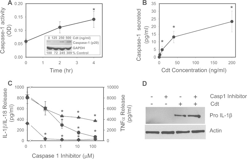
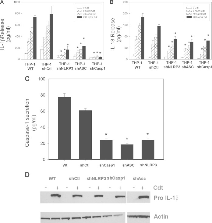
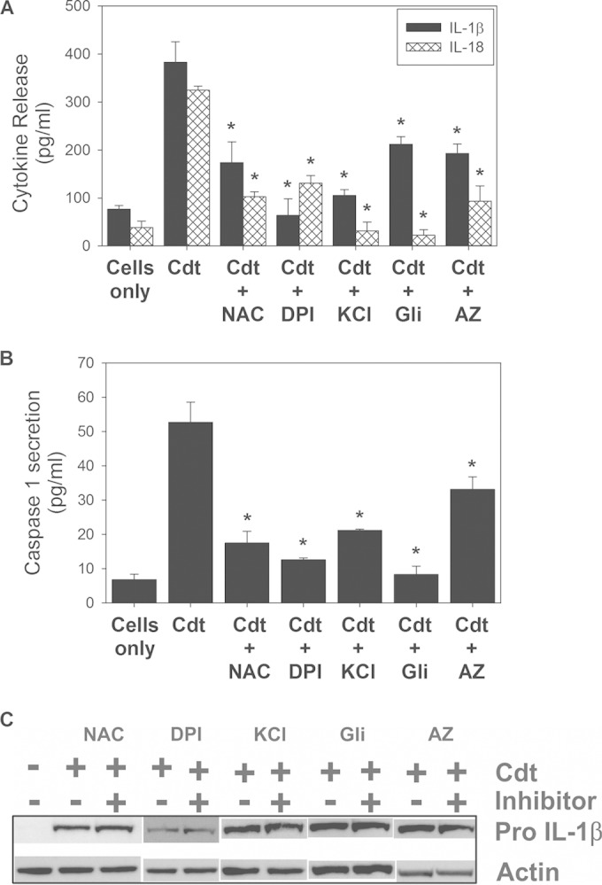
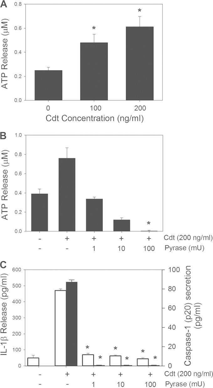
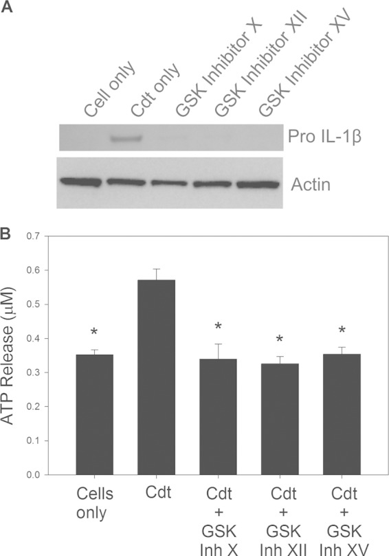
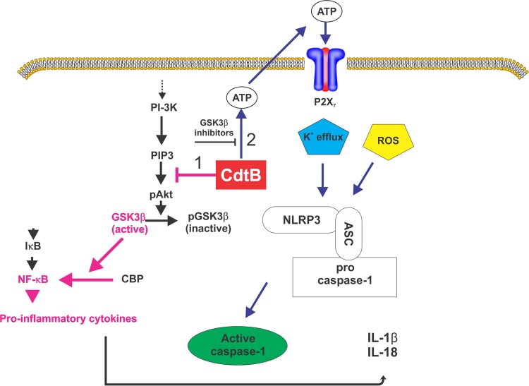
The cytolethal distending toxin (Cdt) is produced from a number of bacteria capable of causing infection and inflammatory disease. Our previous studies with Actinobacillus actinomycetemcomitans Cdt demonstrate not only that the active toxin subunit functions as a phosphatidylinositol-3,4,5-triphosphate (PIP3) phosphatase but also that macrophages exposed to the toxin were stimulated to produce proinflammatory cytokines. We now demonstrate that the Cdt-induced proinflammatory response involves the activation of the NLRP3 inflammasome. Specific inhibitors and short hairpin RNA (shRNA) were employed to demonstrate requirements for NLRP3 and ASC as well as caspase-1. Furthermore, Cdt-mediated inflammasome activation is dependent upon upstream signals, including reactive oxygen species (ROS) generation and Cdt-induced increases in extracellular ATP levels. Increases in extracellular ATP levels contribute to the activation of the P2X7 purinergic receptor, leading to K+ efflux. The relationship between the abilities of the active toxin subunit CdtB to function as a lipid phosphatase, activate the NLRP3 inflammasome, and induce a proinflammatory cytokine response is discussed. These studies provide new insight into the virulence potential of Cdt in mediating the pathogenesis of disease caused by Cdt-producing organisms such as Aggregatibacter actinomycetemcomitans.


Figures

FIG 1
FIG 1
Cdt-induced proinflammatory cytokine release from macrophages is dependent upon caspase-1 activation. (A) THP-1-derived macrophages were treated with 200 ng/ml Cdt for 2 and 4 h. Cell extracts were assessed for caspase-1 activity as described in Materials and Methods. The inset shows Western blot analysis of extracts obtained from THP-1-derived macrophages treated with 0 to 500 ng/ml Cdt; the mature form of caspase-1 (p20 subunit) is shown, along with glyceraldehyde-3-phosphate dehydrogenase (GAPDH) as a loading control. Levels of expression are shown below the blot as a percentage of levels in untreated control cells. OD, optical density. (B) THP-1-derived macrophages were treated with Cdt (0 to 200 ng/ml) for 5 h. Supernatants were then analyzed for the presence of caspase-1 (anti-p20) by an ELISA. Results are the means ± standard errors of the means for three experiments, each performed in triplicate. (C) Effects of caspase-1 inhibition on release of proinflammatory cytokines. Macrophages were exposed to various amounts of the caspase inhibitor Z-WEHD-FMK for 60 min; 50 ng/ml Cdt was then added to the cells for 5 h. Culture supernatants were analyzed for IL-1β (circles), IL-18 (diamonds), and TNF-α (triangles) by an ELISA. Results are plotted as the mean ± the standard error of the mean pg/ml cytokine released versus the Z-WEHD-FMK concentration. Levels of cytokines release from untreated control cells were 9.8 ± 7.8 pg/ml (IL-1β), 38.1 ± 14.0 pg/ml (IL-18), and 120.9 ± 2.6 pg/ml (TNF-α). Asterisks indicate statistical significance (P ≤ 0.05) compared to untreated control cells (A and B) or compared to cells treated with toxin alone (C). (D) Cells were assessed for the expression of pro-IL-1β after 5 h of exposure to Cdt following solubilization, fractionation by SDS-PAGE, and analysis by Western blotting. Actin is shown as a loading control. Western blots are representative of results from three experiments.
FIG 2
FIG 2
Cdt-induced cytokine release from THP-1-derived macrophages involves activation of the NLRP3 inflammasome. THP-1 cells were stably transfected with an shRNA that targets caspase-1, NLRP3, or ASC or with nontargeted control shRNA. (A to C) The effect of protein knockdown in cells exposed to 0 to 200 ng/ml Cdt was determined, and culture supernatants were analyzed for IL-1β (A), IL-18 (B), and caspase-1 (p20) (C) release by an ELISA. Results are the means ± standard errors of the means for three experiments performed in triplicate; asterisks indicate a statistically significant difference from the shRNA control P ≤ 0.05). (D) Cells were also analyzed by Western blotting for the presence of pro-IL-1β. Actin is shown as a loading control. WT, wild type.
FIG 3
FIG 3
Cdt-induced inflammasome activation involves ROS, K+ efflux, and extracellular ATP. NAC (10 mM) and DPI (250 nM) were used to determine the requirement for ROS, elevated extracellular levels of K+ (70 mM) and glibenclamide (Gli) (50 μg/ml) were used to determine the requirement for K+ efflux, and AZ11645373 (AZ) (1 μM) was used to determine the requirement for an ATP-PTXR7 interaction. (A and B) Effects of these agents on Cdt (50 ng/ml)-induced production of IL-1β and IL-18 (A) and on caspase-1 release (B). Results are the means ± standard errors of the means for three experiments, each performed in triplicate; asterisks indicate statistical significance compared to the Cdt-only control (P ≤ 0.05). (C) Cells were also analyzed for the presence of pro-IL-1β by Western blotting. Actin is shown as a loading control.
FIG 4
FIG 4
The proinflammatory response induced by Cdt is dependent upon extracellular ATP. (A) Macrophages were treated with 0 to 200 ng/ml Cdt for 2 h, and supernatants were then analyzed for ATP. (B and C) Cells were also analyzed for the effect of apyrase (Pyrase) on extracellular ATP levels (B) as well as IL-1β release (open bars) and caspase-1 release (solid bars) (C). Results represent the means ± standard errors of the means for three experiments, each performed in triplicate; asterisks indicate statistical significance compared to control cells (P ≤ 0.05).
FIG 5
FIG 5
GSK3β activation contributes to Cdt-induced expression of pro-IL-1β and increased extracellular ATP levels. (A) THP-1-derived macrophages were pretreated with GSK inhibitors for 1 h, followed by the addition of 200 ng/ml Cdt. After 4 h, cells were fractionated by SDS-PAGE and analyzed by Western blotting for pro-IL-1β. Results are representative of three experiments. (B) Macrophages were pretreated with GSK inhibitors as described above and then treated with 200 ng/ml Cdt for 4 h. Supernatants were analyzed for ATP as described in Materials and Methods. Results represent the means ± standard errors of the means for three experiments; asterisks indicate statistical significance compared to control cells treated with Cdt alone (P ≤ 0.05).
FIG 6
FIG 6
Summary diagram depicting the mechanism by which Cdt induces a proinflammatory cytokine response in macrophages. Two signals are proposed. The first signal (pink arrows) involves the upregulation of inflammatory cytokine gene and protein expression; this signal is dependent upon Cdt's abilities to function as a PIP3 phosphatase, block PI-3K signaling, and activate GSK3β, leading to the expression and synthesis of pro-IL-1β and IL-18. The second signal (blue arrows) involves the activation of the NLRP3 inflammasome, which requires the generation of extracellular ATP, an ATP-P2X7 interaction, K+ efflux, and utilization of endogenous ROS. Inhibitors of GSK3β also block ATP generation, suggesting that Cdt-induced activation of this kinase is also critical to ATP-dependent inflammasome activation.

Similar articles

Cited by

References

    1. Comayras C, Tasca C, Peres SY, Ducommun B, Oswald E, De Rycke J. 1997. Escherichia coli cytolethal distending toxin blocks the HeLa cell cycle at the G2/M transition by preventing cdc2 protein kinase dephosphorylation and activation. Infect Immun 65:5088–5095. - PMC - PubMed
    1. Okuda J, Fukumoto M, Takeda Y, Nishibuchi M. 1997. Examination of diarrheagenicity of cytolethal distending toxin: suckling mouse response to the products of the cdtABC genes of Shigella dysenteriae. Infect Immun 65:428–433. - PMC - PubMed
    1. Okuda J, Kurazono H, Takeda Y. 1995. Distribution of the cytolethal distending toxin A gene (cdtA) among species of Shigella and Vibrio, and cloning and sequencing of the cdt gene from Shigella dysenteriae. Microb Pathog 18:167–172. - PubMed
    1. Scott DA, Kaper JB. 1994. Cloning and sequencing of the genes encoding Escherichia coli cytolethal distending toxin. Infect Immun 62:244–251. - PMC - PubMed
    1. Pickett CL, Cottle DL, Pesci EC, Bikah G. 1994. Cloning, sequencing, and expression of the Escherichia coli cytolethal distending toxin genes. Infect Immun 62:1046–1051. - PMC - PubMed

Publication types

MeSH terms

Substances

LinkOut - more resources