. 2015 Apr;83(4):1598-609.
doi: 10.1128/IAI.03122-14. Epub 2015 Feb 2.

Protein A is released into the Staphylococcus aureus culture supernatant with an unprocessed sorting signal

Affiliations

Protein A is released into the Staphylococcus aureus culture supernatant with an unprocessed sorting signal

Dara P O'Halloran et al. Infect Immun. 2015 Apr.

Abstract

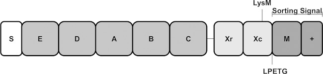
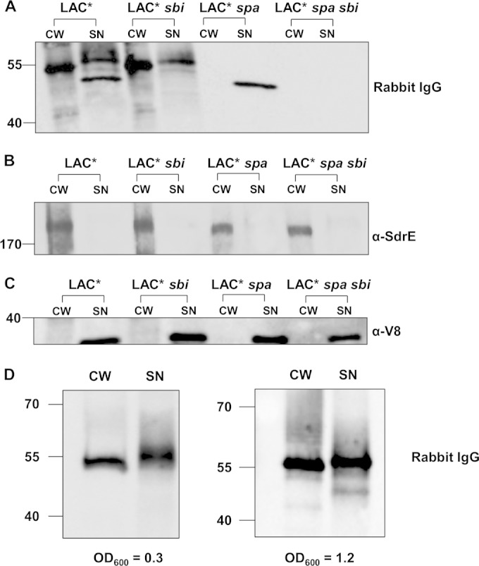
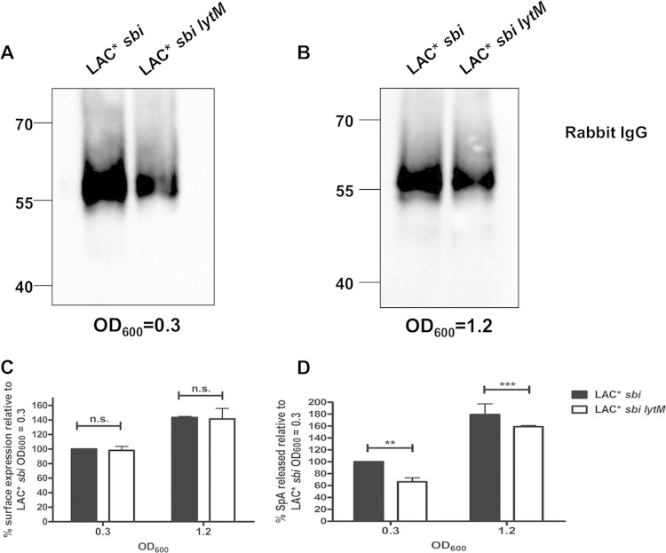
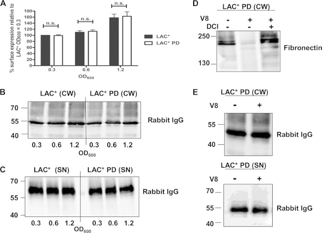
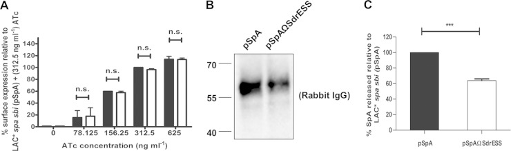
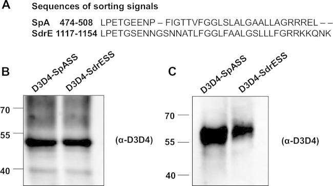
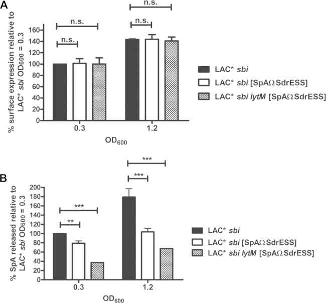
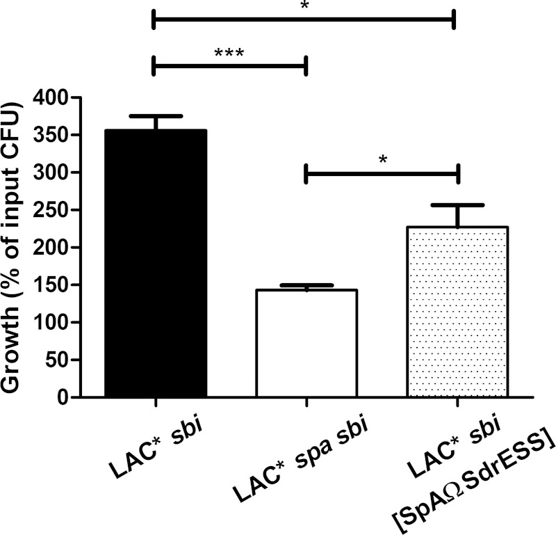
The immunoglobulin binding protein A (SpA) of Staphylococcus aureus is synthesized as a precursor with a C-terminal sorting signal. The sortase A enzyme mediates covalent attachment to peptidoglycan so that SpA is displayed on the surface of the bacterium. Protein A is also found in the extracellular medium, but the processes involved in its release are not fully understood. Here, we show that a portion of SpA is released into the supernatant with an intact sorting signal, indicating that it has not been processed by sortase A. Release of SpA was reduced when the native sorting signal of SpA was replaced with the corresponding region of another sortase-anchored protein (SdrE). Similarly, a reporter protein fused to the sorting signal of SpA was released to a greater extent than the same polypeptide fused to the SdrE sorting signal. Released SpA protected bacteria from killing in human blood, indicating that it contributes to immune evasion.


Figures

FIG 1
FIG 1
Schematic representation of the domain organization of protein A. Protein A consists of an N-terminal signal sequence (S) followed by up to five IgG-binding domains (E to C), an antigenic variable region (Xr), and a cell wall-spanning region (Xc). The Xc region harbors an LysM domain which can mediate noncovalent binding of proteins to peptidoglycan. The sorting signal comprises an LPETG motif, a hydrophobic membrane-spanning region (M), and a positively charged tail region (+).
FIG 2
FIG 2
Staphylococcus aureus LAC* releases SpA. S. aureus was grown to an OD600 of 1.2. Cell wall extracts (CW) were diluted 1:5 prior to loading on the gel, and supernatant samples (SN) were not diluted. Protein A was detected using HRP-conjugated rabbit IgG (A), SdrE was detected using anti-SdrE IgG (B), and V8 was detected using anti-V8 serum (C). S. aureus LAC* sbi was grown to an OD600 of 0.3 or 1.2 as indicated (D). Cell wall extracts were diluted 1:20, and supernatant samples were not diluted. Size markers are indicated (kDa).
FIG 3
FIG 3
LytM contributes to release of protein A by LAC*. (A and B) Culture supernatants from LAC* sbi and LAC* sbi lytM grown to the OD600 indicated were probed with HRP-labeled rabbit IgG in Western immunoblotting. Supernatants harvested at an OD600 of 0.3 were concentrated 8-fold before being loaded on a gel, and supernatants harvested at an OD600 of 1.2 were concentrated 2-fold. Size markers are indicated (kDa). (C) Protein A on the surface of LAC* sbi and LAC* sbi lytM was detected using FITC-labeled rabbit IgG, and the fluorescence intensity was measured by flow cytometry. Values are plotted as a percentage of the mean fluorescence intensity measured for LAC* sbi grown to an OD600 of 0.3. Bars represent the mean values, and error bars indicate the standard errors of the means of three independent experiments. (D) Protein was captured from culture supernatants using chicken anti-SpA polyclonal IgY and detected using biotin-conjugated mouse monoclonal anti-SpA IgG followed by streptavidin-HRP in an ELISA. Values are expressed as a percentage of total released SpA measured for LAC* sbi grown to an OD600 of 0.3. Bars represent the mean percent release from four independent experiments. Error bars represent the standard errors of the means. **, P = 0.006; ***, P = 0.0003; ns, not significant (P > 0.05).
FIG 4
FIG 4
Extracellular proteases are not required for the release of protein A. (A) Protein A on the surface of LAC* and LAC* PD was detected using FITC-labeled rabbit IgG, and the fluorescence intensity was measured by flow cytometry. Values are expressed as a percentage of the mean fluorescence intensity measured for LAC* harvested at an OD600 of 0.3. Bars represent the mean values, and error bars indicate the standard errors of the means of three independent experiments. ns, not significant (P > 0.05). (B and C) Cell wall extracts (CW) and culture supernatants (SN) from LAC* and LAC* PD grown to the OD600 indicated were probed with HRP-labeled rabbit IgG in a Western immunoblot. Supernatants harvested at an OD600 of 0.3 were concentrated 8-fold, those at an OD600 of 0.6 were concentrated 4-fold, and those at an OD600 of 1.2 were concentrated 2-fold before being loaded on a gel. (D) LAC* PD was grown to an OD600 of 0.8 in broth alone or in broth supplemented with V8 (1 U/ml) and DCI (200 μM), and cell wall extracts were probed with biotin-labeled fibronectin in a ligand affinity blot. Bound fibronectin was detected using streptavidin-HRP. (E) The same cell wall extracts and supernatants from the same cultures were probed with HRP-conjugated rabbit IgG to detect protein A. Size markers are indicated (kDa).
FIG 5
FIG 5
Release of protein A into S. aureus culture supernatants can be inhibited by altering the sorting signal. (A) Protein A on the surface of LAC* spa sbi(pSpA) (black bars) and LAC* spa sbi(pSpAΩSdrESS) (white bars) was detected using FITC-labeled rabbit IgG, and the fluorescence intensity was measured using flow cytometry. Values are expressed as a percentage of the mean fluorescence intensity measured for LAC* spa sbi(pSpA) grown in broth supplemented with ATc (312.5 ng/ml). Bars represent the mean of three independent experiments. Error bars represent the standard errors of the means. ns, not significant (P > 0.05). (B) LAC* spa sbi(pSpA) and LAC* spa sbi(pSpAΩSdrESS) were grown in broth supplemented with ATc (312.5 ng/ml), and culture supernatants were probed with HRP-conjugated rabbit IgG. Size markers are indicated (kDa). (C) Quantification of SpA in culture supernatants of LAC* spa sbi(pSpA) and LAC* spa sbi(pSpAΩSdrESS) by ELISA. Bacteria were grown in broth supplemented with ATc (312.5 ng/ml), and SpA was captured from culture supernatants using chicken anti-SpA polyclonal IgY. Bound SpA was detected using biotin-labeled mouse monoclonal anti-SpA IgG and HRP-conjugated streptavidin in an ELISA. The absorbance at 450 nm was measured, and readings from wells incubated with culture supernatants from LAC* spa sbi(pRMC2) were subtracted from the mean readings for LAC* spa sbi(pSpA) and LAC* spa sbi(pSpAΩSdrESS) to account for background. Values for LAC* spa sbi(pSpA ΩSdrESS) are expressed as a percentage of the values measured for LAC* spa sbi(pSpA). Bars represent the mean of three independent experiments, and error bars represent the standard errors of the means. ***, P < 0.0001.
FIG 6
FIG 6
Release of D3D4 reporter protein into S. aureus culture supernatants can be inhibited by altering the sorting signal. (A) Amino acid sequences of SpA and SdrE sorting signals. Amino acid coordinates are indicated. Cell wall extracts (B) and culture supernatants (C) from LAC* spa sbi(pD3D4-SpASS) and LAC* spa sbi(pD3D4-SdrESS) were probed with rabbit anti-D3D4 IgG. Bound antibody was detected using HRP-conjugated goat anti-rabbit IgG. Size markers are indicated (kDa).
FIG 7
FIG 7
Replacing the SpA sorting signal with the sorting signal from SdrE reduces release of SpA by LAC*. (A) Protein A on the surface of LAC* sbi, LAC* sbi [SpAΩSdrESS], and LAC* sbi lytM [SpAΩSdrESS] was detected using FITC-labeled rabbit IgG, and the fluorescence intensity was measured using flow cytometry. Values are expressed as a percentage of the mean fluorescence intensity measured for LAC* sbi grown to an OD600 of 0.3. Bars represent the mean values, and error bars indicate the standard errors of the means of three independent experiments. (B) Protein A was captured from culture supernatants using chicken anti-SpA polyclonal IgY and detected using biotin-conjugated mouse monoclonal anti-SpA IgG followed by streptavidin-HRP in an ELISA. The absorbance at 450 nm was measured, and the mean reading from wells incubated with culture supernatants from LAC* spa sbi were subtracted from the readings for all other wells to account for background. Values are expressed as a percentage of total released SpA measured for LAC* sbi grown to an OD600 of 0.3. Bars represent the mean of four independent experiments. Error bars represent the standard errors of the means. **, P = 0.007; ***, P < 0.0001; ns, not significant (P > 0.05).
FIG 8
FIG 8
Released SpA protects S. aureus from killing in human blood. Washed bacteria were incubated in blood for 3 h at 37°C, and the number of input and recovered bacteria was calculated by viable counting. The percentage increase in CFU counts (growth) of each strain was determined by dividing the mean CFU count after 3 h by the mean input CFU count. Bars represent the mean percentage increase in CFU counts from three independent experiments, and error bars indicate the standard errors of the means. ***, P < 0.001; *, P < 0.05.

Similar articles

Cited by

References

    1. Klevens RM, Morrison MA, Nadle J, Petit S, Gershman K, Ray S, Harrison LH, Lynfield R, Dumyati G, Townes JM, Craig AS, Zell ER, Fosheim GE, McDougal LK, Carey RB, Fridkin SK, Active Bacterial Core surveillance (ABCs) MRSA investigators . 2007. Invasive methicillin-resistant Staphylococcus aureus infections in the United States. JAMA 298:1763–1771. doi:10.1001/jama.298.15.1763. - DOI - PubMed
    1. Lachica RV, Genigeorgis CA, Hoeprich PD. 1979. Occurrence of protein A in Staphylococcus aureus and closely related Staphylococcus species. J Clin Microbiol 10:752–753. - PMC - PubMed
    1. Movitz J. 1976. Formation of extracellular protein A by Staphylococcus aureus. Eur J Biochem 68:291–299. doi:10.1111/j.1432-1033.1976.tb10788.x. - DOI - PubMed
    1. Becker S, Frankel MB, Schneewind O, Missiakas D. 2014. Release of protein A from the cell wall of Staphylococcus aureus. Proc Natl Acad Sci U S A 111:1574–1579. doi:10.1073/pnas.1317181111. - DOI - PMC - PubMed
    1. Forsgren A, Nordstrom K. 1974. Protein A from Staphylococcus aureus: the biological significance of its reaction with IgG. Ann N Y Acad Sci 236:252–266. doi:10.1111/j.1749-6632.1974.tb41496.x. - DOI - PubMed

Publication types

MeSH terms

LinkOut - more resources