. 2015 Feb 3;14(1):19.
doi: 10.1186/s12943-015-0287-3.

Receptor protein tyrosine phosphatase beta/zeta is a functional binding partner for vascular endothelial growth factor

Affiliations

Receptor protein tyrosine phosphatase beta/zeta is a functional binding partner for vascular endothelial growth factor

Marina Koutsioumpa et al. Mol Cancer. .

Abstract

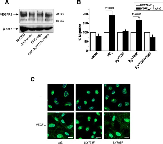
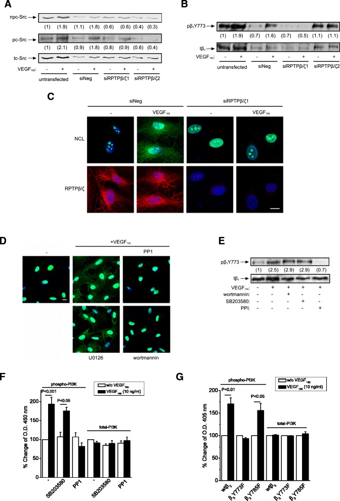
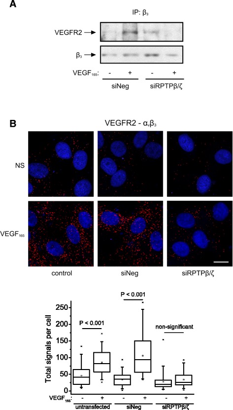
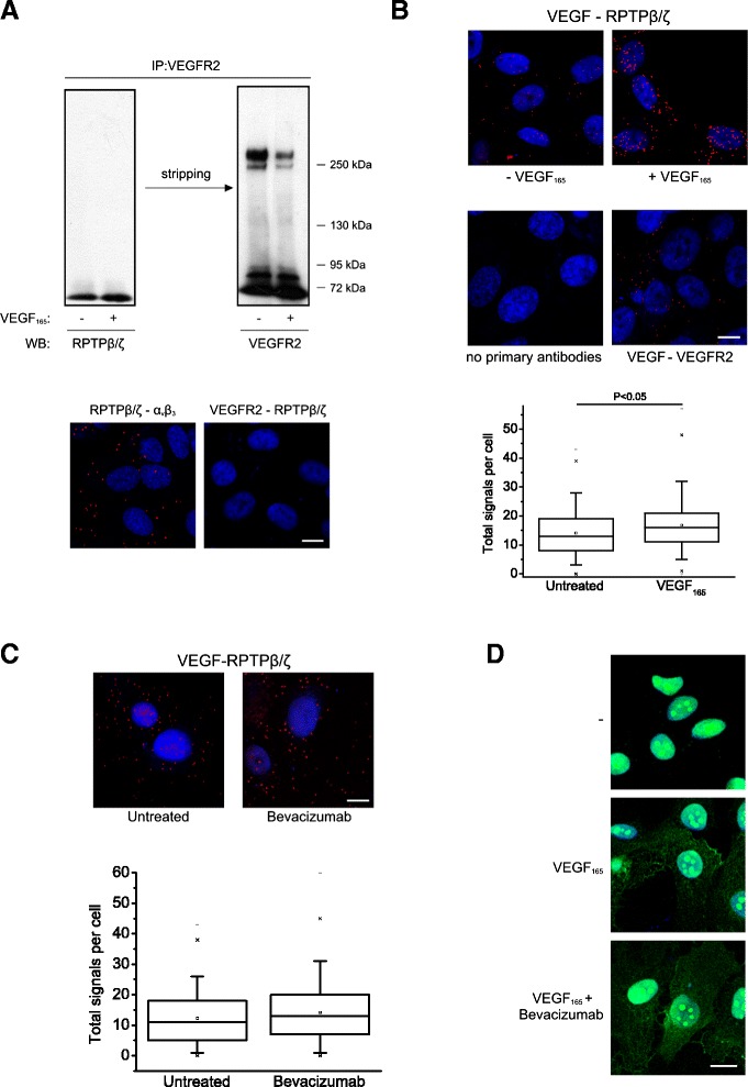
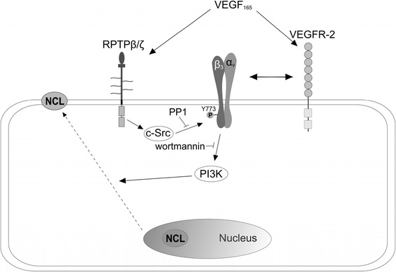
Background: Receptor protein tyrosine phosphatase beta/zeta (RPTPβ/ζ) is a chondroitin sulphate (CS) transmembrane protein tyrosine phosphatase and is a receptor for pleiotrophin (PTN). RPTPβ/ζ interacts with ανβ₃ on the cell surface and upon binding of PTN leads to c-Src dephosphorylation at Tyr530, β₃ Tyr773 phosphorylation, cell surface nucleolin (NCL) localization and stimulation of cell migration. c-Src-mediated β₃ Tyr773 phosphorylation is also observed after vascular endothelial growth factor 165 (VEGF₁₆₅) stimulation of endothelial cells and is essential for VEGF receptor type 2 (VEGFR2) - ανβ₃ integrin association and subsequent signaling. In the present work, we studied whether RPTPβ/ζ mediates angiogenic actions of VEGF.

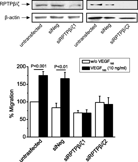
Methods: Human umbilical vein endothelial, human glioma U87MG and stably transfected Chinese hamster ovary cells expressing different β₃ subunits were used. Protein-protein interactions were studied by a combination of immunoprecipitation/Western blot, immunofluorescence and proximity ligation assays, properly quantified as needed. RPTPβ/ζ expression was down-regulated using small interference RNA technology. Migration assays were performed in 24-well microchemotaxis chambers, using uncoated polycarbonate membranes with 8 μm pores.

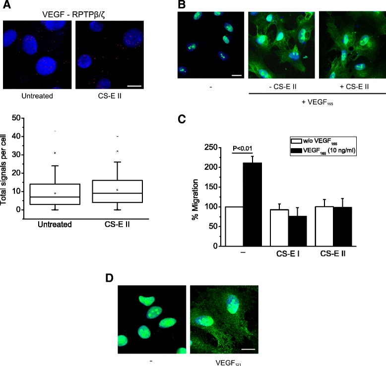
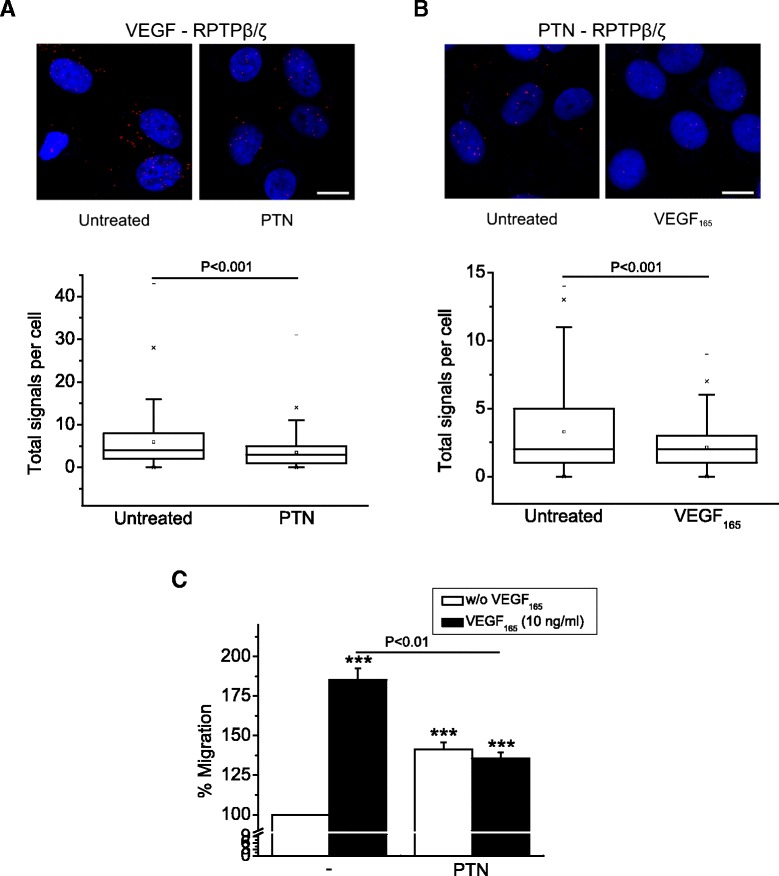
Results: RPTPβ/ζ mediates VEGF₁₆₅-induced c-Src-dependent β₃ Tyr773 phosphorylation, which is required for VEGFR2-ανβ₃ interaction and the downstream activation of phosphatidylinositol 3-kinase (PI3K) and cell surface NCL localization. RPTPβ/ζ directly interacts with VEGF165, and this interaction is not affected by bevacizumab, while it is interrupted by both CS-E and PTN. Down-regulation of RPTPβ/ζ by siRNA or administration of exogenous CS-E abolishes VEGF₁₆₅-induced endothelial cell migration, while PTN inhibits the migratory effect of VEGF₁₆₅ to the levels of its own effect.

Conclusions: These data identify RPTPβ/ζ as a cell membrane binding partner for VEGF that regulates angiogenic functions of endothelial cells and suggest that it warrants further validation as a potential target for development of additive or alternative anti-VEGF therapies.


Figures

Figure 1
Figure 1
Phosphorylation of β 3 Tyr773 is required for VEGF 165 -induced cell migration and cell surface NCL localization. (A) Protein extracts of CHO cells were analysed for expression of VEGFR2. HUVEC were used as a positive control and β-actin as a loading control. (B) Effect of VEGF165 (10 ng/ml) on CHO cell migration. Data are from five independent experiments and are expressed as mean ± s.e.m. percentage change in number of migrating cells compared with the corresponding non stimulated cells (set as default 100). (C) Immunofluorescence images stained for NCL (green) and nucleus (blue) in serum starved CHO cells treated with VEGF165 (10 ng/ml) for 5 h at 37°C. Vector, cells transfected with the plasmid vector; wtβ3, cells over-expressing wild-type β3; β3Y773F, cells over-expressing β3Y773F; β3Y785F, cells over-expressing β3Y785F; β3Y773F/Y785F, cells over-expressing double mutant β3Y773F/Y785F.
Figure 2
Figure 2
RPTPβ/ζ is required for VEGF 165 -induced cell surface NCL localization. Serum starved HUVEC were treated with VEGF165 (10 ng/ml) for 10 min. Cell lysates were analyzed by Western blot for non Tyr530 phosphorylated (npc-Src), Tyr419 phosphorylated (pc-Src) and total (tc-Src) c-Src (A), as well as for phospho-β3Y773 (pβ3Y773) and total β3 (tβ3) integrin (B). Numbers in brackets denote the average-fold change of the ratio npc-Src:tc-Src, pc-Src:tc-Src or pβ3Y773:tβ3 respectively, compared with the corresponding non stimulated, untransfected cells (set as default 1). (C) Representative immunofluorescence images stained for NCL (green), RPTPβ/ζ (red) and nucleus (blue) from serum starved HUVEC treated with VEGF165 (10 ng/ml) for 5 h at 37°C. (D) Representative immunofluorescence images stained for NCL (green) and nucleus (blue) from serum starved VEGF165-stimulated HUVEC in the presence or absence of inhibitors for c-Src (PP1 10 μΜ), PI3K (wortmannin 100 nM) and ERK½ (U0126 20 nM). Scale bars in C and D correspond to 10 μm. (E) Lysates from serum starved VEGF165-stimulated HUVEC in the presence or absence of PP1 and wortmannin, were analyzed by Western blot for pβ3Y773 and tβ3 integrin. Numbers in brackets denote the average-fold change of the ratio pβ3Y773:tβ3 compared with untreated cells (set as default 1). (F and G) Phosphorylation of PI3K in HUVEC and CHO cells respectively. Data are expressed as mean ± s.e.m percentage change in PI3K compared with the untreated cells (set as default 100). In all cases, data come from three independent experiments. siNeg, HUVEC transfected with a negative control siRNA; siRPTPβ/ζ1, HUVEC transfected with siRPTPβ/ζ#1; siRPTPβ/ζ2, HUVEC transfected with si RPTPβ/ζ#2; vector, CHO cells transfected with the plasmid vector; wtβ3, CHO cells over-expressing wild-type β3; β3Y773F, CHO cells over-expressing β3Y773F; β3Y785F, CHO cells over-expressing β3Y785F.
Figure 3
Figure 3
RPTPβ/ζ regulates VEGF 165 -induced VEGFR2-α ν β 3 interaction. Down-regulation of RPTPβ/ζ expression by siRNA was followed by treatment of serum-starved HUVEC with VEGF165 (10 ng/ml) for 10 min. (A) Cells lysates were immunoprecipitated for β3 and analyzed by Western blot for the presence of VEGFR2 and β3. (B) Formation of β3-VEGFR2 complexes as evidenced by in situ PLA. The box plots indicate the median, mean and range of the detected signals (n = 8 image fields with ~4 cells per image per sample type, each sample run in duplicate). Scale bar corresponds to 10 μm. siNeg, HUVEC transfected with a negative control siRNA; siRPTPβ/ζ, HUVEC transfected with siRPTPβ/ζ#1.
Figure 4
Figure 4
RPTPβ/ζ is required for VEGF 165 -induced endothelial cell migration. Effect of VEGF165 on HUVEC migration after down-regulation of RPTPβ/ζ expression by two different siRNAs. Data are from five independent experiments and are expressed as mean ± s.e.m. percentage change in number of migrating cells compared with the non stimulated untransfected cells (set as default 100). Untrasfected, untransfected HUVEC; siNeg, HUVEC transfected with a negative control siRNA; siRPTPβ/ζ1, HUVEC transfected with siRPTPβ/ζ#1; siRPTPβ/ζ2, HUVEC transfected with si RPTPβ/ζ#2. The Western blot on top shows effective down-regulation of RPTPβ/ζ by both siRNA sequences used.
Figure 5
Figure 5
VEGF directly interacts with RPTPβ/ζ in a VEGFR-independent manner. (A) Serum-starved untreated or VEGF165-stimulated HUVEC lysates were immunoprecipitated for VEGFR2 and analyzed by Western blot for the presence of RPTPβ/ζ or VEGFR2 (up). No direct interaction between VEGFR2 with RPTPβ/ζ was observed by performing in situ PLA in HUVEC (down). The RPTPβ/ζ-ανβ3 interaction was used as a positive control. Data are from two independent experiments. (B) Formation of VEGF-RPTPβ/ζ complexes as evidenced by in situ PLA in HUVEC, untreated or after addition of exogenous VEGF165 (10 ng/ml) at 24 h. The VEGF-VEGFR2 interaction was used as a positive control. Data are from five independent experiments. (C) Formation of VEGF-RPTPβ/ζ complexes as evidenced by in situ PLA in HUVEC in the absence or the presence of bevacizumab (250 μg/ml). The box plots in B and C indicate the median, mean and range of the detected signals (n > 20 image fields with ~4 cells per image per sample type, each sample run at least in duplicate) from three independent experiments. (D) Immunofluorescence images stained for NCL (green) and nucleus (blue) in serum starved HUVEC treated for 5 h at 37°C with VEGF165 (10 ng/ml) in the presence or the absence of bevacizumab (250 μg/ml). Representative pictures from three independent experiments. Scale bars in all cases correspond to 10 μm.
Figure 6
Figure 6
Effect of CS-E on VEGF 165 -induced endothelial cell signaling and migration. (A) Formation of VEGF-RPTPβ/ζ complexes as evidenced by in situ PLA in HUVEC in the absence or the presence of CS-E II (100 ng/ml). The box plots indicate the median, mean and range of the detected signals (n > 20 image fields with ~4 cells per image per sample type, each sample run at least in duplicate) from three independent experiments. (B) Immunofluorescence images stained for NCL (green) and nucleus (blue) in serum starved HUVEC treated for 5 h at 37°C with VEGF165 (10 ng/ml) in the absence or the presence of CS-E II (100 ng/ml). Representative pictures from two independent experiments. (C) Effect of CS-E I and II (both at 100 ng/ml) on VEGF165-induced HUVEC migration. Data are from three independent experiments and are expressed as mean ± s.e.m. percentage change in number of migrating cells compared with the non stimulated untransfected cells (set as default 100). (D) Immunofluorescence images stained for NCL (green) and nucleus (blue) in serum starved HUVEC treated for 5 h at 37°C with VEGF121 (10 ng/ml). Representative pictures from two independent experiments. Scale bars in all cases correspond to 10 μm.
Figure 7
Figure 7
PTN and VEGF compete for binding to RPTPβ/ζ. (A) Formation of VEGF-RPTPβ/ζ complexes as evidenced by in situ PLA in HUVEC in the absence or the presence of exogenous PTN (100 ng/ml). (Β) Formation of PTN-RPTPβ/ζ complexes as evidenced by in situ PLA in HUVEC in the absence or the presence of exogenous VEGF165 (10 ng/ml). The box plots in A and B indicate the median, mean and range of the detected signals (n > 20 image fields with ~4 cells per image per sample type, each sample run at least in duplicate) from three independent experiments in each case. (C) Effect of PTN (100 ng/ml) on VEGF165-induced HUVEC migration. Data are from five independent experiments and are expressed as mean ± s.e.m. percentage change in number of migrating cells compared with the non stimulated untransfected cells (control, set as default 100). Asterisks denote a statistically significant difference from control. ***P < 0.001.
Figure 8
Figure 8
Schematic representation of the proposed mechanism that involves RPTPβ/ζ and leads to increased α ν β 3 -VEGFR2 interaction, cell surface NCL localization and stimulation of cell migration by VEGF. Binding of VEGF165 to RPTPβ/ζ on the surface of endothelial or cancer cells leads to c-Src activation, β3 Tyr773 phosphorylation and increased interaction of ανβ3 with VEGFR2, as well as PI3K activation and translocation of NCL from the nucleus to the cell membrane. Both are required for VEGF165-induced endothelial cell migration. For more details see text.

Similar articles

Cited by

References

    1. Koch S, Claesson-Welsh L. Signal transduction by vascular endothelial growth factor receptors. Cold Spring Harb Perspect Med. 2012;2:a006502. doi: 10.1101/cshperspect.a006502. - DOI - PMC - PubMed
    1. Mahabeleshwar GH, Feng W, Reddy K, Plow EF, Byzova TV. Mechanisms of integrin-vascular endothelial growth factor receptor cross-activation in angiogenesis. Circ Res. 2007;101:570–580. doi: 10.1161/CIRCRESAHA.107.155655. - DOI - PMC - PubMed
    1. Somanath PR, Malinin NL, Byzova TV. Cooperation between integrin alphavbeta3 and VEGFR2 in angiogenesis. Angiogenesis. 2009;12:177–185. doi: 10.1007/s10456-009-9141-9. - DOI - PMC - PubMed
    1. Ingley E. Src family kinases: regulation of their activities, levels and identification of new pathways. Biochim Biophys Acta. 2008;1784:56–65. doi: 10.1016/j.bbapap.2007.08.012. - DOI - PubMed
    1. Koutsioumpa M, Papadimitriou E: PG receptors with phosphatase action in cancer and angiogenesis. In Extracellular Matrix: Pathobiology and Signalling. Edited by Karamanos N. Berlin/Boston: Walter de Gruyter GmbH and Co KG; 2012:813–823.

Publication types

MeSH terms

Substances

LinkOut - more resources