. 2015 May;24(5):729-40.
doi: 10.1002/pro.2645. Epub 2015 Mar 10.

Molecular insights into the binding of coenzyme F420 to the conserved protein Rv1155 from Mycobacterium tuberculosis

Affiliations

Molecular insights into the binding of coenzyme F420 to the conserved protein Rv1155 from Mycobacterium tuberculosis

Ellene H Mashalidis et al. Protein Sci. 2015 May.

Abstract

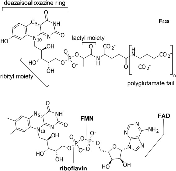
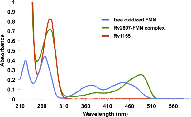
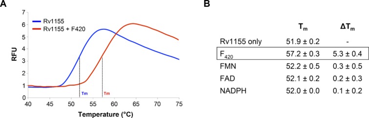
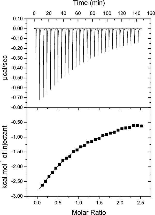
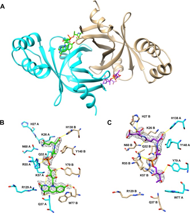
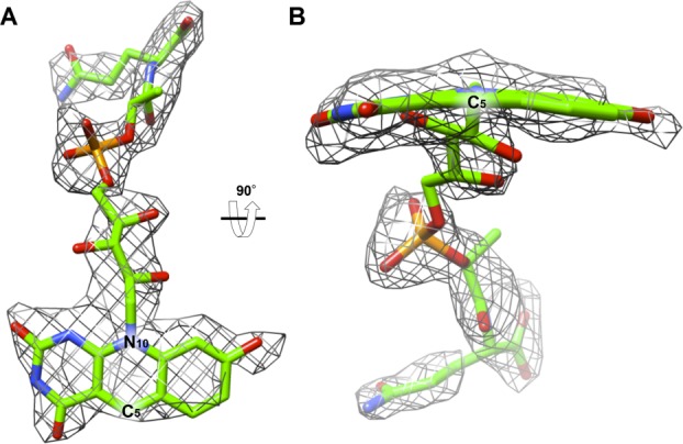
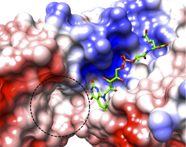
Coenzyme F420 is a deazaflavin hydride carrier with a lower reduction potential than most flavins. In Mycobacterium tuberculosis (Mtb), F420 plays an important role in activating PA-824, an antituberculosis drug currently used in clinical trials. Although F420 is important to Mtb redox metabolism, little is known about the enzymes that bind F420 and the reactions that they catalyze. We have identified a novel F420 -binding protein, Rv1155, which is annotated in the Mtb genome sequence as a putative flavin mononucleotide (FMN)-binding protein. Using biophysical techniques, we have demonstrated that instead of binding FMN or other flavins, Rv1155 binds coenzyme F420 . The crystal structure of the complex of Rv1155 and F420 reveals one F420 molecule bound to each monomer of the Rv1155 dimer. Structural, biophysical, and bioinformatic analyses of the Rv1155-F420 complex provide clues about its role in the bacterium.

Keywords: Mycobacterium tuberculosis; Rv1155; coenzyme F420; conserved hypothetical protein.


Figures

Figure 1
Figure 1
Coenzyme F420 compared to the structures of three other flavins, riboflavin, FMN (flavin mononucleotide), and FAD (flavin adenine dinucleotide). Oxidized F420 (upper) is shown. The predominant F420 isoforms observed in mycobacteria have polyglutamate tails consisting of glutamates (n = 5 or 6) that are linked by γ-glutamyl bonds., In the lower panel are shown the oxidized structures of riboflavin, FMN, and FAD.
Figure 2
Figure 2
UV–visible absorbance spectra of Rv1155 and Rv2607, members of the Mtb PNPOx family, show that Rv1155 lacks the absorbance profile characteristic of an FMN-binding protein. The UV–visible spectrum of free, oxidized FMN (blue trace) has two peaks in ∼350–500 nm region. The UV–visible spectrum of recombinant Rv2607 (green trace) has two peaks in the same part of the spectrum, which is consistent with Rv2607 being a known FMN-binding protein. Rv1155 (red trace) lacks absorbance in this region of the spectrum, indicating that it does not co-purify with a flavin when expressed recombinantly in E. coli. Free oxidized FMN, Rv2607-FMN, and Rv1155 are present at 30 μM.
Figure 3
Figure 3
Rv1155 binds coenzyme F420 as assessed by differential scanning fluorimetry (DSF). (A) DSF melt curves in the presence of F420 (red trace) and in the absence of F420 (blue trace). Melt curves for FMN, FAD, and NADPH (not shown) were almost identical to the Rv1155 curve in the absence of F420. (B) Tm values for Rv1155 in the presence of 50 μM F420, FMN, FAD, and NADPH. The change in TmTm) was calculated relative to the Rv1155 only sample. Each measurement was made in triplicate. RFU, relative fluorescence units.
Figure 4
Figure 4
Isothermal titration calorimetry reveals the binding of F420 to Rv1155. The ligand, F420 (880 μM), was titrated into a solution of the protein, Rv1155 (50 μM as monomer). The top panel shows the ITC thermogram of the raw titration data that were measured for each injection of F420. The data points in the bottom panel form the binding isotherm, in which each integrated peak area in units of kcal/mol is plotted against the ratio of the number of moles F420 that were injected to the number of moles of Rv115 present in the experiment. The solid line represents the best fit of the isotherm data to a one-binding-site model, which yielded a KD value of 170 μM for the binding of F420 to Rv1155.
Figure 5
Figure 5
Crystal structure of Rv1155 bound to F420 at 2.3 Å resolution. (A) Structure of the Rv1155 homodimer with two F420 molecules (green, purple) bound at the interface between chain A (cyan) and chain B (tan). (B) Detailed view of binding site 1. (C) Detailed view of binding site 2. Electron density for the ring system of F420 is not visible in binding site 2 (see text). The 2FoFc electron density for F420 is contoured at 1.0σ. Hydrogen bonds are represented as yellow lines.
Figure 6
Figure 6
The 2FoFc omit electron density of the F420 in the crystal structure of Rv1155 contoured at 1.0σ. (A) F420 modeled into the density found in the binding site 1 viewed from the interface of chains A and B. (B) F420 as viewed after 90° rotation around the horizontal axis relative to panel A. The density indicates that the ring assumes a butterfly conformation, meaning the ring system is not fully planar, but that the pyrimidine and benzene rings of the deazaisoalloxazine ring system are bent below the central ring, which contains C5 and N10. The butterfly angle is 16.5°, defined as the angle between the plane of the pyrimidine ring and the plane of the benzene ring. The angle was measured using Chimera.
Figure 7
Figure 7
Putative active site in Rv1155–F420. Surface representation of the Rv1155 colored by electrostatic potential (red, negative; blue, positive; white, neutral, which corresponds to areas occupied by polar or hydrophobic residues). The dashed line encircles a putative substrate-binding pocket near the F420 deazaisoalloxazine ring system, which has a volume of 130 Å3.

Similar articles

Cited by

References

    1. Koul A, Arnoult E, Lounis N, Guillemont J, Andries K. The challenge of new drug discovery for tuberculosis. Nature. 2011;469:483–490. - PubMed
    1. Raviglione M, Marais B, Floyd K, Lonnroth K, Getahun H, Migliori GB, Harries AD, Nunn P, Lienhardt C, Graham S, Chakaya J, Weyer K, Cole S, Kaufmann SH, Zumla A. Scaling up interventions to achieve global tuberculosis control: progress and new developments. Lancet. 2012;379:1902–1913. - PubMed
    1. Barry CE, III, Boshoff HI, Dowd CS. Prospects for clinical introduction of nitroimidazole antibiotics for the treatment of tuberculosis. Curr Pharm Des. 2004;10:3239–3262. - PubMed
    1. Cheeseman P, Toms-Wood A, Wolfe RS. Isolation and properties of a fluorescent compound, factor 420, from Methanobacterium strain M.o.H. J Bacteriol. 1972;112:527–531. - PMC - PubMed
    1. Bair TB, Isabelle DW, Daniels L. Structures of coenzyme F420 in Mycobacterium species. Arch Microbiol. 2001;176:37–43. - PubMed

Publication types

LinkOut - more resources