. 2015 Apr:81:81-93.
doi: 10.1016/j.yjmcc.2015.01.013. Epub 2015 Jan 30.

Mechanism of automaticity in cardiomyocytes derived from human induced pluripotent stem cells

Affiliations

Mechanism of automaticity in cardiomyocytes derived from human induced pluripotent stem cells

Jong J Kim et al. J Mol Cell Cardiol. 2015 Apr.

Abstract

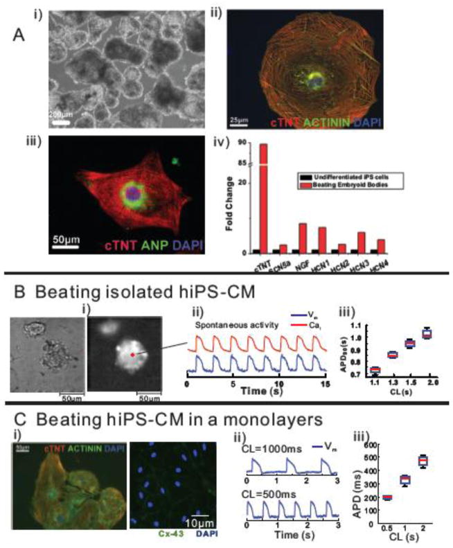
Background and objectives: The creation of cardiomyocytes derived from human induced pluripotent stem cells (hiPS-CMs) has spawned broad excitement borne out of the prospects to diagnose and treat cardiovascular diseases based on personalized medicine. A common feature of hiPS-CMs is their spontaneous contractions but the mechanism(s) remain uncertain.

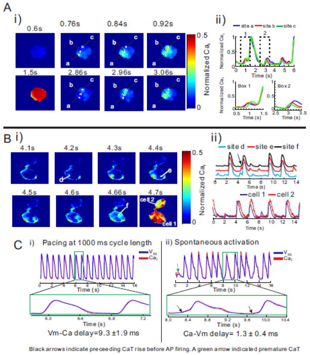
Methods: Intrinsic activity was investigated by the voltage-clamp technique, optical mapping of action potentials (APs) and intracellular Ca(2+) (Cai) transients (CaiT) at subcellular-resolution and pharmacological interventions.

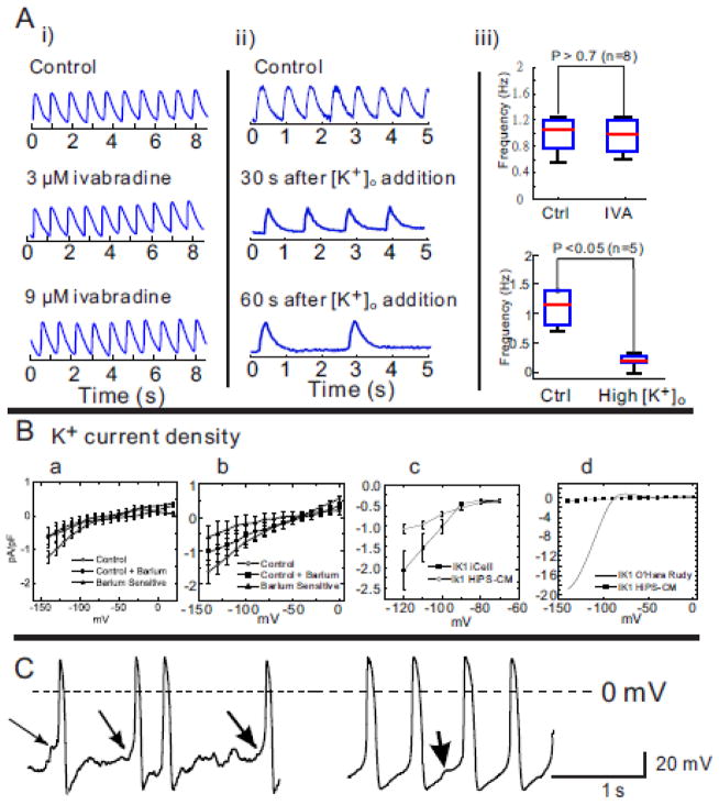
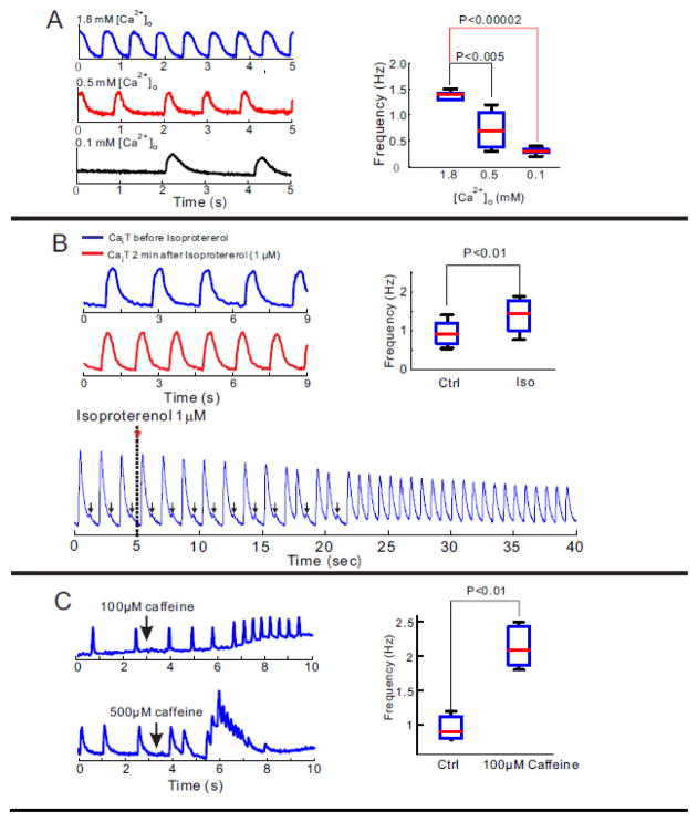
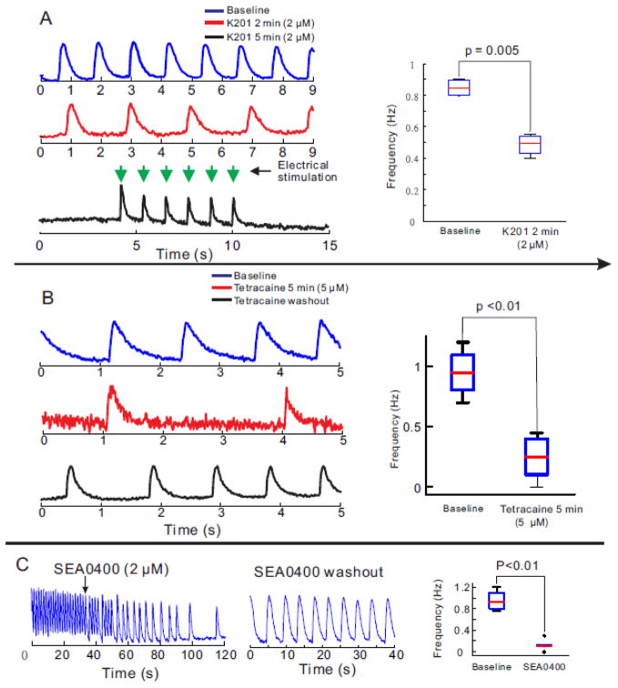
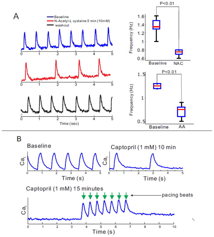
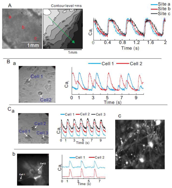
Results: The frequency of spontaneous CaiT (sCaiT) in monolayers of hiPS-CMs was not altered by ivabradine, an inhibitor of the pacemaker current, If despite high levels of HCN transcripts (1-4). HiPS-CMs had negligible If and IK1 (inwardly-rectifying K(+)-current) and a minimum diastolic potential of -59.1±3.3mV (n=18). APs upstrokes were preceded by a depolarizing-foot coincident with a rise of Cai. Subcellular Cai wavelets varied in amplitude, propagated and died-off; larger Cai-waves triggered cellular sCaTs and APs. SCaiTs increased in frequency with [Ca(2+)]out (0.05-to-1.8mM), isoproterenol (1μM) or caffeine (100μM) (n≥5, p<0.05). HiPS-CMs became quiescent with ryanodine receptor stabilizers (K201=2μM); tetracaine; Na-Ca exchange (NCX) inhibition (SEA0400=2μM); higher [K(+)]out (5→8mM), and thiol-reducing agents but could still be electrically stimulated to elicit CaiTs. Cell-cell coupling of hiPS-CM in monolayers was evident from connexin-43 expression and CaiT propagation. SCaiTs from an ensemble of dispersed hiPS-CMs were out-of-phase but became synchronous through the outgrowth of inter-connecting microtubules.

Conclusions: Automaticity in hiPS-CMs originates from a Ca(2+)-clock mechanism involving Ca(2+) cycling across the sarcoplasmic reticulum linked to NCX to trigger APs.

Keywords: Cell–cell coupling; Funny current; Human myocytes from stem cells; Optical mapping of calcium and action potentials; Spontaneous activity; Subcellular calcium waves.


Conflict of interest statement

The authors have no conflicts to declare

Figures

Figure 1
Figure 1. Characterization of iPS-derived cardiomyocytes
A: Immuno-staining of beating embryoid bodies of hiPS-CMs. i) a bright-field image, ii) immune-staining of CTNT (red), Actinin (green) and a nuclear stain, DAPI (blue), iii) immuno-staining of cardiac troponin T (CTNT, red), atrial natriuretic protein (ANP, green), a marker for late stage atrial or fetal CMs, and DAPI (blue) and iv) quantitative real time PCR using RNA isolated from undifferentiated hiPS cells and beating embryonic bodies of cardiac troponin T (cTNT), nerve growth factor (NGF) and the hyperpolarization activated cycling nucleotide gated channels (HCN 1 and 4) that underlie the pacemaker ‘funny’ current. Experiments were performed in triplicates. B: Simultaneous measurement of intracellular Ca2+ (Cai) transients and Vm changes in embryoid bodies of hiPS-CMs. i) a bright-field (left) and a fluorescent image of PGH1 stained spontaneously beating embryoid bodies of hiPS-CMs, ii) simultaneous optical recording of Cai transients and membrane potential (Vm) changes at a marked pixel (pixel resolution=1.5×1.5μm), and iii) the relationship between action potential duration (APD) and cycle length (CL). C: Immunolabeling and optical measurement of Vm changes in a spontaneously beating hiPS-CM monolayer. i) immunostaining of CTNT (red), actinin (green) and a nuclear stain, DAPI (blue) (left) and connexins 43 (Cx-43,green) and a nuclear stain, DAPI (blue), ii) optical measurement of Vm changes, and iii) the relationship between APD and CL.
Figure 2
Figure 2. Pacemaker activity in dispersed hiPS-CMs
A: i) Ivabradine, an inhibitor of If, had no effect on spontaneous activity at concentrations of 3 and 9 μM even though the hiPS-CMs activity slowed down by increasing external [K+]o from 4 to 8 mM. ii) An increase in extracellular K+ from 4–8 mM decreased the rate of spontaneous activity in embryoid bodies (n=5/5). iii) Statistical comparison of spontaneous contraction in hiPS-CMs. B: Early and late currents estimate a very small If and IK1. Panel a: Current-to-voltage (I–V) plot of barium sensitive, insensitive and difference currents at the end of a 2000 ms pulse. Each data point represent the average of the current recorded in going from a holding potential of −40 mV to test potentials between −140 to 0 mV in steps of 10 mV. The cells were then treated with Ba2+ (1 mM) and the Ba2+ sensitive current was taken as the difference between the control and the Ba2+ treated current; n=12 hiPS-CMs (10 ventricular and 2 atrial). Panel b shows data from the same cells for peak inward current. Panel c: Current-to-voltage relationship of background currents surrounding EK for comparing relative magnitude of IK1 currents in iCell (Cellular Dynamics International) prepared hiPS-CMs versus scale preparation of hiPS-CMs in normal Tyrode’s solution. Each data point represents the peak current recorded from −120 mV to −60 mV from a holding potential of −90 mV (n=19 and n=10 respectively). Panel d: Barium sensitive (early, from panel b, IK1) currents in hiPS-CMs were compared to IK1 currents calculated for human ventricular myocytes. The early barium sensitive current density per cell in panel B was plotted against the O’Hara Rudy model [44] for IK1. Current density was normalized to cell capacitance and expressed in pA/pF. C: Two examples of pacemaking activity in hiPS-CMs measured using whole cell recordings in the current-clamp mode. The first hiPS-CM (left) showed the typical “flat” period prior to initiation of an AP, with a preceding abrupt depolarization typical of a spontaneous Ca2+-release event or a delayed afterdepolarization, DAD (arrows), the second (right) occurred less frequently and showed the appearance of pacemaker activity, even in such runs, spontaneous “DAD” behavior was noted (arrow). In this group, there were 11 ventricular, 4 atrial and 3 other cell type.
Figure 3
Figure 3. Subcellular Cai waves and spontaneous contractions in isolated hiPS-CMs
A: i) Maps of normalized Cai taken at different time-points reveal a sequence of subcellular Ca2+ release and local wave propagation occurring during diastole. The first subcellular wave was initiated at at site a (at 0.76s) and propagated to site b (0.84s) then c (0.92s) (see top panels) then a large synchronous SR Ca2+ release filled the cell at 1.5s. The next subcellular Cai elevation started near site c (2.86s), propagated c to b (2.96s) then to a (3.06s). Field of view was 50 × 50 μm2. For GCaMP2 fluorescence, λexcitation was 480±20 nm and λemission was 510±15 nm. ii) Shows the time course of fluorescence traces of Cai recorded from sites a, b, and c at slow (top traces) and fast (bottom traces) sweep speeds. The superposition of Cai signals from sites a, b and c reveal the time-course and relative amplitude of subcellular waves. The small subthreshold local Cai elevations were typically 1/4 of the full amplitude of cellular CaTs. At fast sweep speed (Box 1), the subcellular Cai wave started at site ‘a’ and propagated to ‘b’ then ‘c’ before eliciting a CaT that produced a global Cai elevation throughout the cell (A, panel at 1500 ms). The next two sequences of subcellular Cai elevations started at site c (Box 2) and propagated to b then a; the sequence was repeated twice followed by a full size CaT. B: i) Example of subcellular small amplitude Cai release that can dissipate and self-terminate or amplify to precede a global Cai release. A local Cai wave initiated at site ‘e’ propagated but self-terminated without producing a full size Cai transient. In contrast, a Cai wave initiated at site ‘f’ propagated, was amplified and preceded a global Cai release. Field-of-view was 75 × 75 μm2. ii) Optical traces of Cai recorded at site d, e, and f (top) and in two neighboring cells (bottom). Premature CaTs were unsynchronized but global CaTs were synchronized in these coupled cells with ~100ms time delays. C: Temporal delay between AP and Cai upstrokes. An ensemble of dispersed hiPS-CMs, optical APs and CaTs were simultaneously recorded. In (i), the cells were paced at 1 s cycle length resulting in the expected Vm to Cai coupling where Vm preceded Cai by 9.3±1.9 ms. In contrast when the dispersed cells were not paced but exhibited spontaneous automaticity, Cai preceded Vm by 1±0.4 ms, (n=5 preparations of dispersed cells).
Figure 4
Figure 4. Modification of automaticity in hiPS-CMs by [Ca2+]o titration and changes in RyR2 open probability
A: Frequency of intrinsic contractions is [Ca2+]o dependent. The rate of spontaneous contractions in a cluster of hiPS-CMs is [Ca2+]o dependent. Raising or lowering [Ca2+]o from 0.5 mM, significantly increased or decreased the frequency of spontaneous contractions, respectively. Statistical analysis of the frequency of Cai transients demonstrated a tight correlation between frequency and [Ca2+]o (right panel) B: Chronotropic response of hiPS-CMs to β adrenergic stimulation. Infusion of 1μM of isoproterenol significantly increases in frequency of spontaneous contraction in a cluster of hiPS-CMs (left traces). Statistical analysis of frequency of contractions for 5 preparations of iPS-CMs. Premature CaTs were gradually developed to full transients under 1μM of isoproterenol administration. C: Frequency of spontaneous contraction is caffeine sensitivity. After administration of 100 μM caffeine, spontaneous contraction of hiPS-CMs was gradually increased (left top panel). 500μM of caffeine infusion emptied SR and stopped spontaneous contraction of hiPS-CMs (left bottom panel). Statistical comparison of frequency of spontaneous contraction before and after 100 μM caffeine infusion
Figure 5
Figure 5. Manipulations of RyR2, and NCX and its efficacy on spontaneous contraction in hiPS-CMs
A: K201 (2 μM), a selective RyR2 stabilizer gradually decreased the frequency of spontaneous contraction in a cluster of hiPS-CMs and eventually terminated spontaneous contraction (n=5/5). HiPS-CMs remained excitable by electrical stimulation. After 2 min of K201 exposure, a ~2-fold decrease in frequency of automaticity was statistically significant (right panel) B: Similar effects of 5μM tetracaine infusion on spontaneous contraction in hiPS-CMs C: SEA0400 (2 μM), an inhibitor of the forward mode of NCX suppressed spontaneous contractions in a cluster of iPS-CMs; an effect that was reversible by washing out SEA0400 (right trace) (n=4/4).
Figure 6
Figure 6. Modification of automaticity in hiPS-CMs by RyR2 redox state
A: Anti-oxidant agents, N-Acetyl-L-Cysteine (NALC) and ascorbic acid (AA) significantly reduced automaticity of hiPS-CMs (n=10). B: The membrane permeable thiol reducing agent, captopril (1 mM) slowed-down the rate of spontaneous contractions in 10 min (top trace) and stopped automaticity within 15 min (n=3/6). Contractions of iPS-CMs rendered quiescent by captopril were triggered by electrical stimuli (arrows).
Figure 7
Figure 7. Intercellular communication
A: Propagation of spontaneous contraction in a dense cluster of hiPS-CMs. A left panel is a picture of a cluster. A middle panel is an activation map. A green arrow indicates the direction of propagation. A right panel shows superimposed calcium traces at site a, b, and c. B: Unsynchronized and irregular contraction in remote hiPS-CMs. A picture of sparsely distributed hiPS-CMs (left panel). Superimposed calcium traces from cells 1 and 2 (right panel). C: Intercellular synchronization of contraction between a cluster and satellite hiPS-CMs connected to each other via microfiber. Panel a) shows a picture of remote hiPS-CMs (left panel) and superimposed Cai traces of cells 1, 2 and 3 (right traces). Another example of synchronization is shown through a fluorescence image of membrane-bound voltage-sensitive dye (PGH1) (panel b) and Cai traces of a cluster and a satellite hiPS-CMs. A high-resolution fluorescence image of a cluster of hiPS-CMs using a membrane-bound dye reveals a complex network of microfibers that underlie intercellular coupling among CMs that are far apart (30–50 μm) (panel c). All panels have dimensions of 150 μm ×150 μm.

Similar articles

Cited by

References

    1. Thomson JA, Itskovitz-Eldor J, Shapiro SS, Waknitz MA, Swiergiel JJ, Marshall VS, et al. Embryonic stem cell lines derived from human blastocysts. Science. 1998;282:1145–7. - PubMed
    1. Reubinoff BE, Pera MF, Fong CY, Trounson A, Bongso A. Embryonic stem cell lines from human blastocysts: somatic differentiation in vitro. Nat Biotechnol. 2000;18:399–404. - PubMed
    1. Kehat I, Kenyagin-Karsenti D, Snir M, Segev H, Amit M, Gepstein A, et al. Human embryonic stem cells can differentiate into myocytes with structural and functional properties of cardiomyocytes. J Clin Invest. 2001;108:407–14. - PMC - PubMed
    1. Itskovitz-Eldor J, Schuldiner M, Karsenti D, Eden A, Yanuka O, Amit M, et al. Differentiation of human embryonic stem cells into embryoid bodies compromising the three embryonic germ layers. Mol Med. 2000;6:88–95. - PMC - PubMed
    1. Dolnikov K, Shilkrut M, Zeevi-Levin N, Gerecht-Nir S, Amit M, Danon A, et al. Functional properties of human embryonic stem cell-derived cardiomyocytes: intracellular Ca2+ handling and the role of sarcoplasmic reticulum in the contraction. Stem Cells. 2006;24:236–45. - PubMed

Publication types

MeSH terms