Review
. 2015 Feb 1;29(3):225-37.
doi: 10.1101/gad.255968.114.

Lamins at the crossroads of mechanosignaling

Affiliations
Review

Lamins at the crossroads of mechanosignaling

Selma Osmanagic-Myers et al. Genes Dev. .

Abstract

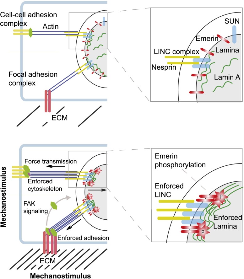
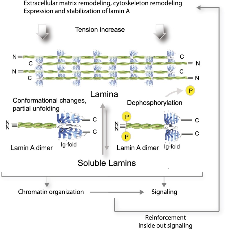
The intermediate filament proteins, A- and B-type lamins, form the nuclear lamina scaffold adjacent to the inner nuclear membrane. B-type lamins confer elasticity, while A-type lamins lend viscosity and stiffness to nuclei. Lamins also contribute to chromatin regulation and various signaling pathways affecting gene expression. The mechanical roles of lamins and their functions in gene regulation are often viewed as independent activities, but recent findings suggest a highly cross-linked and interdependent regulation of these different functions, particularly in mechanosignaling. In this newly emerging concept, lamins act as a "mechanostat" that senses forces from outside and responds to tension by reinforcing the cytoskeleton and the extracellular matrix. A-type lamins, emerin, and the linker of the nucleoskeleton and cytoskeleton (LINC) complex directly transmit forces from the extracellular matrix into the nucleus. These mechanical forces lead to changes in the molecular structure, modification, and assembly state of A-type lamins. This in turn activates a tension-induced "inside-out signaling" through which the nucleus feeds back to the cytoskeleton and the extracellular matrix to balance outside and inside forces. These functions regulate differentiation and may be impaired in lamin-linked diseases, leading to cellular phenotypes, particularly in mechanical load-bearing tissues.

Keywords: LINC complex; cytoskeleton; extracellular matrix; lamins; mechanosensing; mechanotransduction.


Figures

Figure 1.
Figure 1.
The route of mechanosensing and the tension-induced reinforcement response. (Top panels) Cell–cell adhesion and FA complexes that sense tension are physically linked to the nucleus via the cytoskeleton, LINC complexes (SUN and KASH domain proteins) in the nuclear membrane, and the nuclear lamina. Tension forces from the ECM are transmitted into the nucleus via these components and affect mechanoresponsive gene expression. (Bottom panels) In response to a mechanostimulus, such as increase in ECM stiffness, adhesion complexes, the actin cytoskeleton, LINC complexes, and the lamina are reinforced by the assembly of actin filaments, increased recruitment of adhesion complex and LINC complex proteins, and stabilization and assembly of A-type lamins at the lamina, thereby counteracting forces exerted from outside. In addition, the INM protein emerin becomes phosphorylated and contributes to LINC complex reinforcement. Tension also induces the activation of signaling cascades on adhesion complexes, such as FAK signaling, which affects mechanoresponsive gene expression without direct force transmission into the nucleus. Panels at the right depict higher-magnification views of the boxed areas in the nucleus shown in the left panels.
Figure 2.
Figure 2.
Shear stress force induces rearrangement of the cytoskeleton and cell alignment. Lung endothelial cells were exposed to flow shear stress (12 dyn/cm2) for 3 h and processed for immunofluorescence microscopy using actin and VE-cadherin antibodies. Hoechst was used for DNA staining. Shear stress induces increased alignment of actin stress fibers and elongation of nuclei (arrows) in the flow direction. Note “reinforcement” of cell–cell junctions particularly at the posterior and anterior ends of polarized cells as revealed by increased accumulation and “zipper-like” morphology of VE-cadherin junctions and increased cortical actin at these sites (arrowheads). Bar, 20 μm.
Figure 3.
Figure 3.
Lamin A in mechanosignaling. Increasing tension causes partial unfolding and dephosphorylation of lamin A as well as assembly of soluble lamin A into the lamina. These structural and biochemical changes of the protein may affect chromatin organization and cell signaling, thereby activating the reinforcement response, including increased lamin A expression and remodeling of the cytoskeleton and the ECM.

References

    1. Aebi U, Cohn J, Buhle L, Gerace L. 1986. The nuclear lamina is a meshwork of intermediate-type filaments. Nature 323: 560–564. - PubMed
    1. Andres V, Gonzalez JM. 2009. Role of A-type lamins in signaling, transcription, and chromatin organization. J Cell Biol 187: 945–957. - PMC - PubMed
    1. Bank EM, Ben-Harush K, Feinstein N, Medalia O, Gruenbaum Y. 2012. Structural and physiological phenotypes of disease-linked lamin mutations in C. elegans. J Struct Biol 177: 106–112. - PubMed
    1. Barrowman J, Hamblet C, George CM, Michaelis S. 2008. Analysis of prelamin A biogenesis reveals the nucleus to be a CaaX processing compartment. Mol Biol Cell 19: 5398–5408. - PMC - PubMed
    1. Beavan LA, Quentin-Hoffmann E, Schonherr E, Snigula F, Leroy JG, Kresse H. 1993. Deficient expression of decorin in infantile progeroid patients. J Biol Chem 268: 9856–9862. - PubMed

Publication types

LinkOut - more resources