

Dysbiosis gut microbiota associated with inflammation and impaired mucosal immune function in intestine of humans with non-alcoholic fatty liver disease
- PMID: 25644696
- PMCID: PMC4314632
- DOI: 10.1038/srep08096
Dysbiosis gut microbiota associated with inflammation and impaired mucosal immune function in intestine of humans with non-alcoholic fatty liver disease
Abstract
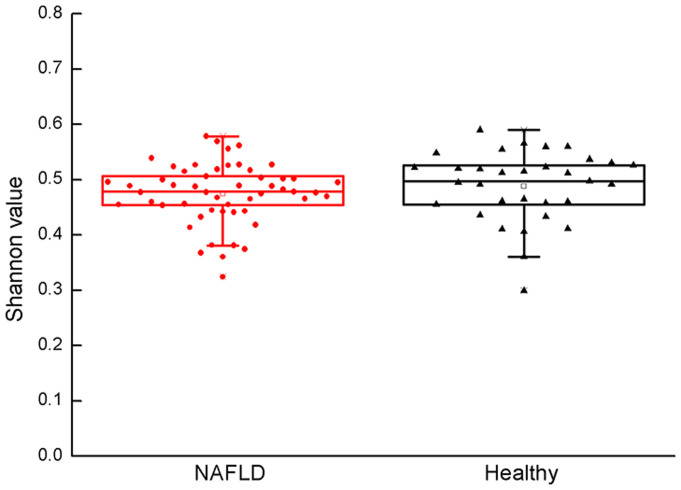
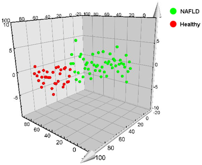
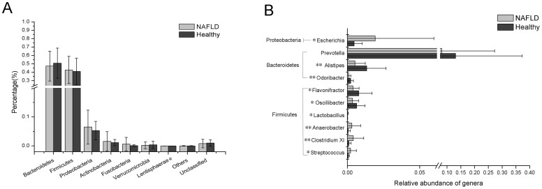
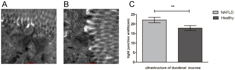
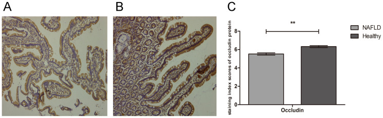
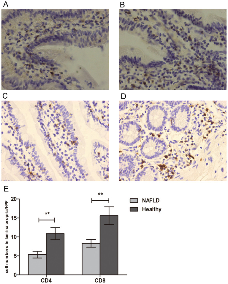
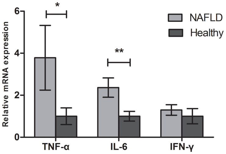
Non-alcoholic fatty liver disease (NAFLD) has recently been considered to be under the influence of the gut microbiota, which might exert toxic effects on the human host after intestinal absorption and delivery to the liver via the portal vein. In this study, the composition of the gut microbiota in NAFLD patients and healthy subjects was determined via 16S ribosomal RNA Illumina next-generation sequencing. Among those taxa displaying greater than 0.1% average abundance in all samples, five genera, including Alistipes and Prevotella, were significantly more abundant in the gut microbiota of healthy subjects compared to NAFLD patients. Alternatively, Escherichia, Anaerobacter, Lactobacillus and Streptococcus were increased in the gut microbiota of NAFLD patients compared to healthy subjects. In addition, decreased numbers of CD4+ and CD8+ T lymphocytes and increased levels of TNF-α, IL-6 and IFN-γ were detected in the NAFLD group compared to the healthy group. Furthermore, irregularly arranged microvilli and widened tight junctions were observed in the gut mucosa of the NAFLD patients via transmission electron microscopy. We postulate that aside from dysbiosis of the gut microbiota, gut microbiota-mediated inflammation of the intestinal mucosa and the related impairment in mucosal immune function play an important role in the pathogenesis of NAFLD.
Figures
References
-
- Torres D. M., Williams C. D. & Harrison S. A. Features, diagnosis, and treatment of nonalcoholic fatty liver disease. Clin Gastroenterol Hepatol 10, 837–858 (2012). - PubMed
-
- Tilg H. & Moschen A. R. Evolution of inflammation in nonalcoholic fatty liver disease: the multiple parallel hits hypothesis. Hepatology 52, 1836–1846 (2010). - PubMed
-
- Abu-Shanab A. & Quigley E. M. The role of the gut microbiota in nonalcoholic fatty liver disease. Nat Rev Gastroenterol Hepatol 7, 691–701 (2010). - PubMed
-
- Moschen A. R., Kaser S. & Tilg H. Non-alcoholic steatohepatitis: a microbiota-driven disease. Trends Endocrinol Metab 24, 537–545 (2013). - PubMed
-
- Turnbaugh P. J. et al. An obesity-associated gut microbiome with increased capacity for energy harvest. Nature 444, 1027–1031 (2006). - PubMed
Publication types
MeSH terms
Substances
Associated data
LinkOut - more resources
Full Text Sources
Other Literature Sources
Medical
Research Materials
