. 2015 Apr;199(4):1159-72.
doi: 10.1534/genetics.114.173963. Epub 2015 Feb 2.

An evolutionarily conserved switch in response to GABA affects development and behavior of the locomotor circuit of Caenorhabditis elegans

Affiliations

An evolutionarily conserved switch in response to GABA affects development and behavior of the locomotor circuit of Caenorhabditis elegans

Bingjie Han et al. Genetics. 2015 Apr.

Abstract

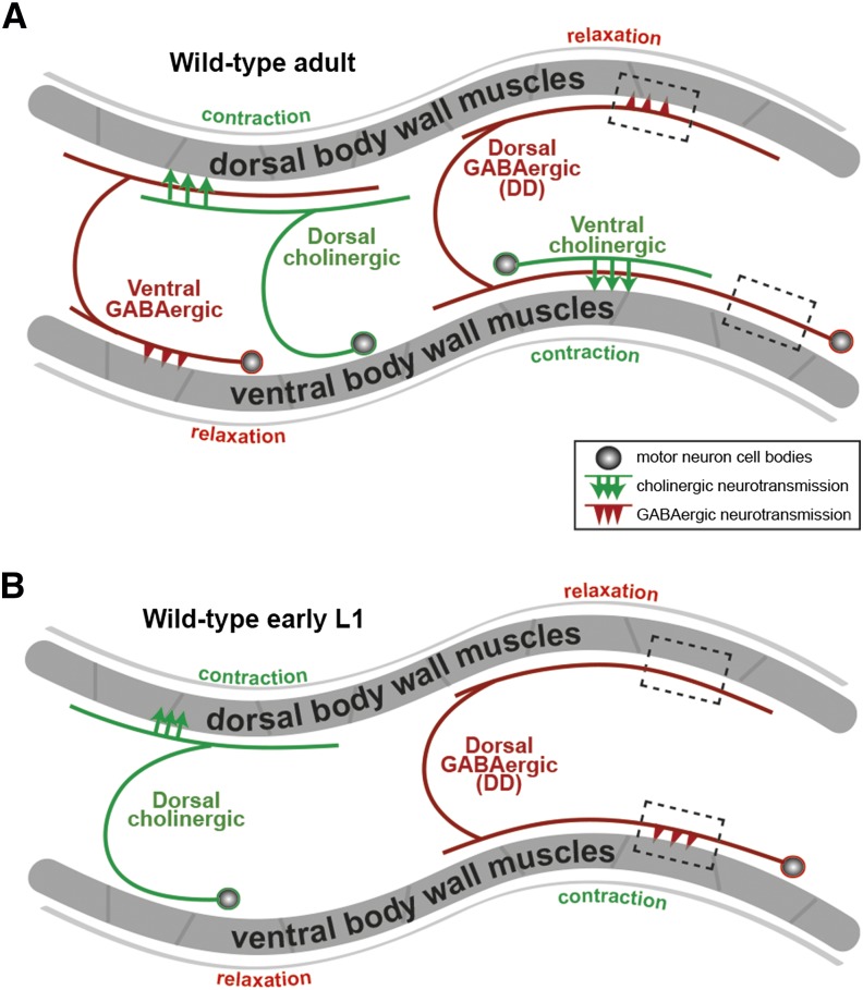
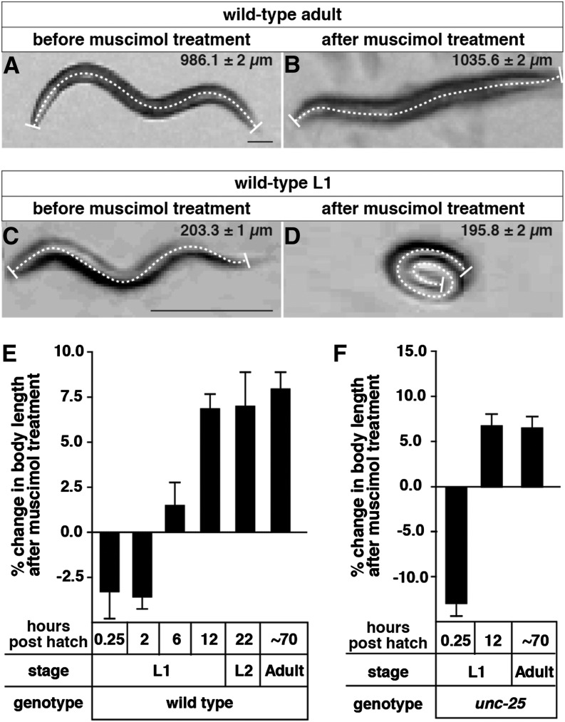
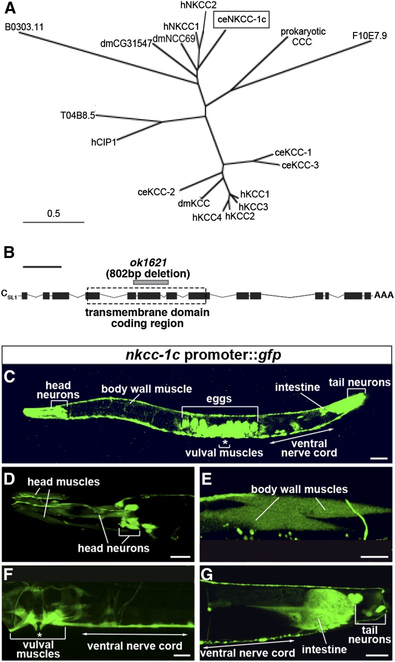
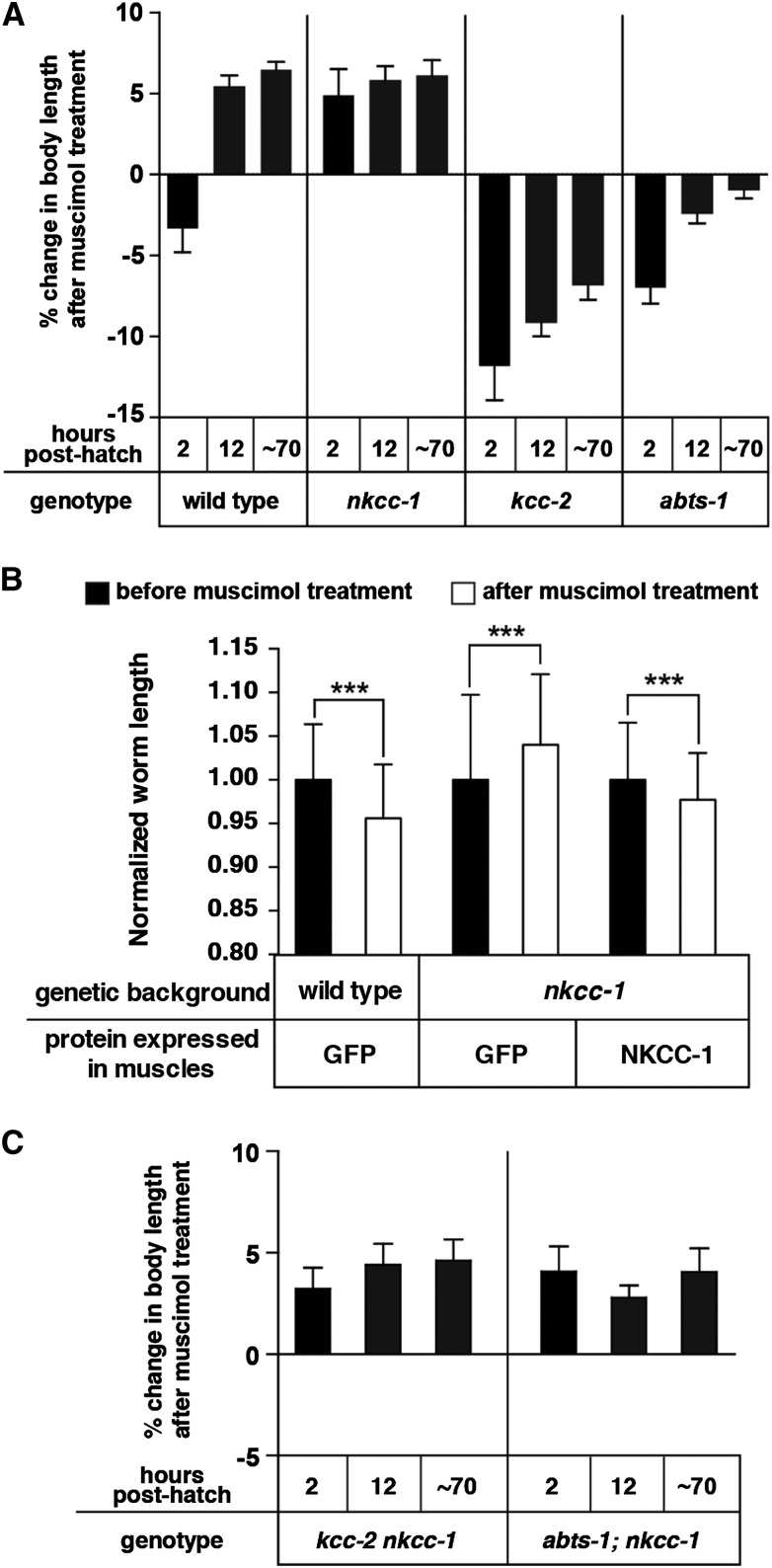
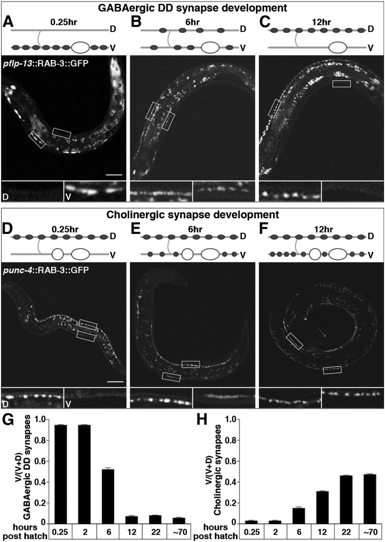
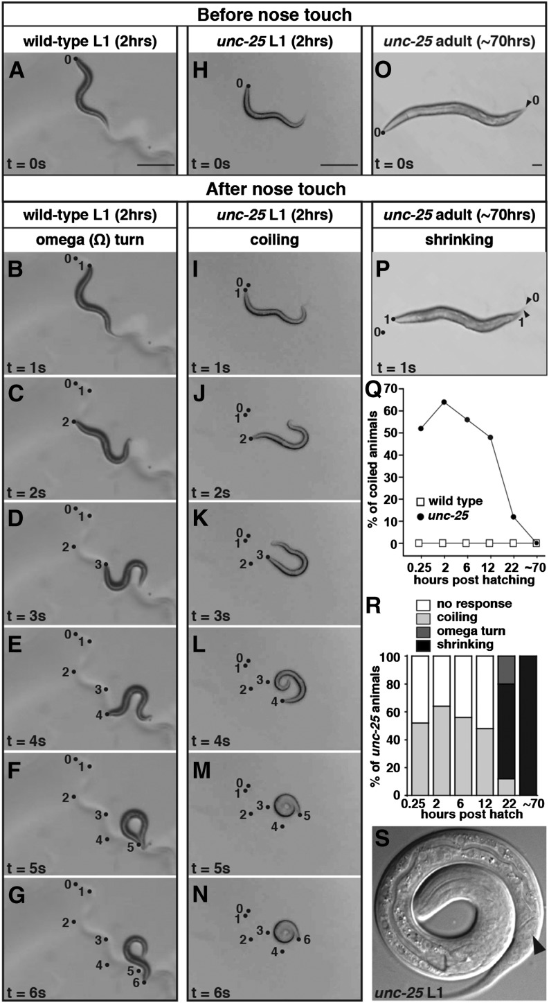
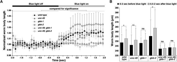
The neurotransmitter gamma-aminobutyric acid (GABA) is depolarizing in the developing vertebrate brain, but in older animals switches to hyperpolarizing and becomes the major inhibitory neurotransmitter in adults. We discovered a similar developmental switch in GABA response in Caenorhabditis elegans and have genetically analyzed its mechanism and function in a well-defined circuit. Worm GABA neurons innervate body wall muscles to control locomotion. Activation of GABAA receptors with their agonist muscimol in newly hatched first larval (L1) stage animals excites muscle contraction and thus is depolarizing. At the mid-L1 stage, as the GABAergic neurons rewire onto their mature muscle targets, muscimol shifts to relaxing muscles and thus has switched to hyperpolarizing. This muscimol response switch depends on chloride transporters in the muscles analogous to those that control GABA response in mammalian neurons: the chloride accumulator sodium-potassium-chloride-cotransporter-1 (NKCC-1) is required for the early depolarizing muscimol response, while the two chloride extruders potassium-chloride-cotransporter-2 (KCC-2) and anion-bicarbonate-transporter-1 (ABTS-1) are required for the later hyperpolarizing response. Using mutations that disrupt GABA signaling, we found that neural circuit development still proceeds to completion but with an ∼6-hr delay. Using optogenetic activation of GABAergic neurons, we found that endogenous GABAA signaling in early L1 animals, although presumably depolarizing, does not cause an excitatory response. Thus a developmental depolarizing-to-hyperpolarizing shift is an ancient conserved feature of GABA signaling, but existing theories for why this shift occurs appear inadequate to explain its function upon rigorous genetic analysis of a well-defined neural circuit.

Keywords: GABA response; inhibitory; signaling; switch.


Figures

Figure 1
Figure 1
The anatomy and development of C. elegans locomotor circuit. Diagrams of neuronal wiring in the locomotor circuit of C. elegans adults (A) or newly hatched first-stage larvae (L1s) (B). The anterior of the animals is to the left. Circles, motor neuron cell bodies; lines extending from circles, neural processes; arrows, acetylcholine release sites; arrowheads, GABA release sites; black dashed boxes, representative regions of DD (dorsal GABAergic) neuron processes adjacent to dorsal and ventral body wall muscles. Each neuron diagrammed represents a class of motor neurons that repeats along the length of the animal. In adults (A), ventral cholinergic neurons release acetylcholine onto the ventral body wall muscles to excite them and produce ventral body bends. Dorsal cholinergic neurons excite dorsal muscles to produce dorsal bends. GABAergic neurons assist body bends by releasing GABA to relax muscles opposite a contraction. Type DD motor neurons relax dorsal muscles, and ventral GABAergic motor neurons relax ventral muscles. In newly hatched L1s (B), however, neuronal processes from neither the ventral cholinergic nor ventral GABAergic motor neurons have developed yet. Instead, the dorsal GABAergic DD neurons make temporary synapses onto the ventral body wall muscles. As ventral motor neurons develop, DD neurons rewire to eliminate ventral synapses and form new dorsal synapses. Diagram adapted from WormAtlas.org.
Figure 2
Figure 2
Muscle response to a GABAA receptor agonist switches from excitatory to inhibitory during C. elegans development. (A and B) Images of a representative wild-type adult in the absence (A) or presence (B) of 1 mM muscimol. (C and D) Images of a representative wild-type L1 at 0.25 hr posthatch in the absence (C) or presence (D) of 1 mM muscimol. A dashed line was drawn down the midline of the worm in each image to measure its body length. The average lengths and standard deviations from 30 animals thus measured are shown at upper right of each image. Bar, 100 µm. (E) Percent changes in body length of wild-type animals in response to 1 mM muscimol throughout development. Muscimol response switches from excitatory to inhibitory at ∼6 hr posthatch. (F) The muscimol response switch was still observed in mutant animals lacking the GABA biosynthetic enzyme UNC-25. n = 30 animals per genotype per time point. Error bars represent 95% confidence intervals calculated using a paired t-test.
Figure 3
Figure 3
NKCC-1 is expressed predominantly in muscles and neurons. (A) Phylogenetic comparison of the C. elegans sodium potassium chloride cotransporter (ceNKCC-1; boxed) to the two human NKCCs (hNKCC-1 and hNKCC-2), two predicted Drosophila melanogaster NKCCs (dmCG31547 and dmNCC69), four C. elegans potassium chloride cotransporters (KCCs; ceKCC-1–ceKCC-4), four human KCCs (hKCC-1–hKCC-4), a human chloride transporter interacting protein (hCIP1), and a predicted C. elegans chloride transporter interacting protein (T04B8.5). A prokaryotic cation chloride cotransporter (CCC) is used to root the comparison. Bar, 0.5 substitutions per 100 amino acids. (B) Structure of the nkcc-1c transcript. Solid black boxes, exons; connecting lines, introns; AAA, the polyadenylation site; dashed box, the predicted transmembrane domain coding region; gray bar, the ok1621 deletion mutation. Bar (black line above the schematic), 1 kb. (C–G) GFP fluorescence in an animal carrying a nkcc-1c promoter::gfp::nkcc-1c 3′ UTR reporter transgene. This reporter transgene was expressed primarily in the body wall muscles (E) and neurons, including the head and tail neurons (D), and the ventral cord neurons (F and G). It was also expressed in the vulval muscles (F) and the posterior intestine (G). Unlaid eggs inside the adults showed high expression (C). Bar, 50 μm. Eggs visible in C are out of the focal plane and thus not visible in F. Body wall muscles are poorly visualized in C but become apparent at the higher magnification in E.
Figure 4
Figure 4
NKCC-1 promotes excitatory signaling by the GABAA agonist muscimol onto body wall muscles. (A) Percent change in body length of the wild type and of Cl transporter mutants after 1 mM muscimol exposure. Pairwise measurements were normalized to the body length of each animal prior to muscimol exposure. n = 30 animals. (B) Rescue of the muscimol response defect of early L1 nkcc-1 mutants by reexpression of nkcc-1 in body wall muscles. Average normalized worm length of transgenic newly hatched L1 animals before and after 1 mM muscimol exposure are shown. A muscle-specific promoter was used to transgenically express cDNAs encoding the control protein GFP or NKCC-1 isoform C. n = 50 animals, ***P = 0.0005. (C) nkcc-1 is epistatic to kcc-2 and abts-1 in the muscimol response assay. Each measurement was normalized to body length of the same animal prior to muscimol exposure. n = 30 animals. Error bars represent 95% confidence intervals.
Figure 5
Figure 5
Presynaptic motor neuron development coincides with the postsynaptic switch in muscle response to muscimol. (A–C) Rewiring of DD presynaptic termini from ventral to dorsal in L1 animals. Wild-type animals expressing GFP::RAB-3 in GABAergic DD motor neuron presynaptic termini under the flp-13 promoter at 0.25 (A), 6 (B), and 12 (C) hr posthatch. (D–F) Development of ventral cholinergic presynaptic termini in L1 animals. Wild-type animals expressing GFP::RAB-3 in cholinergic motor neuron presynaptic termini under the unc-4 promoter at 0.25 (D), 6 (E), and 12 (F) hr posthatch. Cartoons depict a representative DD motor neuron (A–C) or ventral cholinergic motor neuron (D–F) and where their synapses are at each time point. Shaded lines, nerve cords and commissures; “D” and “V”, dorsal, ventral; open ovals, DD motor neuron cell bodies; solid ovals, synapses made from DDs; open circles, ventral cholinergic motor neuron cell bodies; solid circles, synapses made from ventral cholinergic motor neurons. All animals are oriented with head to the left and ventral down. Open boxes outline representative regions of the dorsal and ventral nerve cord that are shown in magnified versions below each image and labeled “D” and “V” for dorsal and ventral in A and D. Magnified images in B, C, E, and F similarly show the dorsal box on the left and the ventral box on the right. Bar, 20 µm. (G and H) Quantification of the proportion of ventral GFP::RAB-3 in DD presynaptic termini (G) or in cholinergic presynaptic termini (H). n = 20 animals.
Figure 6
Figure 6
Endogenous GABA and GABAA receptors relax ventral muscles of early L1 larvae during backing. (A–N) Snapshots of a representative wild type (A–G) or an unc-25 mutant (H–N) L1 at 2 hr posthatch on unseeded NGM plates taken prior to nose touch (t = 0) and every second for 6 sec after nose touch (t = 1 to t = 6). The wild-type L1 responded with backing (B–E), an omega turn (F), and then moving forward again in the direction opposite to that prior to touch (G). The unc-25 L1 curled its tail toward its body (I–K) and eventually coiled its entire body up into a spiral (L and M). This spiral state lasted longer than 1 sec (N). Dots, positions of the tip of the animal’s nose at the corresponding time following nose touch, in seconds, specified by the numbers next to them. The dot labeled 0 represents the nose position one frame before touch. (O and P) Snapshots of an unc-25 adult on an unseeded NGM plate either before (O) or after (P) nose touch. Dots and arrowheads, positions of the tips of the nose and tail, respectively, before nose touch (“0”) and 1 sec after touch (“1”). Contraction of body wall muscles caused the animal to shorten in this response known as “shrinking.” (Q) Quantification of the percentage of animals that coil in response to nose touch as wild-type or unc-25 animals develop. (R) The percentage of unc-25 animals in each category of touch-response behavior during development. The criteria for categorization are described in Materials and Methods. (S) A representative magnified image of a coiled-up unc-25 L1 at 2 hr posthatch. The solid arrowhead points to the ventrally directed opening of the anus, which shows that the animal is coiled with its ventral side in.
Figure 7
Figure 7
Optogenetic activation of GABA release confirms that endogenous GABAA signaling is inhibitory in early L1s. (A) Blue-light-induced change in body length of early L1s expressing channel rhodopsin 2 in GABAergic motor neurons. WormLab software was used to track each animal’s body length in every frame of a 30-frame per second movie recorded for 2 sec before and 2 sec after blue light was applied. For each animal, measurements were normalized to the average body length before blue light was on. Data points plotted represent the average of these normalized length measurements collected for each of 10 animals per genotype over each 0.1-sec interval. Solid and open bars across the top of the graph represent the time periods before and after blue light was on, respectively. Error bars represent SEM. (B) Quantification of the average body lengths measured for animals of each genotype both over intervals 2 sec before and 0.5–2 sec after blue light was on. Statistical results of paired t-tests are as follows: wild type, *P = 0.0137; unc-49, **P = 0.0098; gbb-1, P = 0.1055 (ns, not significant); gbb-2, *P = 0.0195; unc-49; gbb-1, P = 0.0840 (ns); and unc-49; gbb-2, P > 0.9999 (ns). Error bars represent SEM. All animals assayed in this experiment were 2 hr posthatch.

Similar articles

Cited by

References

    1. Akerman C. J., Cline H. T., 2006. Depolarizing GABAergic conductances regulate the balance of excitation to inhibition in the developing retinotectal circuit in vivo. J. Neurosci. 26: 5117–5130. - PMC - PubMed
    1. Bamber B. A., Beg A. A., Twyman R. E., Jorgensen E. M., 1999. The Caenorhabditis elegans unc-49 locus encodes multiple subunits of a heteromultimeric GABA receptor. J. Neurosci. 19: 5348–5359. - PMC - PubMed
    1. Beg A. A., Jorgensen E. M., 2003. EXP-1 is an excitatory GABA-gated cation channel. Nat. Neurosci. 6: 1145–1152. - PubMed
    1. Bellemer A., Hirata T., Romero M. F., Koelle M. R., 2011. Two types of chloride transporters are required for GABA(A) receptor-mediated inhibition in C. elegans. EMBO J. 30: 1852–1863. - PMC - PubMed
    1. Ben-Ari Y., Cherubini E., Corradetti R., Gaiarsa J. L., 1989. Giant synaptic potentials in immature rat CA3 hippocampal neurones. J. Physiol. 416: 303–325. - PMC - PubMed

Publication types

MeSH terms