. 2015 Jun;236(2):210-8.
doi: 10.1002/path.4510. Epub 2015 Mar 3.

Cell cycle deregulation and mosaic loss of Ext1 drive peripheral chondrosarcomagenesis in the mouse and reveal an intrinsic cilia deficiency

Affiliations

Cell cycle deregulation and mosaic loss of Ext1 drive peripheral chondrosarcomagenesis in the mouse and reveal an intrinsic cilia deficiency

Carlos E de Andrea et al. J Pathol. 2015 Jun.

Abstract

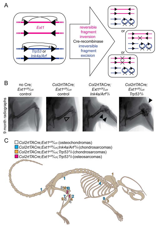
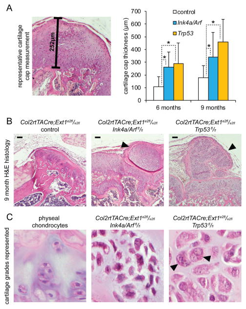
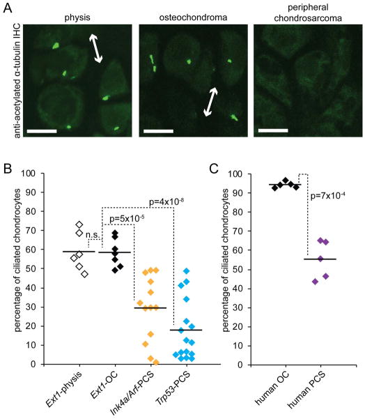
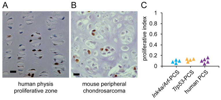
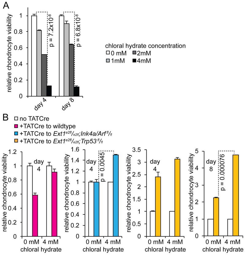
Peripheral chondrosarcoma (PCS) develops as malignant transformation of an osteochondroma, a benign cartilaginous outgrowth at the bone surface. Its invasive, lobular growth despite low-grade histology suggests a loss of chondrocyte polarity. The known genetics of osteochondromagenesis include mosaic loss of EXT1 or EXT2 in both hereditary and non-hereditary cases. The most frequent genetic aberrations in human PCS also include disruptions of CDKN2A or TP53. In order to test the sufficiency of either of these to drive progression of an osteochondroma to PCS, we added conditional loss of Trp53 or Ink4a/Arf in an Ext1-driven mouse model of osteochondromagenesis. Each additional tumour suppressor silencing efficiently drove the development of growths that mimic human PCS. As in humans, lobules developed from both Ext1-null and Ext1-functional clones within osteochondromas. Assessment of their orientation revealed an absence of primary cilia in the majority of mouse PCS chondrocytes, which was corroborated in human PCSs. Loss of primary cilia may be responsible for the lost polarity phenotype ascribed to PCS. Cilia deficiency blocks proliferation in physeal chondrocytes, but cell cycle deregulation is sufficient to rescue chondrocyte proliferation following deciliation. This provides a basis of selective pressure for the frequent cell-cycle regulator silencing observed in peripheral chondrosarcomagenesis. Mosaic loss of Ext1 combined with loss of cell cycle regulators promotes peripheral chondrosarcomagenesis in the mouse and reveals deficient ciliogenesis in both the model and the human disease, explaining biological behaviour including lobular and invasive growth.

Keywords: chondrosarcoma; mouse genetic model; osteochondroma; primary cilium; tumour suppressor.


Conflict of interest statement

Conflict of Interest: The authors declare no conflicts of interest related to this work.

Figures

Figure 1
Figure 1. Modelling peripheral chondrosarcoma in the mouse
(A) Schematic demonstrates the alternative allele design of Ext1e2fl, which results in reversible inversion, and therefore only mosaic loss of Ext1 following Cre-mediated recombination that consistently disrupts both alleles of either Trp53fl or Ink4a/Arffl. This produces chondrocytes lacking the cell cycle regulator and either lacking or retaining Ext1. (B) Radiographs from control and experimental mice demonstrate osteochondroma (open arrow) formation in mice with mosaic loss of Ext1 alone, but formation of larger, more dysmorphic lesions with the addition of cell cycle deregulation (black arrows). (C) Schematic depicting the prevalence and anatomic distribution of osteoschondromas in control mice with only mosaic loss of Ext1 and more aggressive cartilaginous surface lesions in the combination genotype groups.
Figure 2
Figure 2. Mouse tumours match criteria for human peripheral chondrosarcoma
(A) linear measurement of the thickness of cartilaginous caps of the skeletal surface lesions in mice with mosaic loss of Ext1 in chondrocytes either alone or in combination with Ink4a/Arf or Trp53 finds the latter two groups to be increased (n ≥ 5 mice for each assessment, * indicates t-test p-value < 0.0062). (B) Photomicrographs demonstrate the thin cartilaginous cap of a 9 month control osteochondroma, as well as lobules of cartilage (black arrows) extending beyond the thickened cap in both experimental models. (C) Higher power photomicrographs demonstrate the small chondrocytes with tight chromatin in the 9 month physis, while the transformed chondrocytes of peripheral chondrosarcoma have larger cells, more open and coarse chromatin patterns, and even binucleated cells (black arrows). (Magnification bars in B and panel widths in C are each 50μm.)
Figure 3
Figure 3. Both Ext1-functional and Ext1-null chondrocytes can transform
Photomicrographs of immunohistochemistry using the 10E4 antibody against heparan sulphate demonstrate a mix of cells with retained or lost Ext1 function in an osteochondroma (A), but lobules with clonal retention (B) or loss (C) in mouse PCSs (Magnification bars are each 20μm).
Figure 4
Figure 4. Peripheral chondrosarcomas exhibit deficient ciliagenesis
(A) Photomicrographs of immunofluorescence against acetylated alpha-tubulin in a mouse physis, an osteochondroma, and a peripheral chondrosarcoma lobule, each at 9 months, demonstrate misorientation of primary cilia in the osteochondroma compared to the axis of growth (double-headed arrows) and loss of cilia in the peripheral chondrosarcoma lobule. Charts present the percentage of chondrocytes with primary cilia in each group of mice (B) and in human (C) osteochondroma (OC) and peripheral chondrosarcoma (PCS) samples (bars denote mean for each group; magnification bars are each 10μm).
Figure 5
Figure 5. Peripheral chondrosarcomas maintain a relatively low proliferative index
Photomicrographs demonstrating immunohistochemistry against Ki67 to mark proliferating cells in human physis (A) and a mouse PCS nodule (B). (C) Chart presenting the fraction of cells in each specimen staining positively for Ki67. (Magnification bars are each 10μm.)
Figure 6
Figure 6. Cell cycle deregulation rescues the block in chondrocyte proliferation from loss of cilia
(A) Chart presenting relative cell viability of mouse chondrocytes cultured in the presence of increasing concentrations of chloral hydrate. (B) Charts demonstrating that chloral hydrate-blocked proliferation is partially rescued in chondrocytes that have lost Ext1 in mosaic fashion with either Trp53 or Ink4a/Arf. (Each bar presents the mean of 3 replicates with the error bar denoting the standard deviation.)

References

    1. Giuffrida AY, Burgueno JE, Koniaris LG, et al. Chondrosarcoma in the United States (1973 to 2003): an analysis of 2890 cases from the SEER database. J Bone Joint Surg Am. 2009;91:1063–1072. - PubMed
    1. Bovee JV, Cleton-Jansen AM, Kuipers-Dijkshoorn NJ, et al. Loss of heterozygosity and DNA ploidy point to a diverging genetic mechanism in the origin of peripheral and central chondrosarcoma. Genes Chromosomes Cancer. 1999;26:237–246. - PubMed
    1. Lin PP, Moussallem CD, Deavers MT. Secondary chondrosarcoma. J Am Acad Orthop Surg. 2010;18:608–615. - PubMed
    1. Ahmed AR, Tan TS, Unni KK, et al. Secondary chondrosarcoma in osteochondroma: report of 107 patients. Clin Orthop Relat Res. 2003:193–206. - PubMed
    1. Fletcher CDM World Health Organization. WHO classification of tumours of soft tissue and bone. 4. IARC Press; Lyon: 2013. International Agency for Research on Cancer.

Publication types

MeSH terms