. 2015 Feb 3:12:23.
doi: 10.1186/s12974-015-0248-1.

Anti-inflammatory activity of Wnt signaling in enteric nervous system: in vitro preliminary evidences in rat primary cultures

Affiliations

Anti-inflammatory activity of Wnt signaling in enteric nervous system: in vitro preliminary evidences in rat primary cultures

Rosa Di Liddo et al. J Neuroinflammation. .

Abstract

Background: In the last years, Wnt signaling was demonstrated to regulate inflammatory processes. In particular, an increased expression of Wnts and Frizzled receptors was reported in inflammatory bowel disease (IBD) and ulcerative colitis to exert both anti- and pro-inflammatory functions regulating the intestinal activated nuclear factor κB (NF-кB), TNFa release, and IL10 expression.

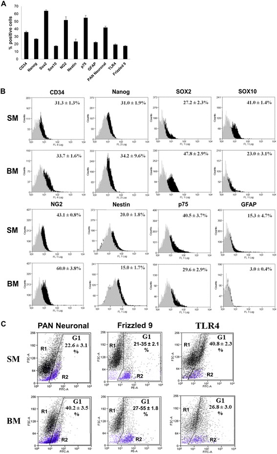
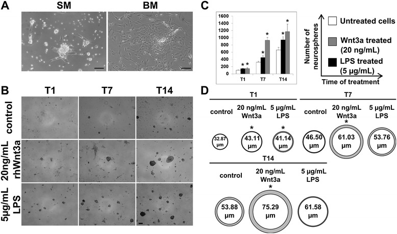
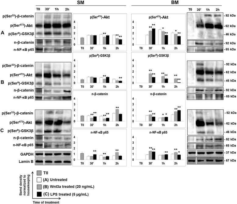
Methods: To investigate the role of Wnt pathway in the response of the enteric nervous system (ENS) to inflammation, neurons and glial cells from rat myenteric plexus were treated with exogenous Wnt3a and/or LPS with or without supporting neurotrophic factors such as basic fibroblast growth factor (bFGF), epithelial growth factor (EGF), and glial cell-derived neurotrophic factor (GDNF). The immunophenotypical characterization by flow cytometry and the protein and gene expression analysis by qPCR and Western blotting were carried out.

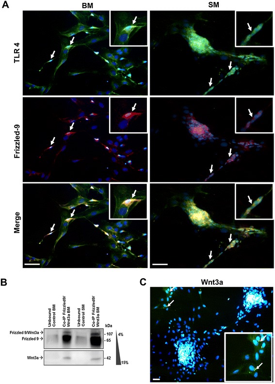
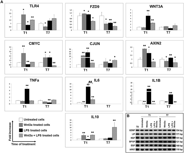
Results: Flow cytometry and immunofluorescence staining evidenced that enteric neurons coexpressed Frizzled 9 and toll-like receptor 4 (TLR4) while glial cells were immunoreactive to TLR4 and Wnt3a suggesting that canonical Wnt signaling is active in ENS. Under in vitro LPS treatment, Western blot analysis demonstrated an active cross talk between canonical Wnt signaling and NF-кB pathway that is essential to negatively control enteric neuronal response to inflammatory stimuli. Upon costimulation with LPS and Wnt3a, a significant anti-inflammatory activity was detected by RT-PCR based on an increased IL10 expression and a downregulation of pro-inflammatory cytokines TNFa, IL1B, and interleukin 6 (IL6). When the availability of neurotrophic factors in ENS cultures was abolished, a changed cell reactivity by Wnt signaling was observed at basal conditions and after LPS treatment.

Conclusions: The results of this study suggested the existence of neuronal surveillance through FZD9 and Wnt3a in enteric myenteric plexus. Moreover, experimental evidences were provided to clarify the correlation among soluble trophic factors, Wnt signaling, and anti-inflammatory protection of ENS.


Figures

Figure 1
Figure 1
FCM analysis of ENSc at T0 and T7. (A) As reported by FCM analysis, freshly isolated ENSc (T0) resulted as a heterogeneous population including stem-like, progenitor, glial, and neuronal cells. For each experimental condition, 104 cells were used for acquisition by FACSCanto II and data were expressed as percentage (%) of positive cells ± standard deviation (SD) where each marker was compared to control samples stained only with isotype or secondary control antibody. The positive expression was quantified using the overton subtraction tool of Summit 4.3 software (Beckman Coulter Inc). Under SM and BM culture conditions, the aspecific modulation of glial and neuronal differentiation was detected. (B) Immunophenotypical characterization by FCM of ENSc cultured for 7 days (T7). Data were expressed as percentage (%) of positives (black profile) ± SD for each marker compared to corresponding staining control (gray profile). (C) FCM analysis of PAN neuronal, Frizzled 9, and TLR4 expression. Data were reported as FSC vs fluorescent marker dot plot where R1 (blue colored) and R2 (black colored) subsets were defined. The positive expression of each target marker ± SD was discriminated in gate G1 defined with respect to staining control.
Figure 2
Figure 2
IF analysis of TLR4 and Frizzled 9. (A) Confocal microscopy analysis evidenced the coexpression of TLR4 and Frizzled 9 on neuronal cells (arrows) either in SM- than BM-treated samples. (B) Co-immunoprecipitation was performed on total protein extracts from ENSc cultured for 7 days in SM or BM. Immunoaffinity purification was carried out using goat anti-rat Frizzled 9 and rabbit anti-rat Wnt3a antibodies pre-immobilized onto Protein A Sepharose. Western blot analysis was assessed using 4%–15% gradient precast polyacrylamide gel. (C) By immunofluorescence assay, ENSc showed cytoplasmic vesicles immunoreactive (arrows) to rabbit anti-rat Wnt3a antibody. Bar: 15 μm.
Figure 3
Figure 3
ENSc and formation of neurospheres. (A) After 1 day of culture, standard medium promoted the formation of neurospheres as evidenced by optical microscopy analysis. In contrast, several fibroblastoid colonies were observed in ENS cultures maintained in BM ENS medium. Bar: 100 μm. (B) Quantitative analysis of number and diameter of neurospheres obtained by LPS (5 μg/mL) or Wnt3a (20 ng/mL) stimulation. In parallel, the analysis was performed on LPS- and Wnt3a-untreated samples that were used as control. Light microscopic pictures reflected the increased number of neurospheres treated with LPS and Wnt3a after 1 (T1), 7 (T7), and 14 (T14) days in comparison to untreated cultures (control). (C) Data were quantified and normalized to controls (100%) (n = 3). (D) Diameter of neurospheres at T1, T7, and T14 was measured (n = 150) using ImageJ software. Statistical significance compared to control; p value ≤0.05*. Bar: 100 μm.
Figure 4
Figure 4
WB analysis of Wnt/NF-κB interplay. A cross talk between TLR4 and Wnt signaling occurred as demonstrated by WB analysis at T0, T30′, T1h, and T2h using specific antibodies for cytoplasmic p(Ser473)-Akt, p(Ser9)-GSK3β, p(Ser33)-β-catenin and nuclear β-catenin (n-β-catenin), and NF-κB p65 (n NF-κB p65) in unstimulated cells; (A) samples treated with Wnt3a (20 ng/mL) (B) and LPS (5 μg/mL) (C) in SM and BM medium. GAPDH and lamin B were considered as housekeeping proteins for the analysis of cytoplasmic and nuclear antigens. The quantification of protein expression levels was performed using ImageJ processing software. Data were reported as ratio within target protein and relative housekeeping protein expression. Statistical significance was calculated by Student’s t-test by comparing to control: p value ≤0.05*, p value ≤0.01**.
Figure 5
Figure 5
WB and Co-IP of β-catenin and p65. (A) Wnt3a interfered with NF-κB p50/p65-mediated inflammatory response as suggested by WB analysis of untreated Wnt3a (20 ng/mL) and/or LPS (5 μg/mL)-treated cells at T0, T30′, T1h, and T2h. The quantification of protein expression levels was performed using the image processing software ImageJ. Data were reported as ratio within target protein and relative lamin B housekeeping protein expression. Statistical significance was calculated by Student’s t-test comparing to control: p value ≤0.05*, p value ≤0.01**. (B) Co-IP assay confirmed a nuclear interplay between Wnt signaling and LPS NF-кB pathway, as previously reported [67]. Immunoaffinity purification was carried out using mouse anti-rat NF-κB p65 and mouse anti rat β-catenin antibodies pre-immobilized onto Protein A Sepharose.
Figure 6
Figure 6
Gene expression study on ENSc stimulated with Wnt3a and LPS. (A) The anti-inflammatory effect was demonstrated to be exerted by Wnt3a using qPCR analysis and ENS cells treated with Wnt3a (20 ng/mL) and LPS (5 μg/mL). The analysis was focused on the expression of β-catenin target genes (AXIN2, CMYC, and CJUN), membrane receptors (TLR4 and FZD9), Wnt3a ligand, and pro- and anti-inflammatory target genes (IL1B, IL6, TNFa, and IL10). The amount of gene products was calculated using linear regression analysis from standard curves, demonstrating the amplification efficiencies ranging from 90% to 100%. Data were reported as a fold increase of gene expression that is defined as the cDNA ratio between target gene and reference gene (HPRT) normalized to untreated sample. Statistical significance was calculated using Student’s t-test, comparing to untreated cells: p value ≤0.05*, p value ≤0.01**; samples compared to LPS-treated cells: p value ≤0.01 (two black triangles). (B) To detect the gene expression of typical ENS growth factors (GDNF, EGF, BFGF, NGF, and LIF), RT-PCR was performed using Qiagen One Step RT-PCR Kit on samples untreated (T0) and stimulated with Wnt3a (20 ng/mL) and LPS (5 μg/mL) for 1 (T1) and 7 (T7) days. RT-PCR products were electrophoresed on 2% agarose gel and stained by GelRed™. HPRT was used as housekeeping gene.
Figure 7
Figure 7
The interplay model of Wnt/β-catenin, LPS/TLR4, and growth factors in ENS. After the interaction of Wnt ligands with a G protein-coupled receptor (Frizzled), GSK3β is inhibited and β-catenin is released from a “scaffolding” complex consisting of Axin, adenomatous polyposis coli (APC), casein kinase 1 (CK1), and glycogen synthase kinase 3β (GSK3β). Consequently, the stabilized β-catenin shuttles to the nucleus and the transcription process of its target genes is promoted. LPS-mediated activation of TLR4 pathway or growth factor signaling involves phosphatidylinositol 3 kinase (PI3K) pathway. PI3K inactivates GSK3β through Akt, and nuclear accumulation of β-catenin occurs. In steady conditions, β-catenin is trapped at the plasma membrane by cadherins or is tagged by phosphorylation for ubiquitin-mediated degradation. In parallel, PI3K and TRAF6 inhibit IκBα and the activated heterodimer NF-κB p50/p65 translocates to the nucleus for promoting the specific gene expression.

References

    1. De Santa Barbara P, Van den Brink GR, Roberts DJ. Development and differentiation of the intestinal epithelium. Cell Mol Life Sci. 2003;60(7):1322–32. - PMC - PubMed
    1. Stuhlmiller TJ, García Castro MI. Current perspectives of the signalling pathways directing neural crest induction. Cell Mol Life Sci. 2012;69(22):3715–37. - PMC - PubMed
    1. Gregorieff A, Pinto D, Begthel H, Destrée O, Kielman M, Clevers H. Expression pattern of Wnt signaling components in the adult intestine. Gastroenterology. 2005;129(2):626–38. - PubMed
    1. Wodarz A, Nusse R. Mechanisms of Wnt signalling in development. Annu Rev Cell Dev Biol. 1998;14:59–88. - PubMed
    1. Bhanot P, Brink M, Samos CH, Hsieh JC, Wang Y, Macke JP, et al. A new member of the frizzled family from Drosophila functions as a wingless receptor. Nature. 1996;382(6588):225–30. - PubMed

Publication types

MeSH terms

LinkOut - more resources